 CrossRef
 CrossMark
 Mendeley
 Scholar
Introduction
Caseous Lymphadenitis (CL) is a chronic infectious and contagious disease that affects various hosts, being particularly relevant for sheep and goat farming and veterinary medicine [1]. It is characterized by the formation of abscesses in cutaneous or visceral lymph nodes, with manifestations in lymph nodes, liver, kidney, and lungs, among other organs [2]. Additionally, atypical clinical presentations have been described, including neonatal toxemia, arthrosynovitis, endometritis, epididymitis, mastitis, and orchitis [1].
CL has been reported and recognized as a cause of economic losses, reflected in decreased wool, meat, and milk production, reproductive disorders, and condemnation of carcasses and viscera [1,4,5].
Its etiological agent is Corynebacterium pseudotuberculosis, which affects sheep and goats (biovar ovis), as well as horses, camels, and buffaloes (biovar equi), depending on the biovar. It can also infect humans, causing various forms of lymphadenitis [6-8]. CL has been classified as an emerging occupational zoonosis by many authors, and its true prevalence is likely underestimated, as it may not be diagnosed early and can present subclinically or asymptomatically. Furthermore, it is not a notifiable disease in sheep farming, and the highest-risk group consists mainly of male rural workers [9].
C. pseudotuberculosis is a pleomorphic bacillus with a coccobacillary shape and palisade arrangement. It is non-motile, non-sporulating, non-encapsulated, facultatively anaerobic, and grows on enriched culture media at 37 °C and pH 7-7.2. [10-12].
This microorganism produces several virulence factors, among which three are particularly important: its cell wall structure (mycolic acids), its intracellular persistence within macrophages, and the production of phospholipase D (PLD) as an exotoxin. Other factors that may contribute to its pathogenicity have also been described, including adherence factors, biofilm formation, and production of extracellular enzymes [3,4].
In the Chilean Andes, cases of CL have been reported in Hippocamelus bisulcus, commonly known as the huemul, an endangered species whose abscess management and treatment must be carried out under strict protocols. In the Argentine Andes, possible cases of CL in the same species have been detected through direct observation (Figure 1).
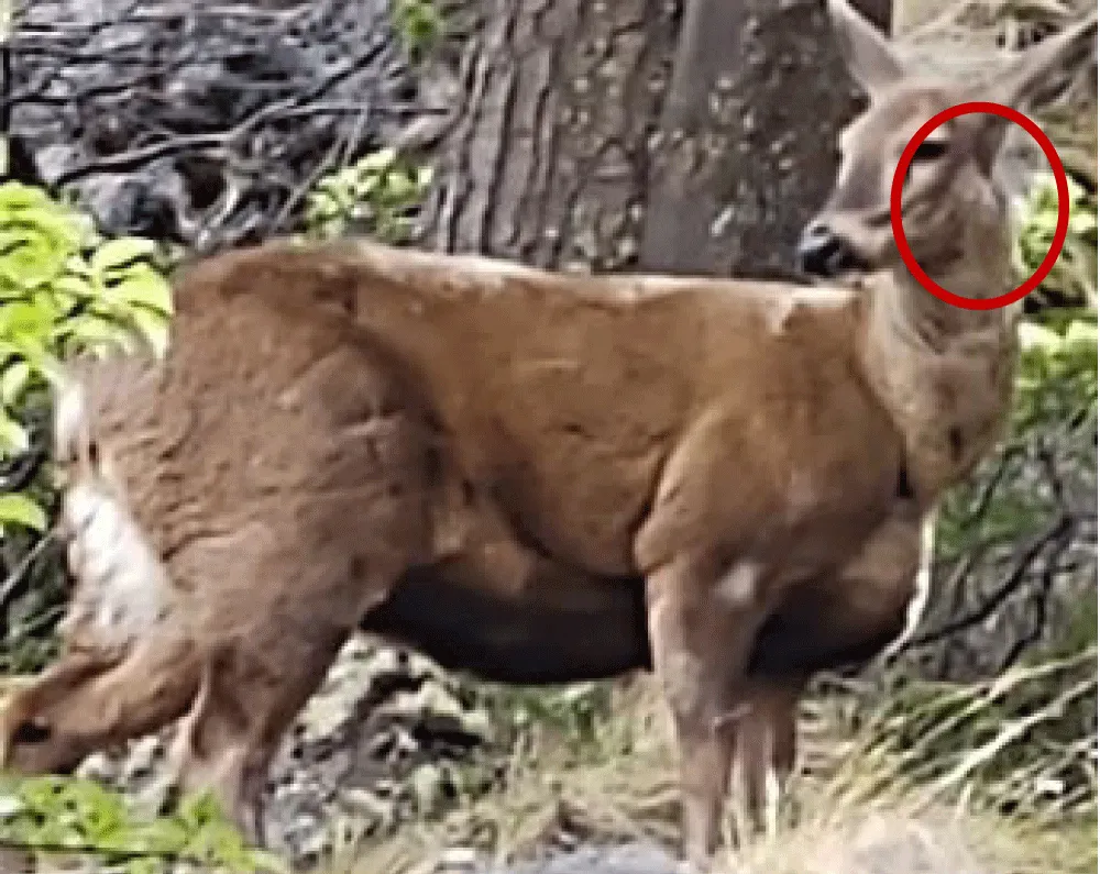
This environmental persistence becomes particularly concerning at the livestock–wildlife interface. Reports of suspected cases in the huemul (Hippocamelus bisulcus) in Argentina warrant a more rigorous evaluation of the available evidence. To strengthen the epidemiological status of this species, it is imperative to categorize findings according to their level of confirmation: from clinical observations of regional lymphadenopathy in live individuals (e.g., photographic documentation or ranger sightings) to necropsy-based diagnoses demonstrating caseous pyogranulomas. In the absence of confirmation through microbiological culture or molecular techniques (PCR) capable of identifying the causative strain, the significance of caseous lymphadenitis for huemul conservation remains within the realm of clinical suspicion, underscoring the urgent need for standardized health monitoring protocols and epidemiological surveillance in protected areas where domestic livestock and native wildlife coexist.
Antibiotic treatment is challenging because encapsulated lesions hinder drug penetration, making therapies often prolonged and ineffective, frequently leading to the culling of animals to prevent disease spread. In the case of cutaneous CL, a post-surgical wound treatment following lymph node excision using a cream based on biogenic silver nanoparticles has been developed, which not only accelerates wound healing but also prevents disease recurrence without causing side effects in treated animals [16,17].
In the Argentine Patagonian region, this disease is highly prevalent, with significant sanitary and economic impacts on sheep farming, substantially affecting both domestic and international markets [18]. Moreover, it has begun to spread to other wildlife species.
Materials and Methods
The study was conducted through random sampling across farms located at the following geographic coordinates: Astra (−45.734244, −67.491368), Puerto Madryn (−42.768480, −65.038270), Comodoro Rivadavia (−45.859400, −67.471400), Puerto Deseado (−47.750000, −65.916700), Sierra Chaira (−45.091350, −68.338640), and Esquel (−42.909700, −71.310600). A total of 1,832 animals were examined, comprising a systematically selected cohort designed to represent the productive variability of the Patagonian region. Clinical inspection focused on the identification of lesions consistent with Caseous Lymphadenitis (CL), including the assessment of superficial lymphadenomegaly, encapsulated abscesses, and scarring in key lymph nodes (parotid, submandibular, and precapsular). To ensure reproducibility, diagnostic criteria based on lesion consistency and anatomical location, as described by Estevao, et al. [18] were applied to differentiate these findings from other nonspecific lymphadenopathies. Lesions consistent with caseous lymphadenitis were detected at all sites of origin of the sampled animals, suggesting widespread endemicity across the region and thereby ruling out the hypothesis of isolated focal outbreaks.
A total of 224 samples were collected from lesions compatible with CL from these locations. Samples were isolated on Tryptic Soy Agar (TSA) supplemented with 5% sheep blood and incubated at 37 °C for 48 h.
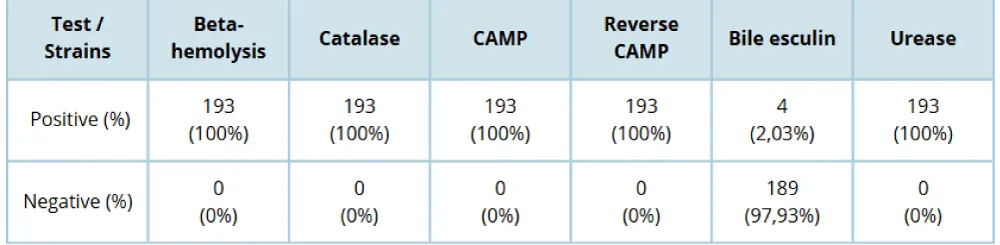
From these cultures, microbiological identification of the isolates was performed using light microscopy with Gram staining and the following biochemical tests for preliminary identification: β-hemolysis, catalase, urea, nitrate reduction, bile esculin, CAMP, and reverse CAMP tests, with incubation for 48 h at 37°C (Table 1).
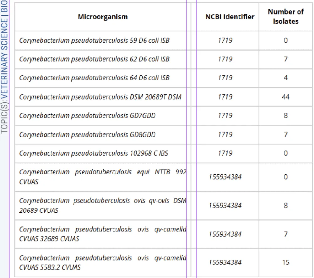
Each microbiologically identified strain was confirmed using a MALDI-TOF Biotyper Sirius system. Analyses were performed in duplicate, taking a single colony, homogenizing it with formic acid, and subsequently applying a matrix. The identified strains were cross-referenced against two complementary databases. The Bruker database provided the following strains: Corynebacterium pseudotuberculosis 59 D6 coll ISB, 62 D6 coll ISB, 64 D6 coll ISB, DSM 20689T DSM, GD7GDD, GD8GDD, and 102968 C IBS. The CVUA-BW database (Food Chemistry and Veterinary Analysis Agency) included: Corynebacterium pseudotuberculosis equi NTTB 992 CVUAS, C. pseudotuberculosis ovis qv-ovis DSM 20689 CVUAS, C. pseudotuberculosis ovis qv-camelid CVUAS 32689 CVUAS, and C. pseudotuberculosis ovis qv-camelid CVUAS 5583.2 CVUAS.
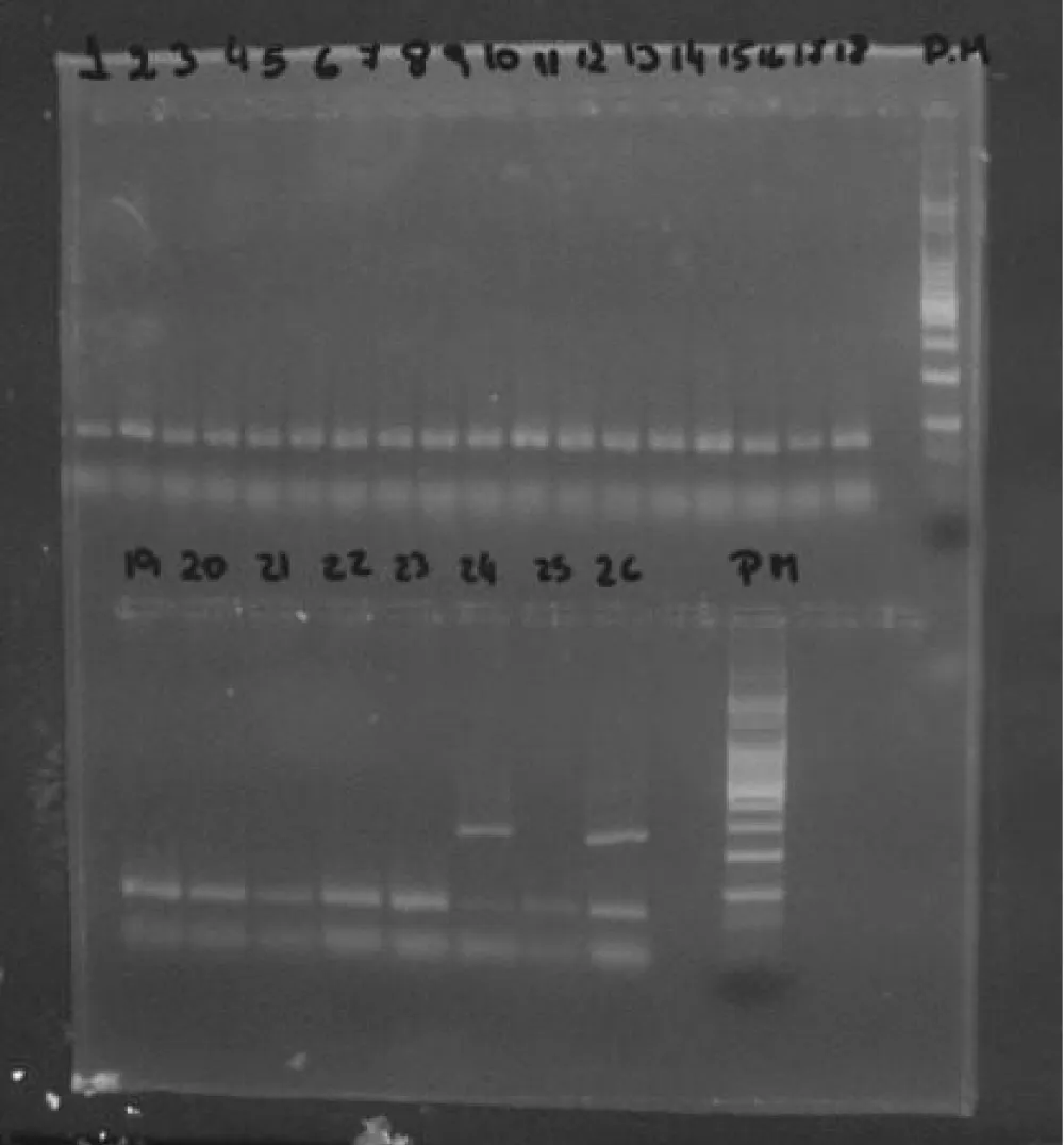
Additionally, the strains were subjected to PCR detection of the diphtheria toxin gene. A bacterial suspension was prepared in mQ water, DNA was extracted by heating at 101°C for 15 minutes, centrifuged at 13,000 rpm for 5 minutes, and the supernatant was collected for the PCR reaction. The forward (F) and reverse (R) primers used generate an amplification product of 248 bp corresponding to: F: 5´-ATCCACTTTTAGTGCGAGAACCTTCGTCA-3´ R: 5´-GAAAACTTTTCTTCGTACCACGGGACTAA-3´
The PCR protocol followed procedures established by the INEI-ANLIS Carlos G. Malbrán Institute, using as positive controls DNA from C. diphtheriae 510 NCTC 10648 and 512 NCTC 3984, and as a negative control DNA from C. diphtheriae 511 NCTC 10356. Amplification products were visualized on 2% agarose gel (UltraPure™ INVITROGEN) supplemented with GelRed dye (1000×, Biotium).
Results
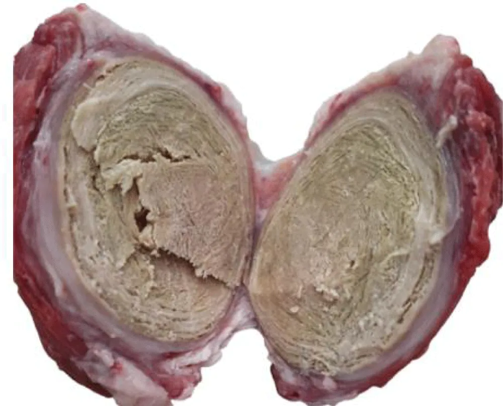
Through visual inspection and palpation, lesions compatible with CL were identified (Figure 2). The number of cases recorded per location was as follows: Astra (n = 46), Puerto Madryn (n = 17), Comodoro Rivadavia (n = 48), Puerto Deseado (n = 65), Sierra Chaira (n = 8), and Esquel (n = 40), totaling 224 samples.
Optical microscopy revealed the presence of Gram-positive coccobacilli with a palisade arrangement, characteristic of C. pseudotuberculosis, in 193 (86%) of the analyzed samples. Additionally, biochemical tests yielded results that allowed the identification of this microorganism (Table 1).
Of the 193 biochemically identified strains, 100 (52%) were analyzed at the ANLIS Malbrán Institute for confirmatory identification using the MALDI-TOF method (Matrix-Assisted Laser Desorption/Ionization – Time of Flight). The results confirmed C. pseudotuberculosis as the sole microorganism in these isolates, based on comparison with the available databases (Table 2).
The 100 strains confirmed by MALDI-TOF were subjected to screening for the diphtheria toxin gene. All isolates (100 %) tested negative for this gene when compared with the corresponding positive and negative controls (Figure 3). Although C. pseudotuberculosis can harbor and express this gene within its genome, such occurrence is rare. Nevertheless, its potentially toxigenic capacity underscores the importance of screening for this determinant, given its zoonotic potential in the Patagonian region.
Conclusion
This study not only confirms the persistence of caseous lymphadenitis in Patagonia but also reshapes the current understanding of its epidemiology by demonstrating widespread endemicity rather than isolated outbreaks. These findings substantially update previous prevalence estimates and highlight the significant latent economic impact on regional livestock production, arising not only from productivity losses and carcass condemnation but also from the chronic deterioration of animal welfare in extensive systems.
A critical aspect emerging from this study is the identification of infectious foci in areas adjacent to the habitat of the huemul, which raises concern regarding transmission risk at the domestic–wildlife interface and the potential threat to the survival of this critically endangered species. For the proposed One Health approach to be effective, it must move beyond a theoretical framework through the implementation of specific measures, such as the development of standardized diagnostic protocols for both domestic and wild species and the establishment of integrated surveillance systems involving coordinated sampling in shared grazing areas.
Ultimately, this work provides a foundation for health management strategies aimed at mitigating losses in the productive sector while safeguarding the sanitary integrity of Patagonian biodiversity.
Acknowledgement
The author would like to thank the Special Bacteriology and Clinical Bacteriology Services at the ANLIS Malbrán Institute for their collaboration and guidance during the author’s training period, during which instruction was provided on the use of the MALDI-TOF system and work was conducted using PCR techniques.