 CrossRef
 CrossMark
 Mendeley
 Scholar
Introduction
Chondrolipoma of the breast is a benign lipoma-like tumor formed with mature adipose tissue and islands of hyaline cartilage, although its histogenesis remains vague and indistinct [1,2]. It is an exceptionally rare tumor with a modest potential for misdiagnosis. Its clinical presentation is like that of a fibro adenoma but on mammography it may show calcifications favouring carcinoma [3]. Thus, even with triple assessment the major bulk of diagnosis relies on the histopathological examination. In concordance with the rarity of this tumor, its exact incidence worldwide is difficult to determine but as per literature only a few such cases have been reported so far, with numerous histological variations [4].
Case Presentation
A middle-aged postmenopausal female presented to the breast clinic in our tertiary care centre with a left breast lump for two years. She had been diagnosed with hypothyroidism five years ago and was on medication since then. The lump was insidious in onset and gradually progressive in size. She had no history of fever, trauma, pain, skin changes or nipple discharge. There was no significant obstetric or family history. On examination, the lump was 6 cm x 5 cm in size, mobile, nontender, with an irregular surface, involving majorly the upper inner quadrant. The clinical impression was phyllodes tumor.
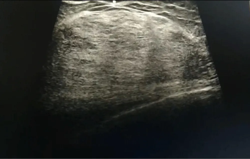
An ultrasound of both breasts with axilla was done which showed a 6.9 cm x 8 cm X 6.8 cm hyperechoic well defined smooth lump at 12 o’clock position in left breast, 1cm from the skin and 1.2 mm from NAC (Nipple Areolar Complex), with no infiltration of the pectoralis muscle. Few sub centimetric reactive lymph nodes were seen in both the axillae with maximum SAD (Short Axis Diameter) 5 mm. This was suggestive of a BIRADS 2 (Breast Imaging Reporting and Data System) lesion.
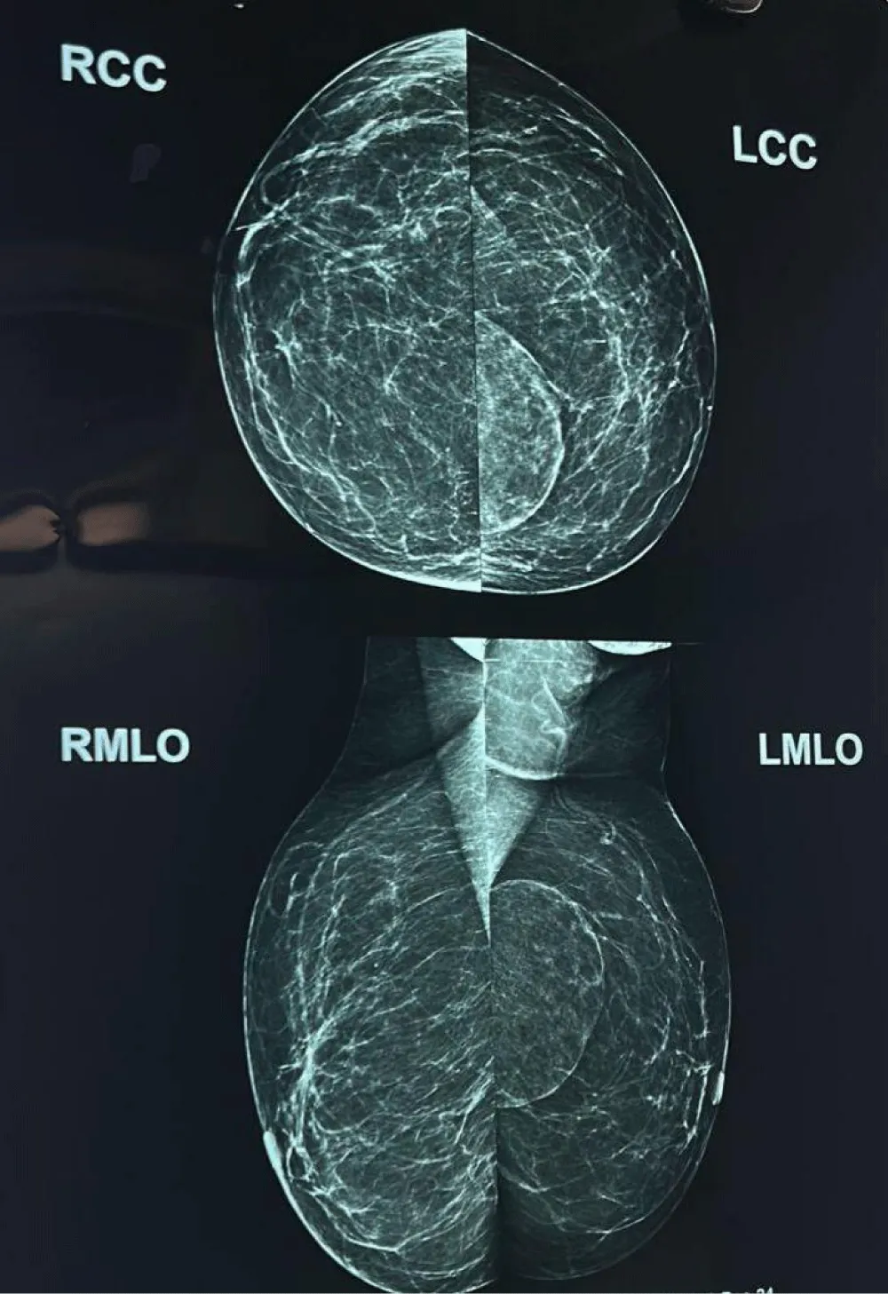
On Mammography a well-defined, large, rounded radio-opacity surrounded by a thin capsule in the upper inner quadrant of the left breast was seen, suggestive of a BIRADS 4A lesion, with a typical breast in a breast appearance.
Based on the clinical examination and radiological investigations, the differential diagnoses given for this lump were:
- Hamartoma,
- Atypical phyllodes tumor, and
- Low-grade mucinous carcinoma of the breast.
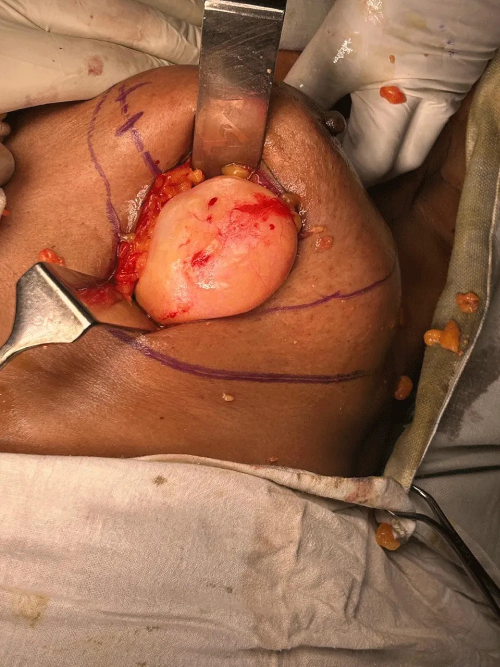
The yield of core biopsy was poor. Thus, surgical excision of the mass was planned. The patient underwent a wide local excision of the left breast lump. The Histopathology report of the excision biopsy depicted features of a hamartomatous proliferation favoring benign chondrolipoma of the left breast, measuring 6.2 cm x 5.3 cm x 2.1 cm in size and weighed around 600 gm.
Discussion
There are two common types of hamartomas of breast: chondrolipomas and adenolipomas, with chondrolipomas being a rarer entity in the lipomatous breast tissue [3]. As per Agarwal P, et al. [10] only nine such cases have been reported till date including their case repot. This could be the tenth one.
Chondrolipomas are frequently encountered in young females, mostly in their third decade of life and are benign [6]. Agarwal P, et al. [10] has quoted that chondrolipomas can exist in females across a wide age range from 14 to 70 years, with a median age of 35 years, supporting the fact that it is not uncommon in a middle-aged post-menopausal female.
Regarding correlation with thyroid disorders, although there is no direct association between the two, but some studies suggest that there might be an increased risk of developing benign breast diseases in a patient with hypothyroidism [11,12].
Chondrolipoma is a cartilaginous metaplasia. Appearance of cartilage is common in malignant neoplasms of breast but quite uncommon in benign breast tumors [5]. The pathophysiology of chondrolipomas is not very clear but it is said to be originating from mesenchymal tissues within the breast.
Giant chondrolipomas are those that measure more than 5cm in any one dimension and weigh more than 500 gm [6,7]. It is characterized by the presence of peripheral compressed mammary parenchyma which gets transformed into a true capsule for the tumor and, on the other hand, ductal structures and mammary stroma may not be present at the tumor level [4,6,8].
A chondrolipoma, belonging to the breast hamartoma family, presents as a well-circumscribed lesion with varying echogenicity as compared to the surrounding fat, on ultrasound imaging. A ‘Breast within the breast’ sign, also called a ‘cut sausage appearance’ in a mammogram may be noted in such well circumscribed masses having fat and soft tissue density surrounded by a capsule, as was seen in our case [14]. This typical sign is more commonly seen in fibroadenolipomas as compared to chondrolipomas [13].
Management of such giant chondrolipomas of breast involve complete surgical excision [6,9]. After confirming the benign nature of the lump via histopathological examination, a simple enucleation may serve the purpose. In case of a giant chondrolipoma of the breast, this process may end up being a total mastectomy [6]. In our case, a wide local excision was preferred as the benign nature of the lesion was not well established before the surgery.
The histopathological features of chondrolipomas include the presence of mature adipocytes and lobules of hyaline cartilage within a fibrous capsule. In a study conducted by Shintaku M, et al. [1] they found that the chondrocytes within the chondrolipomas were positive for ER and PgR (Estrogen and Progesterone Receptors) indicating an origin from the metaplasia of hormone-sensitive spindle cells, which also readily express the receptors. The concept of receptor positivity is not very significant in these cases as much as it in breast cancer patients.
Even with high sensitivity and specificity of triple assessment for breast lumps, making a diagnosis of chondrolipoma relies greatly on the histopathological analysis [10]. Owing to its rarity, chondrolipomas commonly pose a diagnostic challenge as they mimic other benign and malignant breast diseases. In our case report, clinically the left breast lump gave the impression of a phyllodes tumor and on mammography a BIRADS 4A lesion, tilting more towards a malignant disease. Misdiagnosis may lead to unnecessary surgery and overtreatment and potential complications thereafter [6] (Figures 1-3).
Conclusion
Chondrolipomas in the breast are rare. The benign nature of these lesions and a simple non-radical excision of the mass as part of its management is well established. The need for familiarity with this entity is emphasized by the fact that it is frequently misdiagnosed. Accurate diagnosis with a high level of suspicion to consider it as a differential is required for appropriate management of breast masses and to avoid needless surgical complications.