 CrossRef
 CrossMark
 Mendeley
 Scholar
Introduction
The innate and adaptive immune systems function in a coordinated manner r to protect the host from pathogenic invasion and maintain immunological homeostasis [1]. The body's first line of defence is its innate immunity, which is also known as its inborn immunity. It turns on to fight infection as soon as possible, which helps keep the body healthy. It has cells like macrophages, NK cells, dendritic cells, and mast cells that find harmful pathogens and make the immune system work to get rid of them. This immune responses are transient, evidence suggests that innate immune cells can undergo epigenetic and metabolic reprogramming following exposure to specific stimuli, resulting in enhanced responsiveness upon subsequent challenge and this phenomenon is called trained innate immunity. Proinflammatory cytokines like IL-1β, which are regulated by inflammasome signaling pathways, plays the vital role in mediating responses in innate immunity. Pattern Recognition Receptors (PRRs) are present on various immune and non-immune cells detects pathogens by recognizing Pathogen-Associated Molecular Patterns (PAMPs). Certain Pattern Recognition Receptors (PRRs), such as nucleotide-binding oligomerization domain and Leucine-Rich Repeat (LRR)-containing receptors (NLRs) and AIM2-Like Receptors (ALRs), respond to infection by assembling a multiprotein cytosolic complex known as the inflammasome [2].
When PRRs find patterns in microbes, initiates the inflammasomes, which activates the inflammatory caspase-1, which is a cysteine protease. This also modulates proinflammatory cytokines like pro-interleukin-1 and pro-interleukin-18 into their biologically active forms, IL-1β and IL-18. In addition to cytokines maturation, inflammasomes activation induces pyroptosis, a form of inflammatory programmed cell death [2,3]. The NLRC4 inflammasome activation is a key component of this signalling pathway implicated in the pathogenesis of both communicable and non-communicable diseases. This review aims to provide comprehensive overview of the structural organisation, functional domains, and signalling pathways of NLRC4, with emphasis on its role in infectious and inflammatory disorders, such as Urinary Tract Infections (UTIs), Auto Inflammation with Infantile Enter Colitis (AIFEC), cancer, and Metabolic Dysfunction-Associated Steatotic Liver Disease (MASLD), as well as in host defense. In addition, current available genetic tools used to investigate NLRC4 function across diverse pathological conditions are discussed. Furthermore, the potential association of NLRC4 with trained innate immunity is also examined to highlight its potential emerging relevance in immune memory and disease modulation.
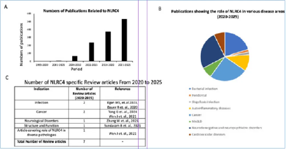
To contextualize the expanding research landscape surrounding NLRC4, a bibliometric overview of publications related to NLRC4 across different disease domains is presented in figure 1. The observed increase in NLRC4-focused studies over recent years highlights its emerging relevance across multiple pathological conditions. However, existing reviews remain largely confined to specific disease contexts, suggesting the need for a comprehensive understanding of the involvement of NLRC4 across a wide range of infectious and inflammatory disorders.
This review is unique in that it examines NLRC4 across multiple disease contexts while integrating its structural biology, activation mechanisms, and downstream signalling pathways in both infectious and non-infectious conditions. It also explores the emerging association between NLRC4 and trained innate immunity. To date, these aspects have not been comprehensively addressed together within a single publication.
Methodology
A thorough literature search was performed to identify relevant studies investigating the structural organization, activation mechanisms, signaling pathways, and disease-specific roles of the NLRC4 inflammasome. Electronic databases, such as PubMed, Scopus, and Web of Science, were systematically searched for peer-reviewed articles that were published between January 1995 and December 2025. The search strategy incorporated combinations of the following keywords and Medical Subject Headings (MeSH) terms: “NLRC4 inflammasome," “NAIP-NLRC4 signaling," “inflammasome activation," "pyroptosis," “auto inflammatory diseases," “trained innate immunity," “metabolic inflammation," "neuroinflammation," "cancer," "cardiovascular," and “host-pathogen interactions." Databases.
Structure and Components of the NLRC4 Inflammasome
Poyet and colleagues were the first to describe NLRC4 as a proapoptotic protein [6]. During their search for the structural equivalent of APAF1, scientists found a protein that can turn on caspase-1 (CASP1, also called IL-1 converting enzyme, ICE). This protein is called ICE-Protease-Activating Factor (IPAF). Later, it was discovered that its domain structure fit into the NLR protein family. Because it also has the amino-terminal CARD domain, it was given the name NLRC4 [7]. However, studies on NLRC4-Deficient Bone Marrow-Derived Macrophages (BMDMs) have shown that NLRC4 is unable to activate caspase-1 and induce pyroptosis after exposure to S. typhimurium. It was subsequently shown that flagellin from S. typhimurium and Legionella pneumonia can activate NLRC4, followed by activation of caspase-1. Collectively, these findings established NLRC4 as a critical cytosolic sensor of bacterial virulence-associated components [4].
NLRC4 has three domains: an N-terminal CARD, a middle nucleotide-binding domain called NACHT, and a C-terminal Leucine-Rich Repeat (LRR) domain [4]. A distinguishing structural feature of NLRC4 relative to other inflammasome sensors such as NLRP3 and AIM2 is the presence of an N-terminal Caspase Recruitment Domain (CARD). It is essential for downstream signaling and its interaction with other proteins, such as pro-caspase-1. The NACHT domain functions as the nucleotide-binding and oligomerization region required for ATP-dependent NLRC4 activation. The LRR domain, on the other hand, maintains protein dormancy until activation signals are detected. It is also involved in ligand recognition and autoinhibition [5] (Figure 2).
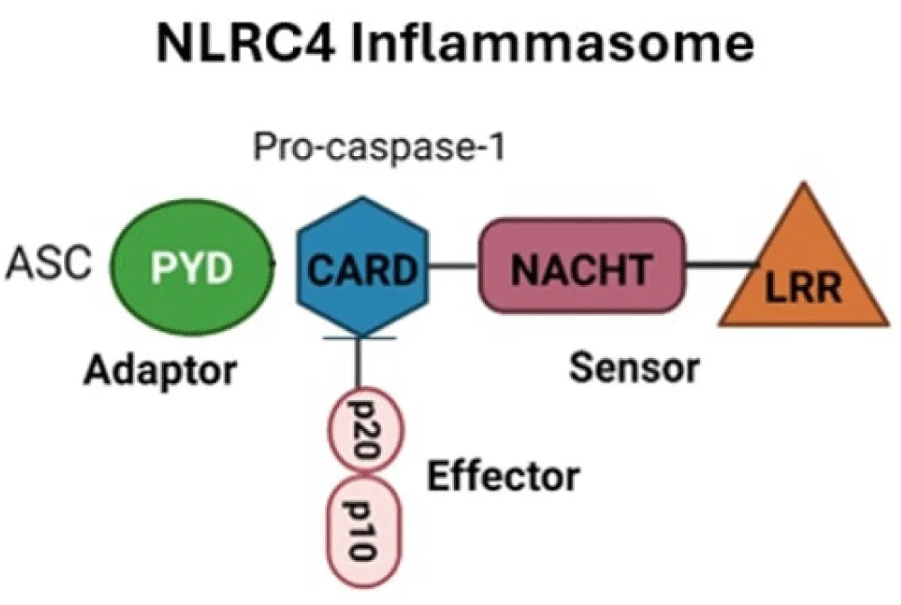
NLRC4 has three domains: an N-terminal CARD, a middle nucleotide-binding domain called NACHT, and a C-terminal Leucine-Rich Repeat (LRR) domain [4]. A distinguishing structural feature of NLRC4 relative to other inflammasome sensors such as NLRP3 and AIM2 is the presence of an N-terminal Caspase Recruitment Domain (CARD). It is essential for downstream signaling and its interaction with other proteins, such as pro-caspase-1. The NACHT domain functions as the nucleotide-binding and oligomerization region required for ATP-dependent NLRC4 activation. The LRR domain, on the other hand, maintains protein dormancy until activation signals are detected. It is also involved in ligand recognition and auto inhibition [5] (Figure 2).
Domain structure of NLRC4
NLRC4 consists of three principal domains: an N-terminal CARD domain, a central NACHT domain, and a C-terminal Leucine-Rich Repeat (LRR) domain. The CARD domain mediates downstream signaling through protein-protein interactions with effector molecules, including pro-caspase-1. The NACHT domain, which is in the middle, consists of seven preserved motifs, including the P-loop, which is specific to ATPase or GTPase; the Mg²⁺ ion binding site; and five more specific motifs that have dNTPase activity and oligomerization. The third, C-terminal LRR domain, on the other hand, is responsible for binding to ligand or for activator sensing [8].
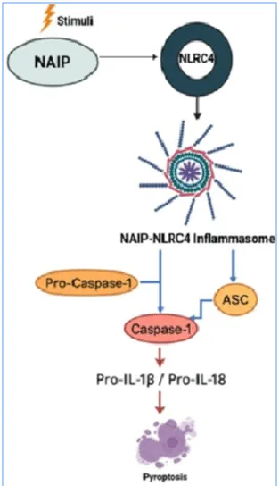
The NLRC4 inflammasome follows an inflammasome pattern: flagellin is the stimulus, NAIP is the upstream sensor, NLRC4 is the nucleator, ASC is the adaptor, and CASP1 is the effector [7]. NLRC4 inflammasomes are cytosolic complexes made up of multiple proteins, including NLRC4, ASC (apoptosis-associated speck-like protein containing a CARD), the adaptor protein, and procaspase-1 [4]. NAIPs serve as immune sensor proteins to activate the NLRC4 inflammasome. They are composed of three domains: an N-terminal Baculovirus IAP-Repeat (BIR) domain, a central NACHT domain, and a C-terminal LRR domain. NAIPs play an important role in identifying bacterial virulence factors in the cytosol, and their association with NLRC4 leads to the activation of the NAIP-NLRC4 inflammasome [4]. The transcription factor IRF8 (interferon regulatory factor 8) controls the expression of NAIPs and NLRC4 during the bacterial infection [3] (Flow chart 1).
Activation of the NLRC4 inflammasome
When NLRC4 is activated, it forms a multi-subunit structure composed of many NLRC4 molecules in the form of a wheel or disk, followed by its binding to pro-caspase-1 using a bipartite adapter molecule called apoptosis-associated speck-like protein, which has a caspase recruitment domain (ASC). This leads to the formation of a multiprotein complex, which facilitates the downstream signaling [7].
Binding of NLRC4 by its respective ligands leads to its activation and interaction with NLR Family Apoptosis Inhibitory Proteins (NAIPs), followed by a change in its conformation and the creation of a new catalytic surface. Activated NLRC4 molecules then recruit and activate other NLRC4 molecules and promote self-propagating oligomerization into a disk-like supramolecular complex Hu Z, et al. [7]. The NAIP-NLRC4 inflammasome is a multi-subunit complex composed of nine to eleven NLRC4 molecules and one NAIP molecule.
The interaction between pro-Caspase-1 and the NLRC4 inflammasome complex is primarily dependent on the CARD domain of NLRC4. When NAIPs sense various stimuli, NLRC4 changes its conformation and activates the CARD domain. Although ASC is not strictly required for pro-caspase-1 activation by NLRC4, it enhances inflammasome assembly by facilitating CARD-CARD interactions between NLRC4 and pro-caspase-1 [7-9]. In certain instances, Caspase-1 activation necessitates the ASC adapter to link NLRC4 and pro-Caspase-1, which is essential for the formation of a large multiprotein complex [7] (Figure 3).
Signaling pathways
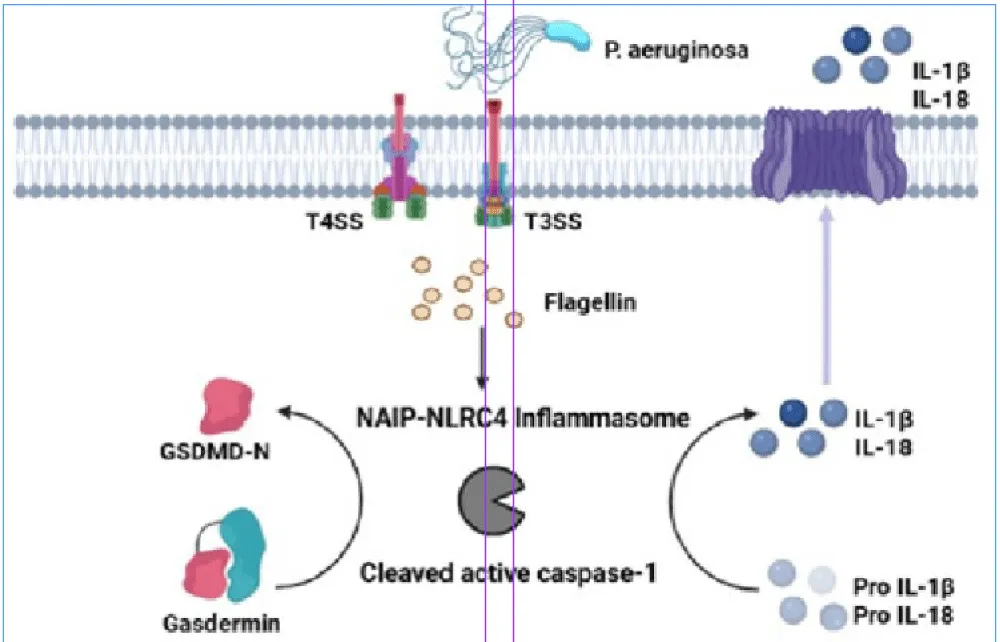
The NAIP protein identifies various bacterial secretions that act as ligands. While murine NAIP1 and NAIP2 can identify ligands, like needles and inner rods, from gram-negative bacteria that use the Type 3 Secretion System (T3SS), murine NAIP5 and NAIP6 can identify flagellin. Human NAIP (HNAIP), on the other hand, can identify all T3SS proteins, including needles, inner rod proteins, and flagellin. When the NAIP senses the T3SS as a ligand, it associates with NLRC4 to initiate the activation of the NAIP/NLRC4 inflammasome. During this process, NLRC4 can directly associate with procaspase-1 through a CARD-CARD interaction. NLRC4 also associates with the adaptor molecule ASC (apoptosis-associated speck-like protein containing a CARD) through its CARD domain. This leads to the formation of NLRC4, ASC, and pro-caspase 1 complex, followed by activation of Caspase-1 (CASP-1). This further leads to the activation of Gasdermin-D (GSDMD), leading to pyroptosis and the formation of pores in the host cell membrane. Caspase-1 then cleaves proinflammatory cytokines such as IL-1β and IL-18 and the release of their active forms through the pores made by Gasdermin-D [4]. NLRC4 also leads to activation of caspase-8 (CASP8), which further leads to activation of caspase-3/7 (CASP3/7) and initiation of apoptosis. Transcriptional regulation of NAIPs is induced by Interferon Regulatory Factor 8 (IRF8) [4,7,10] (Figure 4) (Flow chart 2).
Interactions of NAIP proteins with bacterial ligands lead to a change in their conformation and stabilization. Studies in mouse NAIP chimeras have shown that the α-helical region of the NBD, not the LRR domain, is responsible for ligand recognition [11].
Activated NAIPs attach to latent NLRC4 monomers, leading to change in their conformation and activation of NLRC4. Structural studies have shown that ADP-mediated interactions in the NBD and WHD domains stabilize inactive NLRC4, whereas the LRR domain remains autoinhibited [11]. Mutations that disrupt these connections can lead to persistent activation in the absence of flagellin. A single NAIP is enough to make NLRC4 oligomers because it has a catalytic surface instead of a receptor interface. This catalytic mechanism permits inflammasome assembly even under conditions of limited ligand availability [7,12].
Role of NLRC4 in Innate Immunity and Inflammatory Cell Death
NLRC4 functions as a cytosolic pattern recognition receptor involved in the detection of intracellular bacterial components, including Salmonella, Shigella, Legionella and Pseudomonas species. When it is activated, it leads to inflammation, which helps to eliminate pathogens. NLRC4 dysregulation or hyperactivation has been associated with numerous inflammatory and infectious diseases [13]. A recent study demonstrated that Panoptosis, a unique, physiologically significant, and inflammatory programmed cell death mechanism, is the method by which various infectious agents and sterile insults circumvent inflammatory cell death. Research illustrating the flexibility and interrelation of various cell death mechanisms in preventing intracellular infections supports this idea [14]. The PANoptosome is the chemotype that is required for the simultaneous engagement of potential molecules from pyroptosis, apoptosis, and necroptosis, and it initiates and regulates Panoptosis [15].
Biochemical characterization is employed to define Panoptotic compounds. Immunoprecipitation confirmed that Receptor-Interacting Serine/Threonine-Protein Kinase 1 (RIPK1), Z-DNA-Binding Protein 1 (Z-DNA-ZBPI), and Receptor-Interacting Serine/Threonine-Protein Kinase 3 (RIPK3) all interact with NLRP3. RIPK3 also immunoprecipitates with caspase 8, RIPK1, NBP1, ASC, and NLRP3, which makes Panoptotic components coimmunoprecipitate and turn on a Panoptosome molecule. The harmony of these molecules helps to balance different kinds of cancer, inflammation, and infection [16].
Emerging evidence suggests a functional interplay between Panoptotic signaling and NLRC4 inflammasome activation. Studies examining the relationship between NLRC4 and apoptosis indicate that caspase-mediated cleavage of the DNA damage sensor poly (ADP-ribose) Polymerase 1 (PARP1) is an essential aspect of apoptosis and breaks down to 89 kDa when the NLRP3 and NLRC4 inflammasomes are activated. Since NLRP3, CASP1, NLRC4, and PYCARD are important for the apoptotic and pyroptotic cell death pathways, macrophages lacking these proteins cannot degrade this molecule [17].
Recent research has shown that Panoptosis initiated by S. typhimurium infection eventually connects cell pathways via apoptosis, pyroptosis, and necroptosis. They activated caspase-1 and GSDMD (pyroptosis), caspase-8, caspase-7, and caspase-3 (apoptosis), Mixed Lineage Kinase Domain-Like Pseudokinase (MLKL) (necroptosis), and protected macrophages by inhibiting Panoptotic molecules such as Casp1, Casp8, Casp11, and Ripk8 [18]. Nevertheless, removing individual aspects of cell death does not provide complete protection against cell death, indicating that PANoptosis plays an important role during S. typhimurium infection [15]. More experimental data are needed to fully understand the role of NLRC4 in PANoptosis for host defense mechanisms, as well as to explain how NAIP/NLRC4 interacts with PANoptosome complex molecules.
Role of Nlrc4 in Different Diseases
Since NLRC4 is linked to inflammation, it plays a crucial role in causing certain autoinflammatory diseases in humans, which are characterized by systemic or organ-specific inflammation that is not attributed to infection, malignancy, or antigen-specific autoimmunity [19]. NLRC4 evokes an immune response during bacterial infection through the process of inflammation. This immune response is essential, but hyperactivation of NLRC4 leads to abnormal cell death and cytokine release. Mutations in NLRC4 also cause overactivation of NLRC4, which is harmful and deleterious and can cause autoinflammatory disease [4] (Flow chart 3).
Role of Nlrc4 in infectious diseases
NLRC4 in host defence during bacterial infection: The role of the NLRC4 inflammasome has been identified in foodborne illnesses such as those caused by Salmonella typhimurium. These studies indicate that NLRC4 is essential for protecting the host from S. typhimurium infection, as animals lacking NLRC4 exhibit heightened vulnerability to infection and increased bacterial loads in the cecum, liver, and spleen relative to control animals [4]. A comparable study was conducted utilizing mice deficient in NLRP3, NLRC4, and CASP1. This study showed that these mice had serious health problems and were more likely to get sick when they were exposed to C. rodentium, an enteric bacterial pathogen. This shows how important NLRC4 is for protecting the host from C. rodentium [4].
NLRC4 not only defends against enteric bacteria but also protects the host from various non-enteric bacteria [4]. Recent studies have clarified the interplay between NLRC4 signaling and the cellular immune response, demonstrating that the activation of inflammasomes and the recruitment of CCR2-mediated monocyte-derived dendritic cells collaboratively mitigate L. pneumophila infection [20]. The interaction between NLRC4 and NAIP changes the shape of NLRC4, which helps CASP1 find Pathogen-Associated Molecular Patterns (PAMPs) and then turn on. These findings are considered the structural basis for the formation of the human NAIP-NLRC4 inflammasome [21].
The flagella of the pneumonia-causing bacteria L. pneumophila activate the NAIP-NLRC4 inflammasome. Another study showed that NLRC4-deficient mice were unable to eliminate L. pneumophila during nasal infection, whereas wild-type mice were successful in removing the pathogen [4]. During P. aeruginosa infection, NLRC4-absent macrophages are resistant to cell death caused by P. aeruginosa and secrete less IL-1β [9].
When bacteria infect a cell, active NLRC4 starts the process that activates CASP1, which helps IL-1β and IL-18 mature. This starts inflammatory responses and pyroptosis. When Salmonella typhimurium infects cells, macrophages that don't have NLRC4 can't activate CASP1. This shows how important the inflammasome is for immune defense. Comparative studies of human and mouse NAIP-NLRC4 complexes have shown that they recognize and activate ligands at different levels. This suggests that the amount of NLRC4 activation depends on the type and amount of bacteria present [22]. Moderate activation can stop bacteria from replicating, but too much activation can kill cells and cause inflammation in the tissue. Neutrophils infected with S. typhimurium, on the other hand, do not undergo pyroptosis but still release IL-1β, which adds to inflammation.
In Shigella flexneri infection, the Type III Secretion System (T3SS) drives NLRC4 activation and caspase-1-dependent pyroptosis. It uses virulence factors like OspF to suppress rapid p38-dependent priming of the NAIP-NLRC4 inflammasome [23], which is a sophisticated immune evasion mechanism. In support of this, mice lacking NAIP-NLRC4 show heightened vulnerability to shigellosis [24]. In Pseudomonas aeruginosa infections, NLRC4 activation is induced by flagellin and other Pathogen-Associated Molecular Patterns (PAMPs), contributing to inflammation. According to recent research, Achromobacter species can cause immunopathology and pyroptosis by independently activating the NLRP3 and NLRC4 inflammasomes [25], extending the range of known pathogens that NLRC4 can detect.
In addition to traditional Enterobacteria, NLRC4 also helps protect against infections that are not enteric. The activation of NAIP-NLRC4 in macrophages after Trypanosoma cruzi infection promotes CASP1 cleavage through a cathepsin-dependent mechanism [26], indicating a broader antimicrobial function of this inflammasome beyond bacterial infections. Moreover, Helicobacter pylori-associated gastric illness has been linked to the downregulation of NLRC4, with the emergence of peptic ulcers associated with diminished NLRC4 expression and heightened AIM2 activity [27]. A recent study has shown that Long Non-Coding RNA (lncRNA) controls the activation of NLRC4. For instance, LNCGM1082 increases NLRC4-dependent antimicrobial resistance and strengthens the host's defence [28]. In infections induced by Listeria monocytogenes, the bacterium's escape from phagosomes activates NLRC4, which then activates caspase-1 and pyroptosis. These findings indicate that the NLRC4 inflammasome serves as a significant sensor for various bacterial species. It regulates inflammatory responses and equilibrates immune defense with the potential to damage host cells [29] (Table 1).
| Bacterial Infection | Ligand | Inflammasome activation | Effects | Clinical Insight |
| Salmonella typhimurium | T3SS needle/rod proteins, Flagellin | NAIP5/6 (flagellin), NAIP2 (rod), NAIP1 (needle) activate NLRC4 | Caspase-1 activation, IL-1β/IL-18 secretion, pyroptosis | NLRC4-deficient mice are highly vulnerable to infection, exhibiting increased bacterial load in the spleen and liver. |
| Legionella pneumophila | Flagellin via the Type IV secretion system | NAIP5 → NLRC4 | Initiation of caspase-1-dependent apoptosis and cytokine release | NLRC4-deficient mice fail to clear infection and have elevated inflammation. |
| Pseudomonas aeruginosa | Flagellin, T3SS effectors | hNAIP → NLRC4 | IL-1β release and Pyroptosis | NLRC4-deficient macrophages exhibit reduced cell death and inflammation. |
| Note: Abbreviation: T3SS: Type 3 Secretion System; NAIP5/6: NLR family, Apoptosis Inhibitory Protein 5/6; NAIP2: NLR family, Apoptosis Inhibitory Protein 2; NLRC4: Nucleotide-binding oligomerization domain-like receptor family caspase recruitment domain containing 4; IL-1β: Interleukin 1β; IL-18: Interleukin 18; NAIP: NLR family apoptosis inhibitory proteins. | ||||
Urinary tract infections: Most commonly, UTIs are caused by Escherichia coli. In addition to E. coli, it is caused by Klebsiella pneumoniae, Proteus mirabilis, Enterococcus faecalis, and Staphylococcus saprophyticus. UPEC (uropathogenic Escherichia coli) is the main cause of two predominant types of UTIs: uncomplicated UTIs and complicated UTIs [30]. The innate immune system serves as a trigger for inflammatory responses during UTIs. Studies on UPEC-infected UTI patients have shown that at both the mRNA and protein levels, the expression of NLRC4 is upregulated [31].
Periodontal diseases: A study was performed on individuals with periodontal diseases such as gingivitis and periodontitis and individuals who were periodontally healthy for the expression of NLRC4. The findings demonstrated that patients with gingivitis and periodontitis presented higher levels of salivary NLRC4 than healthy individuals did. These findings indicate that NLRC4 is elevated in these diseases [32].
Shigellosis infection: Shigella is a gram-negative Enterobacteriaceae that causes Shigelosis, which manifests itself through symptoms such as fever, abdominal cramps, and inflammatory diarrhea in the presence of neutrophils. In severe cases, blood can also be observed in this inflammatory diarrhea. Shigella causes disease through invasion and subsequent replication, after which it spreads to Intestinal Epithelial Cells (IECs). Shigella infects the host cell and injects its virulence factors into it, which work as effectors via the Type 3 Secretion System (T3SS) [33]. In humans, Shigella infection has been found to cause inflammasome activation and pyroptosis in macrophages, which aids in disease via the induction of inflammation and the release of bacteria from the cell, therefore permitting bacteria to invade the basolateral side of IECs. However, in mice, the generation of NAIP-NLRC4 and CASP11 inflammasomes provides a defence for the intestinal epithelium against Salmonella. Shigella fails to antagonize the inflammasome in mice, and the intestinal microbiota of these mice resists Shigella colonization, suggesting high and inborn resistance to Shigelosis in these mice [33]. Studies have also been performed on NLRC4-deficient mice and revealed that NLRC4-deficient mice are highly vulnerable to oral Shigella infection. NLRC4-/- (NLRC4-deficient) mice also presented shrinkage of the cecum, thickening of the intestinal mucosa, high MPO (myeloperoxidase) levels in feces, bloody diarrhea, and weight loss when infected with Shigella, but no such case was found in NLRC4+ Shigella-infected mice. These findings demonstrated that NLRC4 functions as a protective barrier against Shigella in mice [33].
NLRC4-associated autoinflammatory diseases
NLRC4-associated auto inflammatory diseases are abbreviated as NLRC4-AID, and they are autosomal dominant conditions that can include Macrophage Activation Syndrome (MAS) and severe Enterocolitis [34].
Macrophage activation syndrome: Macrophage activation syndrome is caused by the hyperactivation of immune cells as a result of an imbalance in the immune system. The symptoms are high fever, enlargement of the liver and spleen (hepatosplenomegaly), swelling of the lymph nodes (lymphadenopathy), and hemorrhagic manifestations. In the MAS, there is continuous immune activation, mainly by CTLs (cytotoxic T cells) and macrophages, due to the high activity of infectious agents and autoimmune diseases [35]. MAS results from a gain-of-function mutation in NLRC4, through which the NLRC4 immune complex becomes hyperactivated and releases proinflammatory cytokines such as IL-1β and IL-18 via caspase-1 activation, as observed in the NLRC4-mediated signaling Elevated IL-18 levels can provide a diagnosis of MAS.
Autoinflammation with Infantile Enterocolitis: AIFEC is an early-onset autoinflammatory disease that manifests early in infancy and affects patients throughout adulthood. It is caused by gain-of-function mutations in the NLRC4 gene, which lead to inflammation and damage to healthy tissues. Enterocolitis is a term used to describe the inflammation of the digestive tract that causes diarrhea and vomiting [36].
The NLRC4 protein is activated because of a mutation in the NLRC4 gene. This protein is a component of an individual's immune system. NAIP proteins are receptors that are present on both intestinal and immune cells. These proteins detect other proteins that are present on certain bacteria, such as Salmonella and Pseudomonas. This recognition of bacteria by NAIP activates NLRC4, which then forms a complex with other proteins known as the NLRC4-NAIP inflammasome. This complex generates inflammatory cytokines, such as IL-1β and IL-18, to kill infected cells. In the intestine, this complex enters the gut lumen, where it may lead to diarrhea. Mutations in NLRC4, on the other hand, lead to its continuous activation and modulation of immune response even in the absence of bacteria. This hyperactivation damages an individual's healthy cells, resulting in AIFEC. In intestinal cells, when NLRC4 is always active, it causes constant shedding of the lining of the GI tract, resulting in Enterocolitis (diarrhea) [36].
Recent studies have linked new NLRC4 polymorphisms to various autoinflammatory manifestations, such as enterocolitis and early-onset recurrent panniculitis. Alongside the traditional AIFEC, the p.Ser445Pro NLRC4 mutation has been identified as detrimental, broadening the clinical spectrum of NLRC4-associated disorders [37]. Recent reports of NLRC4 mutations, including the inaugural mutation in the CARD domain, associated with severe systemic inflammation and infantile enterocolitis [38], furnish more proof that the gene is pivotal in modulating the immune system within the intestines.
Mouse models have also yielded mechanistic insights. The NLRC4V341A mutation increases IL-18 production irrespective of microbiota; yet, it fails to replicate human autoinflammatory symptoms accurately in murine models [39]. Additionally, common traits between ulcerative colitis and other intestinal inflammatory illnesses have been observed in recessive forms of NLRC4-autoinflammatory disease, indicating a similar mechanism involving dysregulated inflammasome signaling [40]. A few more review articles [19,41,42] suggest that diseases mediated by NLRP3, Pyrin, or NLRP1 inflammasomes frequently overlap with NLRC4-driven disorders, highlighting the significance of cytokines IL-1β and IL-18 in their pathogenesis.
Familial cold auto inflammatory syndrome: The chief manifestations of FCAS encompass arthralgia, rashes, and fever in response to cold stimuli. Research found that a missense mutation in NLRC4 leads to the substitution of histidine with proline at position 443, suggesting that NLRC4 is the causative factor for FCAS [43] (Table 2).
| NLRC4-associated autoinflammatory diseases | Trigger | Activation pathway | Downstream effects | Clinical uses |
| AIFEC (Autoinflammation with infantile enterocolitis) | NLRC4 gain-of-function mutation | Constitutively active NLRC4 | Chronic IL-1β and IL-18 release → intestinal epithelial shedding | NLRC4 hyperactivation leads to epithelial damage, diarrhea, and systemic inflammation. |
| Macrophage Activation Syndrome (MAS) | Infection, autoimmunity | Gain-of-function NLRC4 mutations | Excessive IL-18 and IL-1β secretion | Elevated IL-18 is diagnostic and linked to NLRC4 mutations. |
| Note: Abbreviation: NLRC4: Nucleotide-Binding Oligomerization Domain-Like Receptor Family Caspase Recruitment Domain Containing 4; IL-1β: Interleukin 1β; IL-18: Interleukin 18. | ||||
Cancer
NLRC4 levels change much from one type of cancer to another. The levels of NLRC4 mRNA are elevated in breast cancer, stomach cancer, and glioma [44]. Obesity-related breast cancer progresses through the activation of NLRC4 via the Interleukin 1 (IL-1) signaling pathway, which drives the disease through the expression of Vascular Endothelial Growth Factor A (VEGFA) mediated by adipocytes and the formation of new blood vessels (angiogenesis) [44]. Chronic inflammation is associated with gastric malignancies. This is characterized by the elevated expression of proinflammatory cytokines such as IL-1β and IL-18, which contribute to their tumour-promoting actions [45]. Recent studies demonstrate that peptic ulcer disease and the inflammatory environment conducive to stomach carcinogenesis are associated with reduced NLRC4 expression and increased AIM2 gene expression in response to Helicobacter pylori infection [46]. Conversely, colorectal cancer has less NLRC4 expression in infected cells compared to nearby normal tissues, suggesting a potential tumor-suppressive function of NLRC4 inside the intestinal epithelium [47].
Glioma, a cancer of the central nervous system, has been shown in studies to exhibit elevated NLRC4 expression in patients. Glioma patients with high NLRC4 expression had lower survival rates than those with low NLRC4 expression [48,49] (Table 3). It has been demonstrated that chronic IL-1β-induced inflammation regulates Epithelial-To-Mesenchymal Transition (EMT) memory phenotypes via epigenetic modifications in Non-Small Cell Lung Cancer (NSCLC), offering a mechanistic link between inflammasome-derived IL-1 signaling and tumor progression. NLRC4-driven IL-1β production can also mechanistically promote malignant phenotypes beyond these tumor types [50].
| Cancer | Ligand | Effects | Downstream Pathway | Clinical Insight |
| Breast cancer (obesity-associated) | IL-1 signaling | Obesity → increased NLRC4 via adipocyte inflammation | VEGF-A-driven angiogenesis and tumor progression | High NLRC4 expression promotes tumor growth. |
| Colorectal cancer | Chronic inflammation | Decreased NLRC4 expression | Potential loss of inflammasome-driven antitumor immunity | Lower NLRC4 in tumors as compared to adjacent normal tissue. |
| Note: Abbreviation: NLRC4: Nucleotide-Binding Oligomerization Domain-Like Receptor Family Caspase Recruitment Domain Containing 4; VEGF-A: Vascular Endothelial Growth Factor-A.-A. | ||||
In addition to the central nervous system, NLRC4 affects tumor immunity and the course of various cancers. NLRC4 regulation has been identified as a potential therapeutic target in lung cancer [51]. Additionally, increased plasma levels of NLRC4 and NLRP3 have demonstrated diagnostic value in non-small cell lung cancer (NSCLC), especially in patients concurrently suffering from pulmonary tuberculosis [52]. Genetic polymorphisms in NLRC4, NLRP3, and NLRP7 have been linked to a heightened risk of lung cancer, further demonstrating their involvement in inflammation-related carcinogenesis [53].
Researchers discovered that the Hino-Fe chelate molecule inhibits the proliferation of osteosarcoma cells by inducing ferroptosis and NLRC4-mediated pyroptosis concurrently. This is a new technique to treat the condition [52]. In prostate cancer, increased expression of NLRC4 and NLRP1 has been noted, associated with immune modulation and disease advancement [53]. In head and neck squamous cell cancer (HNSCC), transcriptomic studies have shown that age-related variability in NLRC4 regulation affects immune cell infiltration and prognosis [53].
A comprehensive analysis revealed that human NLRC4 expression enhances carcinogenic potential by facilitating cancer cell survival via type I interferon signaling and immune infiltration [54]. NLRC4 interacts with many inflammasomes, including NLRP3, AIM2, and Pyrin, to form multiprotein complexes that induce PANoptosis [55].
Finally, advances in molecular biology have enabled the construction of NAIP5-NLRC4 inflammasome complexes in a laboratory setting. This has given us new understanding about how they work, which could help us design cancer-fighting drugs that target the inflammasome [56].
Metabolic Dysfunction-associated Steatotic Liver Disease
etabolic Dysfunction-Associated Steatotic Liver Disease (MASLD) is characterized by the accumulation of hepatic fat, which can lead to type 2 diabetes mellitus and, in certain cases, hepatic inflammation that can progress to Metabolic Dysfunction-associated Steatohepatitis (MASH). In MASLD, TNF-α levels rise, which turns on the NLRC4 inflammasome. This process increases the levels of IL-1β and IL-18, which kill inflammatory cells by pyroptosis [57]. A recent study employing liver organoid models from both healthy individuals and MASLD patients highlighted the substantial role of oxidative stress in aggravating inflammation, a key feature of MASLD [58,59].
A recent study showed that eriocitrin inhibits inflammation and fibrosis in the liver through PPARα-dependent regulation of the NLRP1/NLRC4 signaling pathway [60]. Another study found that overexpression of FXR leads to inhibition of cholestasis and bile acid homeostasis by inhibiting the activation of the NLRC4 inflammasome [60]. Oxidative stress is strongly linked to insulin resistance, metabolic problems, and DNA repair mechanisms that don't work properly in MASLD [61]. Chronic oxidative and inflammatory stress may make it easier for innate immune sensors like the NLRC4 inflammasome to turn on. This could exacerbate liver inflammation and disease progression in MASLD.
Neurodegenerative and neuropsychiatric disorders
Several studies have demonstrated the presence of the NLRC4 sensor protein in several types of brain cells. Microglia, astrocytes, cerebral endothelial cells, and pericytes are all types of brain cells that contain the NLRC4 sensor protein. Pathological conditions also influence the expression levels of NLRC4. In a specific study, increased levels of NLRC4 were observed in the microglia of persons diagnosed with Alzheimer’s disease [62].
Stroke: Inflammation is a key component of stroke. Microglia, which are the immune cells in the brain, are the main line of defense in the central nervous system following a stroke or reperfusion injury. Cellular damage caused by ischemic stroke leads to the release of Damage-Associated Molecular Patterns (DAMPs), which may facilitate the activation of the NLRC4 inflammasome. Studies indicate that lysophosphatidyl choline can simultaneously activate both NLRC3 and NLRC4 inflammasomes. This process increases the release of proinflammatory cytokines and causes neuroinflammation [63].
A recent study identified TRIM29 (Tripartite Motif Containing 29) as a mediator of NLRC4 inhibition during ischemic stroke. This led to reduced inflammation and neuronal damage, along with the mitigation of NLRC4 inflammasome-related cerebral injury [64]. Microglia under ischemic conditions can significantly reduce pyroptosis. Erythropoietin, a TGF-β-activated kinase 1 (TAK1) regulator, inhibits NLRC4 expression, thus reducing inflammation and neuronal apoptosis in stroke-affected brains. Studies on Sprague-Dawley rats revealed the activation of the NLRC4 inflammasome following Intracerebral Hemorrhage (ICH), increased accumulation of glial cells, neurological injury, brain edema, neuronal death, and damage to the blood‒brain barrier [65].
Traumatic brain injury: Traumatic Brain Injury (TBI) refers to any brain injury caused by an external mechanical force. A prospective cohort study of 140 severe TBI patients and control subjects revealed a strong link between the NLRC4 inflammasome and the severity of brain injury. TBI destroys the cells at the trauma's epicenter, spilling their contents into the extracellular space as DAMPs. This results in breakdown of the Blood‒Brain Barrier (BBB), causing iron infiltration from peripheral immune cells and blood vessels and causing oxidative stress. Trauma also induces depolarization, resulting in the uptake of Na+ and Ca2+, the efflux of K+ and inflammasome activation [66].
Alzheimer’s disease: Alzheimer's Disease (AD) is a degenerative neurological illness, and it is the primary cause of dementia in the elderly population. The clinical features of this disease are insoluble amyloid-β plaques and neurofibrillary tangles. The RT-PCR analysis of microglia derived from the brain tissues of Alzheimer's disease mice revealed significant fluctuations in NLRC4 mRNA expression. Further data shows that the activation of the NLRC4 inflammasome in the microglia of Alzheimer's disease brains promotes a proinflammatory response, characterized by increased mRNA expression and elevated cytokine protein synthesis, including IL-6 and TNF-α [62].
Parkinson’s disease: Parkinson's Disease (PD) is another neurodegenerative condition that is defined by the specific loss of dopaminergic neurons in the Substantia nigra. In Parkinson's Disease (PD), astrocytes become activated due to the buildup of misfolded proteins such as α-synuclein and increase the production of NLRC4, which keeps neuroinflammation going and makes neuron loss worse. A study demonstrated increased levels of TWEAK (tumor necrosis factor-like weak inducer of apoptosis) in Parkinson's disease patients via the PKC-δ/STAT3 signaling pathway. Inhibition of PKC-δ effectively dampens NLRC4 activation, thus protecting dopaminergic neurons from degeneration [63]. Recent research also shows that DJ-1 deletion reduces microglia-mediated neuroinflammation by encouraging autophagy-lysosomal breakdown of inflammasome components like NLRP3, indicating that NLRC4 activity may also be regulated by similar processes [64].
Amyotrophic lateral sclerosis: Lou Gehrig's disease, or ALS, is a deadly neurological disease that damages motor neurons in the brain and spinal cord. NLRC4 is a pattern recognition receptor that, when constantly activated, causes pyroptosis, which makes ALS neuroinflammation worse. A study showed that NLRC4 mRNA and protein levels were higher in SOD1 (G93A) model mice and sporadic ALS (sALS) patients. Immunofluorescence studies demonstrated NLRC4 labeling in large motor neurons within both murine and human lumbar spinal cord tissues. In ALS cells, the blood-spinal cord barrier breaks down, letting immune cells into the CNS. This process causes motor neurons to die.
Multiple sclerosis: Multiple sclerosis is an autoimmune demyelinating disease that affects the CNS. Studies have shown the presence of NLRC4 in regions of demyelination in MS patients, suggesting its oligomerization and caspase-1 activation [65].
Glioma: Glioma is the most common intracranial brain tumor that originates from neuroglial cells. NLRC4 is highly expressed in astrocytes and microglia in gliomas, and its activation promotes the release of IL-1β and IL-18. A positive correlation exists between NLRC4 expression and the immune checkpoint molecules Tim-3 and Gal-9, which promote glioma immune suppression and inflammation [66-68].
Anxiety and depression: Inflammasomes play an important role in neuropsychiatric disorders such as anxiety and depression. According to a recent study, Malvidin-3-O-Glucoside (MG), an anthocyanin derived from grapes, was found to inhibit activation of inflammasomes by decreasing the cleavage of caspase-1 and IL-1β production in microglia and brain tissue. These findings suggest the harmful role of NLRC4, in addition to NLRP3 and AIM2, in neuroinflammation [69,70] (Table 4).
| Neurodegenerative diseases | ||||
| Diseases | Trigger | Pathway | Effects | Clinical insights |
| Alzheimer’s disease, Parkinson’s disease and Amyotrophic lateral sclerosis (ALS) | Protein aggregates, DAMPs | NLRC4 is upregulated in microglia and astrocytes. | Chronic neuroinflammation and neuronal pyroptosis | High NLRC4 is correlated with disease severity in Alzheimer's, Parkinson’s, and ALS models. |
| Traumatic Brain Injury (TBI) | DAMP release, ionic imbalance | NLRC4 inflammasome is activated in brain cells. | IL-1β/IL-18 secretion, pyroptosis | Elevated NLRC4 expression correlated with TBI severity. |
| Multiple Sclerosis (MS) | Autoimmune demyelination | Oligomerized NLRC4 in demyelinated regions. | Caspase-1 activation, cytokine release | Suggests involvement in MS pathology and a potential inflammatory marker. |
| Familial Cold Autoinflammatory Syndrome (FCAS) | Cold-induced mutation-driven activation | Missense mutation in NLRC4. | Uncontrolled inflammation upon cold exposure | Histidine→proline substitution in NLRC4 is linked to the FCAS phenotype. |
| Metabolic Dysfunction-Associated Steatotic Liver Disease (MASLD) | Metabolic stress, TNF-α | TNF-α induces NLRC4 activation. | IL-1β/IL-18 release → hepatic inflammation, pyroptosis | Upregulation in patient liver models and organoids is observed. |
| Glioma | Unknown (possibly chronic inflammation, oncogenic stress) | Elevated NLRC4 expression | High IL-1β/IL-18; immune suppression via Tim-3/Gal-9 checkpoints pathways | Poor prognosis with high NLRC4 expression. |
| Shigellosis | T3SS needle protein | NAIP1 → NLRC4 | Pyroptosis in macrophages | Mice with NLRC4 knockout exhibit susceptibility to Shigella, while normal mice remain resistant. |
| UTIs (Uropathogenic E. coli) | Flagellin | NAIP → NLRC4 | Pyroptosis, IL-1β release | Increased NLRC4 mRNA and protein levels in UTI patients were observed. |
| Periodontitis | Oral microbial dysbiosis | Upregulated NLRC4 in gingival tissues | Enhanced IL-1β secretion → inflammation | Elevated salivary NLRC4 levels in affected individuals were seen. |
| Myocardial Infarction | TNF-α | Promotes inflammation and cardiomyocyte apoptosis. | TNFR1/NF-κB and caspase pathway | Anti-TNF agents have been studied, although with limited success in clinical trials. |
| Note: Abbreviation: DAMP: Damage-Associated Molecular Pattern; NLRC4: Nucleotide-Binding Oligomerization Domain-Like Receptor Family Caspase Recruitment Domain Containing 4; IL-1β: Interleukin 1β; IL-18: Interleukin 18; TNF-α: Tumor Necrosis Factor-α; T3SS: Type 3 Secretion System; NAIP: NLR family Apoptosis Inhibitory Proteins; TNFR1: Tumor Necrosis Factor Receptor 1; NF-κB: Nuclear factor kappa light chain enhancer of activated B cells. | ||||
Cardiovascular diseases
Recent studies have demonstrated that the NLRC4 inflammasome has a significant role in the pathogenesis of cardiovascular diseases, alongside its established functions in immunology and infection. NLRC4 expression was found to be significantly elevated in the early postinjury phase of acute Myocardial Infarction (MI) in male rats, independent of the NLRP3 inflammasome. These findings suggest its role in inflammation and cardiac remodeling [71,72]. In support of this statement, a study on Diabetic Cardiomyopathy (DCM) suggested that NLRC4 serves as a downstream effector linking metabolic stress with inflammatory cardiac damage via the NLRC4-IRF1 axis and in synergy with AIM2-mediated macrophage activation [73]. Additionally, increased NLRC4 inflammasome expression in both cardiac and non-cardiac tissues has been validated by multiorgan investigations in rat models of severe heart failure, indicating systemic implications in the pathogenesis of heart failure [74]. These findings bolster the notion that the NLRC4 inflammasome is a potential targeted therapeutic candidate in metabolic cardiomyopathies.
Inflammatory biomarkers and the challenge of NLRC4 specificity: Biomarkers such as serum amyloid A (SAA), IL-18, and S100 have been found to be helpful to the clinicians for the assessment of the severity of autoimmune diseases [73]. In addition to this, cellular microparticles, such as alarmins and self-antigens, offer novel perspectives on immunological and vascular dysregulation in autoimmunity. Although IL-6, IL-18, and IFN-γ are significant indicators of inflammation, these cytokines are not exclusive to NLRC4-dependent inflammation and may be upregulated due to the activation of other inflammasomes as well. Recent progress in inflammasome biology has underscored the necessity for innovative pathway-specific biomarkers to enhance diagnostic accuracy and inform individualized therapeutic approaches. High-throughput technologies, such as protein arrays, immunoglobulin and T-cell receptor sequencing, and mass cytometry, may further be utilized to improve biomarker identification for diagnosis, prognosis, and treatment in autoinflammatory diseases [75].
Disease-Specific Regulation of the NLRC4 Inflammasome in Infectious and Metabolic Contexts
As discussed above, NLRC4 has been implicated in a wide range of conditions, from infections to metabolic and auto inflammatory disorders and even neurodegeneration. What becomes clear, however, is that NLRC4 does not behave the same way in all these settings. Its effects depend very much on context. The outcome depends on several factors, including the nature of the trigger, whether it is microbial or sterile, the tissue in which activation occurs, the intensity and duration of signaling, and the overall metabolic or immune status of the host. This phenomenon helps explain why NLRC4 may be protective in certain diseases yet harmful in others [76,77].
During a bacterial infection, the NAIP- NLRC4 inflammasome acts as an early sensing mechanism. It recognizes flagellin and components of the type III secretion system and quickly activates caspase 1 [78]. This results in the processing of IL-1β and IL-18 and the cleavage of gasdermin D, leading to cytokine release and pyroptotic cell death [77]. Although this response promotes inflammation, it serves a clear purpose. By removing infected cells and exposing intracellular bacteria, NLRC4 helps limit pathogen spread and strengthens the overall immune defence [79,80]. When tightly regulated, this response is clearly beneficial.
Unlike infections, in metabolic diseases such as MASLD, obesity, and type 2 diabetes [81-82], nutritional excess plays an important role. The constant metabolic load induced by lipotoxicity, oxidative stress, and ongoing inflammation molds tissue homeostasis, leading to persistent inflammation in metabolic tissues such as the liver and adipose tissue. This further results in the development of insulin resistance and progressive organ dysfunction [83,84]. High-fat diet-induced endotoxemia has also been linked to NLRC4 inflammasome activation in visceral adipose tissue, and obesity-associated NLRC4 signaling has been found to promote tumor progression in some cases [85].
These findings suggest that NLRC4 is not exclusively a bacterial sensor, as it has been found to play an important role in various non-infectious and inflammatory pathologies [86]. These studies suggest that non-infectious metabolic signals, such as free fatty acids and oxidative stress, may independently activate NLRC4 in the absence of pathogens. Experimental studies indicate that free fatty acids can induce NLRC4 activation in a TNF-α-dependent manner, resulting in the production of IL-1β and IL-18, as well as the mitochondrial redistribution of NLRC4. Unlike the short-lived activation seen during infection, this form of signaling may be more prolonged and less controlled. Accordingly, NLRC4 activation contributes not only to antimicrobial host defense but also to the pathogenesis of chronic inflammatory disorders [87].
Therefore, NLRC4 occupies a pivotal position at the intersection of immunity and metabolism. The physiological context in which it triggers largely determines whether its activation is protective or harmful. A detailed understanding of this context-dependent behavior is therefore important to clarify its role across different diseases and target NLRC4 for developing therapeutic interventions.
Genetic Tools to Understand the Role of NLRC4 in Different Diseases
Mutations in NLRC4
The formation of the NLRC4 inflammasome is essential for initiating an immune response in the host organism against pathogens. However, the NLRC4 inflammasome can sometimes be activated more than necessary to eliminate the pathogen. This overactivation can cause autoinflammatory diseases like enterocolitis and MAS. Mutations in the NLRC4 gene cause NLRC4 to be hyperactive. These mutations are deadly and cause cells to die in strange ways and cytokines to be released through the NLRC4 pathway [88].
Studies on transgenic mice (mu-NLRC4-mice)
A study employing transgenic mice demonstrated that the introduction of a mutant NLRC4 gene, specifically mu-NLRC4 mice, resulted in markedly elevated expression of this transgene in the spleen. The mice got dermatitis (red, swollen, sore skin) and swollen joints when they were 3 weeks old. The microscopic examination of cells demonstrated infiltration within the joints and skin, resulting in joint swelling and dermatitis, respectively. Bone erosion was also seen. The spleens of Mu-NLRC4 mice were enlarged (splenomegaly) and five times bigger than those of control mice. The count of CD11b+Gr1+F4/80- cells in the spleen was also 15 times higher than that observed in control mice. These results show that changes in the NLRC4 gene cause autoinflammatory disease in mice. The splenocytes in mu-NLRC4 mice produced more IL-1β, IL-17A, and G-CSF than those in control mice [89].
Studies on NLRC4-deficient mice
The NAIP-NLRC4 inflammasome is an important part of a person's natural immune system. Researchers used mice that lacked NLRC4 to see how infections affect hosts when this defense mechanism isn't there. Mice deficient in NAIP-NLRC4 are susceptible to shigellosis [90]. Further investigation is required to elucidate the disease-specific role of NLRC4 signaling. in inflammatory diseases.
NLRC4 gene silencing
Small interfering RNA, or siRNA for short, is a type of molecule that is used to stop genes from working. SiRNAs can lower the expression of specific genes by breaking down the target mRNA in a specific way (cleaving the mRNA). This RNA type works by attaching to mRNA through Watson-Crick base pairing. Researchers looked at NLRC4 gene silencing in septic shock patients and found that it affected lung tissue damage, inflammatory responses, NLR pathway regulation, and DC (dendritic cell) apoptosis [90].
In mouse models, silencing NLRC4 lessened the damage to lung tissue caused by septic shock. This suggests that silencing the NLRC4 gene could lessen the damage to lung tissue caused by septic shock.
- NLRC4 helps control and release proinflammatory cytokines like IL-1β and IL-18. NLRC4 silencing stops IL-1β, which means that silencing the NLRC4 gene could lower the inflammatory responses that happen during septic shock.
- NOD1 or NLRC1, is a cytosolic receptor that is part of the NLR family. When it is activated, it causes septic shock and damages or abnormally functions several organs in animal models. When the NLRC4 gene is silenced, it lowers the expression of NLR pathway members like NOD1, NOD2, RIP2, and NLRC4 itself. This stops the NLR pathway from working properly.
- Dendritic cells are a type of Antigen-Presenting Cell (APC) that takes in antigens and shows them to T cells. Previous research has shown that the NLRC4 inflammasome is active in DCs in the spleen. NLRC4 gene silencing stops DCs from growing and maturing, and it also stops their population from growing.
- These results suggest that siRNA-mediated NLRC4 gene silencing reduces damage to lung tissue caused by septic shock by blocking the NLR signaling pathway and slowing down the maturation of DCs, which in turn lowers the immune response [90].
CRISPR-Cas and NLRC4
CRISPR stands for clustered regularly interspaced short palindromic repeats and CRISPR-associated systems. In prokaryotes, it protects against foreign genetic material and acts as adaptive immunity to the pathogen. A study using a strain of Pseudomonas aeruginosa deficient in CRISPR-Cas indicated that the ability of immune cells to perform autophagy in response to CRISPR-Cas-deficient bacteria was diminished post-infection. Less autophagy stops damaged mitochondria from being cleared out on time. As a result, this causes the inflammasome to become overly active and make more proinflammatory molecules. Mice infected with the CRISPR-Cas deletion strain exhibited heightened inflammation and an excess of IL-1β compared to those infected with the wild-type PA14 strain. NLRC4 was recognized as the primary inflammasome activated during PA14 strain infection [91] (Flow chart 4).
NLRC4 inflammasome in trained innate immunity
Recent evidence indicates that NLRC4 may play a role in Trained Innate Immunity (TII), a process whereby innate immune cells enhance their responsiveness to novel threats. While the mechanistic function of NLRC4 in TII remains inadequately defined, various indications imply a potential association. The NLRC4-dependent activation of L-1β [90,91] induces metabolic reprogramming in macrophages. This occurs due to the accumulation of succinate in macrophages via the HIF-1α proinflammatory signaling pathway [92]. This subsequently induces epigenetic alterations, including a reduction in active histone modifications (H3K9ac and H3K4me3) and an elevation in repressive marks (H3K27me3). These processes are implicated in the epigenetic and metabolic reprogramming underlying trained innate immunity with the increased expression of pro-inflammatory genes [93,94]. In this context, the NLRC4 inflammasome activation may have an indirect effect on the epigenetic markers that support TII (Figure 5).
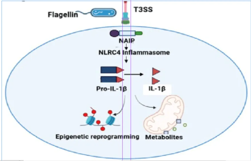
Metabolic adaptability is a principal factor influencing trained immunity [95]. The activation of the inflammasome correlates with transitions to aerobic glycolysis, the accumulation of succinate, and the generation of mitochondrial reactive oxygen species [95-97]. NLRC4 activation in response to bacterial flagellin or type III secretion system proteins may initiate metabolic adaptations that augment or perpetuate inflammatory responses [91]. Direct studies on NLRC4 are scarce; however, analogous findings from NLRP3-mediated trained immunity corroborate this potentiality.
Lastly, the effects of NLRC4-mediated TII probably depend on the situation. In infectious contexts, heightened responsiveness may aid in the elimination of pathogens upon re-exposure; conversely, in chronic metabolic disorders like MASLD or obesity, sustained NLRC4 activation may intensify pathological inflammation [45,97]. In summary, despite the preliminary nature of current knowledge, these findings underscore a possible function of NLRC4 in modulating trained innate immunity via epigenetic and metabolic pathways. Additional research is required to comprehensively elucidate these processes and their effects on both infectious and non-infectious diseases.
Conclusion
The NLRC4 inflammasome plays a significant role in innate immune responses by detecting intracellular microbial components and activating caspase-1-mediated inflammatory pathways. Beyond its role in antimicrobial defense, NLRC4 is increasingly recognized as a context-dependent regulator of inflammatory responses in both infectious and non-infectious diseases. Accumulating evidence indicates that its function is strongly influenced by the nature of the trigger, the tissue microenvironment, and the metabolic-immune status of the host.
Emerging data also suggest a link between NLRC4 signaling and trained innate immunity through metabolic and epigenetic reprogramming. Findings from genetic mutations, gene-silencing approaches, and transgenic models further highlight its complex and disease-specific roles. However, the precise molecular mechanisms governing its differential activation and downstream signaling remain incompletely understood across distinct disease states. Further studies are needed to clarify its regulatory networks and determine whether NLRC4 acts as a driver of pathology, a protective mediator, or a modulatory checkpoint in inflammatory pathophysiology. A deeper mechanistic understanding will be essential for evaluating NLRC4 as a potential therapeutic target in infectious and inflammatory disorders.
Supplementary Materials
[M2] No supplementary material has been added to this manuscript.
Author’s Contribution
Dr. Ruchi Tandon conceived the article and designed its structure. Literature search and data analysis were performed by Ms. Ashwarya Goswami, Ms. Sanchari Roy. Mr. Pallerla Naveen Reddy, and Ms Deepali Siwan. The first draft was written by Ms. Ashwarya Goswami, and critically revised by Dr. Ruchi Tandon, Mr. Pallerla Naveen Reddy, Ms. Sanchari Roy, Ms. Deepali Siwan, Prof. Mukesh Nandave, Prof. Divya Vohora, and Prof. Naranjan S. Dhalla. Material preparation, figures, and manuscript improvements were contributed by Ms. Deepali Siwan and Ms. Sanchari Roy. All authors read and approved the final manuscript. All authors have read and agreed to the published version of the manuscript.
Funding
This research received no external funding.
Data Availability Statement
No new data were created.
Conflicts of Interest
The authors declare no conflict of interest.
Ethics approval and consent to participate: Not applicable. This article is a review and does not contain any studies with human participants or animals performed by any of the authors.
Disclaimer/Publisher’s Note
The statements, opinions and data contained in all publications are solely those of the individual author(s) and contributor(s) and not of the MCB and/or the editor(s). The MCB and/or the editor(s) disclose responsibility for any injury to people or property resulting from any ideas, methods, instructions, or products referred to in the content.