 CrossRef
 CrossMark
 Mendeley
 Scholar
Introduction
Schistosomiasis is a snail-borne trematode disease prevalent in poor communities with inadequate sanitation and unsafe waters [1]. Schistosomiasis is more prevalent in tropical and subtropical countries such as Yemen [2]. Published reports of the schistosomiasis effects on re-productive outcomes had limited to case reports and descriptive series suggesting increased risk of prematurity and of low birth weight [3]. Urogenital schistosomiasis causes infertility in both males and females [4]. In males, seminal vesicles, prostate, epididymis, and vas deferens had mainly affected [5]; however, manifestation of severe schistosomiasis can lead to severe granulomatous epididymitis and inhibition of spermatogenesis, causing male factor infertility [6,7]. Recently, Urinary Schistosomiasis had been associated with the onset of prostatic adenocarcinoma. This study reported a case of genital schistosomiasis showed during semen analysis of a 36-year-old man, Ibn-Sina SP. Hospital. Sana'a, Yemen.
Case Report
A 36-year-old, Yemeni male patient was referred for urine & semen analysis with a history of living and coming from a Schistosoma haematobium endemic rural area in Al-Mahweet governorate. He reported normal urinalysis with no haematuria and pain with intercourse. A semen analysis was performed 30, 90 minutes’ post collection in the clinical laboratory according to criteria of World Health Organization [9].
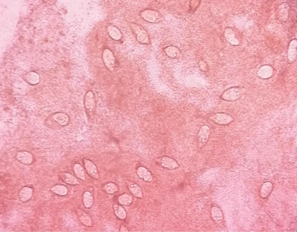
The patient collected a 1.8 mL sample of liquid creamy coloured semen. The progressive sperm motility and non-motile-died was 50% (R 40% and R 32%, respectively). The sperm count was 50.25 million/mL (R > 20 million/mL). Microscopic semen analysis showed slight sperm agglutination, many number of Schistosoma haematobium ova, extensive debris, and many pus cells under high power field (x45) indicating an inflammatory response (Figure. 1). The sperm viability was low, and only 50% of the sperm were viable. The sperm morphology was 81% normal (R > 70% normal), and most abnormal sperm had small head in addition to other abnormalities. The patient treated with two courses of Praziquantel. He moved after this point, and came back after one month to repeat a semen analysis again. His semen and urinalysis was normal.
Discussion
Urinary Schistosomiasis is a public health problem in Yemen, second in importance only to malaria [10]. The first report hematospermia and the presence of Schistosoma haematobium egg in the semen was reported in 1949 [11].
Many studies had observed that ejaculate quality changes when Schistosoma haematobium ova primarily infect the male genital tract. Large autopsy study on 300 cadavers revealed that the ova of Schistosoma haematobium were present mainly in the urinary bladder, seminal vesicles, and vas deferens in 55%, 54%, and 39% of cases, respectively, whereas the affected prostate was only 20% of cases [12]. It was found that seminal vesicles and prostate regarded the main sites of infection [5], and the testicles are not a prime target of Schistosoma haematobium due to differences in anatomy and vasculature.
Infection often affects ejaculate gross appearance and volume. Alterations in ejaculate color in Schistosoma haematobium patients differ, ranging from no colour change to yellow or brown in appearance [13]. Both seminal vesicles & prostate had affected by ova-induced inflammation in Schistosoma haematobium infection, causing apoptosis of sperm and seminal fluid reduction [14].
We found, in the presented case, creamy coloured with reduction in ejaculate semen volume. This indicates that the seminal vesicles and prostate gland may had been slightly affected. Moreover, the sperm count had not affected which similar with previous studies [15,16].
Regarding total and motility of sperms, a significant decrease had found in this case, which contrasts with previous study findings where no decrease in sperm motility was found [15,17]. The presence of debris, pus cells, and Schistosoma ova in the ejaculate indicate an inflammatory condition, which possibly resulted in decreased sperm-motility and viability and which can explain this presented clinical case (Figure 1).
The sperms are very sensitive to hypo-osmotic or hyperosmotic conditions, which can cause membrane damage and irreversible loss of motility. This is due to Schistosoma haematobium infection.
The moderate sperm viability (30%) evaluated using supra-vital stain along with the poor sperm motility observed in this case supports our hypothesis. Overall, multiple semen parameters can be affected by chronic Schistosoma infection.
The presented case emphasizes the importance of request a semen analysis with other diagnostic test in male genital schistosomiasis especially when there is a history or when fertility had suspected.
Therefore, it is very important to train all testing clinical staff on rare semen clinical samples, particularly Schistosoma haematobium ova in semen. This will be educational to the clinical laboratory staff and rewarding for the referring physician and importantly the patient.