 CrossRef
 CrossMark
 Mendeley
 Scholar
Abbreviations
CHIP: Hematopoiesis of Indeterminate Potential
CCUS: Clonal Cytopenia (s) Undetermined Significance
MDS: Myelodysplastic Syndrome
ICUS: Idiopathic Cytopenia of Undetermined Significance
IDUS: Idiopathic Dysplasia of Uncertain Significance
CH: Clonal Hematopoiesis
VAF: Variant Allelic Frequency
MGUS: Monoclonal Gammopathy of Unknown Significance
MBL: Monoclonal B-Cell Lymphocytosis
HMs: Hematologic Malignancies
MN: Myeloid Neoplasms
SMM: Smoldering Multiple Myeloma
MPN: Myeloproliferative Neoplasms
FISH: Fluorescence in Situ hybridization
BMPC: Bone Marrow Plasma Cell
AML: Acute myeloid leukaemia
MM: Multiple Myeloma
uMRD: Undetectable Measuring Residual Diseases
KRd: Carfilzomib, lenalidomide, and dexamethasone
SLiM: SixtyLightchain MRI
FLC: Free Light Chain
AL: Amyloid light-chain
CVD: Cardiovascular Disease TTP: Time to Progression
eGFR: estimated Globular Filtration Rate
HDM-ASCT: High-Dose Melphalan Autologous Stem-Cell Transplantation
DSS: Durie Salmon Stage
Introduction
Hematologic precursor diseases, or Hematologic malignancies, include clonal cytopenia of unknown importance, monoclonal gammopathy of unclear relevance, monoclonal B-cell lymphocytosis, and clonal hematopoiesis. Because these conditions exhibit clonal proliferation of myeloid, lymphoid, or plasma cells without meeting the requirements for active hematologic malignancy or precancerous lesions, they are categorized as premalignant [1,2]. The magnitude and/or persistence of the clone populations are the main features that distinguish premalignant proliferations from indolent neoplasms of lymphoid origin. The number or magnitude of clonal proliferations is not yet sufficient to differentiate premalignant clonal myeloid proliferations from chronic myeloid neoplasms; instead, hematological marrow impairment (Resulting in cytosis or cytopenias) is usually required. Additionally, the World Health Organization now needs morphologic abnormalities and/or specific chromosomal aberrations in order to diagnose a myeloid neoplasm. They are similar to intestinal adenomatous polyps in that they are clonal proliferations that are not currently considered malignant but may develop into such a condition. Similar genetic defects and carcinogenic variables are often present in these "premalignant" clonal hematopoietic proliferations as in their malignant counterparts. The latter are now distinguished from their antecedent lesions by cytomorphology, cell distribution within the damaged tissue (preservation or disruption of the underlying tissue architecture), and the overall load or number of clonal cells. Generally speaking, unchecked hematopoietic clone multiplication is thought to be a necessary condition for a malignant tumor [3]. When blood, bone marrow, lymph node, or solid tumor samples are sequenced using a panel that contains genes often altered in myeloid neoplasms, premalignant clonal myeloid proliferations are most frequently seen. The most frequently mutated genes that underlie CHIP and CCUS are TET2, DNMT3A, SF3B1, ASXL1, TP53, and JAK2. These genes are also often altered in malignant myeloid neoplasms and even lymphoid malignancies [3-6]. It is sometimes difficult or one of challenges to discriminate cancer from non-cancer, and experts will disagree in some cases on whether a particular sample is cancerous or pre-malignant [7]. Although studies conducted to elucidate the role of thrombosis and bleeding as indicators of malignant hematology [8-10], but this review attempted to elucidate the connection between malignant hematology therapy and two clinical indicators of premalignant hematology, thrombosis and bleeding, as well as the difficulties in diagnosing them.
Methods and Materials
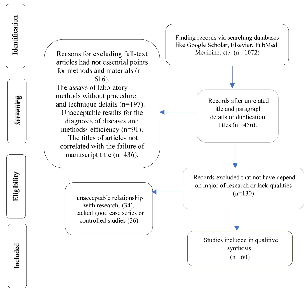
All the information used in this systematic review study was taken from articles that were published between 2009 and 2025 in international journals such as Elsevier, PubMed, Google Scholar, and others. The publications were examined by hematologists, and the abstract, findings, and discussion studies were the main focus of the data analysis. The papers are sorted as shown in figure 1.
Monoclonal B-cell lymphocytosis
The existence of small B-cell clones in asymptomatic individuals has been known for apporxcimnetally two decades [11,12]. A monoclonal B-cell count of less than 5 × 10⁹/L in peripheral blood in people without associated lymphadenopathy, organomegaly, other extramedullary involvement, or any other characteristic of a B-cell lymphoproliferative disorder was the final recognition of the entity of MBL in 2005. Since then, MBL has been divided into three groups according to its immunophenotype: (I) non-CLL type, [13] atypical CLL type, and (III) CLL type [3,14].
Notably, low-level peripheral blood involvement is a common feature of small B-cell lymphomas and leukemias, therefore care should be used when identifying any clonal B-cell. It was unclear whether the MYD88 mutation was specific to the MBL or the IgM MGUS, or if it was present in both clonal B cells and IgM κ+ plasma cells. By flow sorting the MBL and MGUS populations, some scientists have demonstrated that some of these instances reflect two distinct premalignant clonal proliferations, only one of which has the MYD88 mutation [3,14].
Diagnosis of MBL
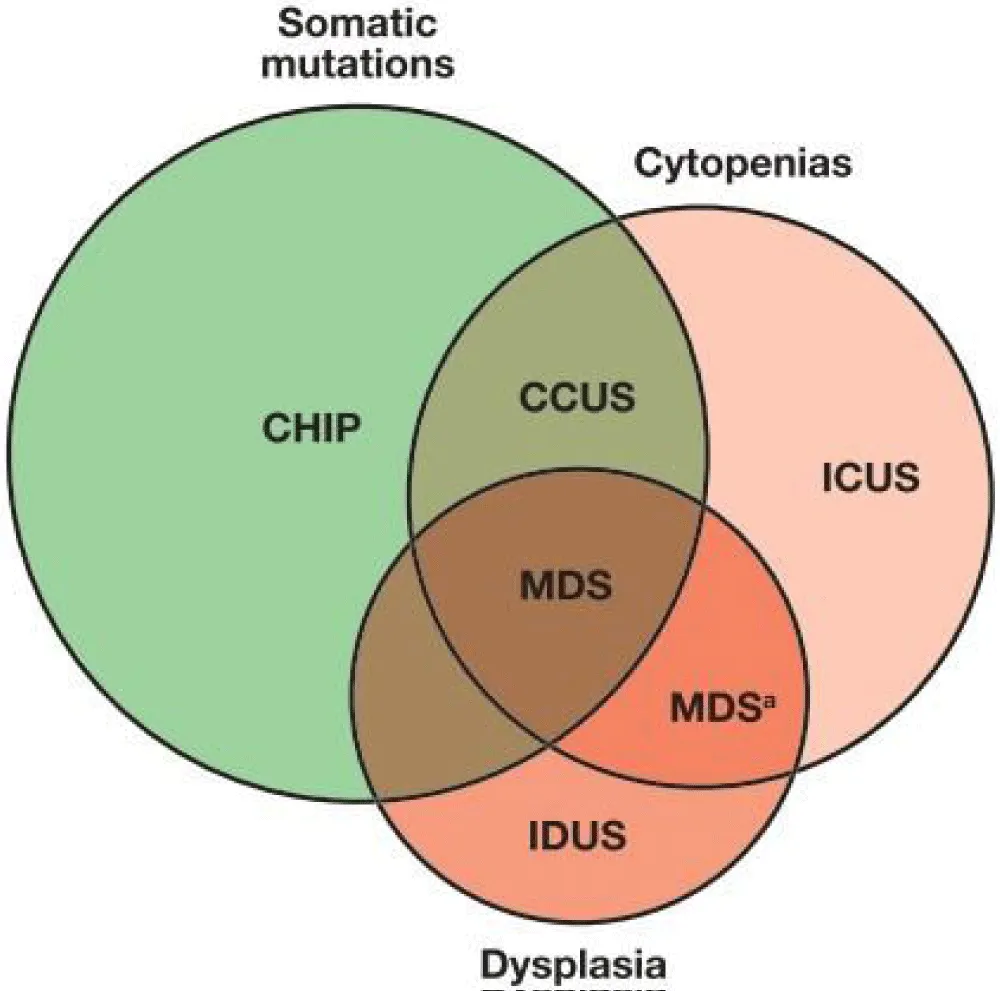
The diagnosis of MBL now requires the presence of a monoclonal B-cell clone < 5 ×10⁹ cells/L without cytopenia, adenopathy, or organ involvement attributable to the clonal B cells, similar to that of Small Lymphocytic Lymphoma (SLL) or Chronic Lymphocytic Leukemia (CLL) [15,16]. Therefore, in otherwise healthy, asymptomatic people, MBL is frequently discovered by chance during regular bloodwork [1]. Clonality testing for B- and T-cells can inadvertently detect premalignant lymphoid proliferations, despite the fact that it is frequently insensitive to small populations of clonal B or T cells. The majority are clonal myeloid proliferations that are premalignant. frequently found when samples of solid tumors, blood, bone marrow, or lymph nodes are sequenced using a panel that contains genes frequently mutated in myeloid neoplasms. The most frequently mutated genes underpinning CHIP and CCUS are TET2, DNMT3A, SF3B1, ASXL1, TP53, and JAK2. These genes are also often altered in malignant myeloid neoplasms and even lymphoid malignancies [4-6]. Karyotype or FISH testing can occasionally identify the existence of an undetected clonal myeloid expansion, as demonstrated in a few examples discussed in this session. There is a lot of overlap between premalignant and malignant clonal myeloid proliferations, making it difficult to distinguish between the two CCUS and MDS figure 2 [3].
Monoclonal gammopathy of undetermined significance
Monoclonal gammopathy is defined by the presence in serum and/or urine of a monoclonal immunoglobulin produced by an abnormal B-cell clone. The clone usually consists of plasma cells when the Monoclonal Immunoglobulin (MIg) is immunoglobulin G (IgG.), IgA, IgD, or Light Chain (LC.) only and lymphoplasmacytic when producing an IgM. It may remain quiescent over a prolonged period, defining MGUS [17]. Moreover, MGUS is characterized by the presence of a monoclonal protein M protein in the serum at less than 30 g/L, without any evidence of end-organ damage (Hypercalcemia or hyper viscosity, anaemia, bone lesions, renal insufficiency), and less than 10% clonal plasma cells in the bone marrow [18].
In actuality, MGUS was the first premalignant hematopoietic clonal proliferation to be identified as a distinct entity. It is characterized by the absence of the usual laboratory and radiologic signs of plasma cell myeloma or lymphoplasmacytic lymphoma and the presence of fewer than 10% clonal plasma cells or clonal lymphoplasmacytic cells in the bone marrow [19]. MGUS is rare in patients under 40, although it occurs in 3% to 4% of people 50 years of age or older, 5% of people 70 years of age or older, and rises to 7.5% in people 85 years of age or older [20].
Notably, two separate entities with seemingly diverse genetic etiologies and varying rates of malignant development have been identified: IgM MGUS (15% of all MGUS) tends to proceed to lymphoplasmacytic lymphoma, other B-cell neoplasms, or AL amyloidosis; non-IgM MGUS tends to develop to plasma cell myeloma, solitary plasmacytoma, or AL amyloidosis. Over the course of the patient's life, the risk of advancement is around 1% annually for non-IgM MGUS and 1.5% for IgM MGUS. Prognostic factors include the type and size of M protein, the serum free light chain ratio, and the identification of a MYD88 p. L265p mutation [3]. In the spectrum of plasma cell dyscrasias, MGUS and asymptomatic or considered precursor Plasma Cell Disorders (PCDs) to active multiple myeloma. On rare occasions, monoclonal gammopathies do not meet active multiple myeloma criteria, but cause clinically significant symptoms affecting organs, such as nerves, kidneys, or skin, and are termed monoclonal gammopathy of clinical significance [21].
Smoldering multiple myeloma
SMM is a plasma cell disorder that has the potential to progress to active multiple myeloma [22,23]. Patients with MGUS and SMM usually have excessive circulating quantities of monoclonal antibodies, and elevated serum and urine macroglobulin levels without the presence of clinical symptoms [24]. SMM is generally asymptomatic and people are often unaware of having the condition until they are diagnosed during a routine laboratory examination [22]. SMM is defined as serum M protein (IgG or IgA) ≥ 3 g/dl or urinary M protein ≥ 500mg/24 h, and/or Bone Marrow Plasma Cells (BMPCs) of 10% to 60% and the absence of myeloma defining events (Hypercalcemia, anemia, lytic bone lesions, or renal insufficiency) or amyloidosis [19].
Moreover, Asymptomatic or SMM are considered precursor Plasma Cell Disorders (PCDs) to active multiple myeloma. On rare occasions, monoclonal gammopathies do not meet active multiple myeloma criteria, but cause clinically significant symptoms affecting organs, such as kidneys, nerves, and skin, and are termed MGUS of clinical significance [21,25].
Clonal cytopenias of undetermined significance
ICUS is the term used to describe patients who have one or more chronic (At least 4 months) and unexplained blood cytopenias (typically anemia), in whom the bone marrow exhibits no dysplasia or tumor and no karyotypic abnormalities [26-29]. CCUS refers to unexplained cytopenia (s) arising in the context of myeloid-associated somatic mutations [30]. and that do not meet diagnostic criteria for defined MN. CCUS is often preceded by clonal hematopoiesis, with an associated high probability of disease progression, along with significant morbidity, particularly CVD. Various selection pressures create clonal composition and propagation, leading to clonal diversification, dominance, and the genomic landscape of neoplastic phenotypes [31]. These people may have any of a number of nonmalignant etiologies leading to their cytopenia or cytopenias, including immune/autoimmune cytopenias, toxic, metabolic, and nutritional variables, which may not yet have received clinical attention. ICUS is not a disease entity. CCUS is the term used to describe chronic unexplained cytopenia without dysplasia but with indications of a clonal genetic defect [26,28,29,32].
Therefore, CCUS is just CHIP plus a chronic, inexplicable cytopenia. Although a proportion of CCUS patients have been known to proceed to myeloid neoplasms, similar to CHIP, the risk of progression has not been well understood. The presence of one somatic mutation with a VAF equal to or greater than 0.10 or the presence of two or more mutations had a positive predictive value for progression to a myeloid neoplasm equal to 0.86 and 0.88, respectively. according to a recent study that sheds light on risk factors in CCUS progression. There was also predictive value for specific mutation patterns, in that spliceosome gene mutations and co-mutations involving TET2, DNMT3A, or ASXL1 had a positive predictive value for developing myeloid neoplasia ranging from 0.86 to 1.0 [3,22]. Correlation of bleeding and thrombosis with premalignant hematology as studies of articles shown in table 1 The findings of an analysis of literature that suggested there were insufficient justifications for accepting thrombosis and bleeding in premalignant hematological kinds were displayed.
| Premalignant cells | Characteristic | Bleeding | Thrombosis | Ref. |
|---|---|---|---|---|
| MBL | CD5 and CD19 expression in people over 40. MBL count in peripheral blood is less than 5 × 10⁹/L in people without organomegaly, extramedullary involvement, lymphadenopathy, or any other characteristic of a B-cell lymphoproliferative disease. | - | - | [1,3] |
| CH | chronic diseases, including solid tumor risk and mortality, autoimmune conditions, osteoporosis, and chronic kidney disease, are associated with mutations that start most frequently in genes involved in epigenetic modification, such as DNMT3A, TET2, and ASXL1. Other mutations arise as a late acquisition and occasionally result in MN. | -/+ | -/+ | [1,5,33] |
| CHIP | Acquired mutations are typically found in myeloid driver genes with variable allele frequency ≥ 2% in bone marrow or peripheral blood without cytopenia or hematologic malignancy. After acquiring a | - | - | [1,37,38] |
| RHOA mutation, three distinct truncating TET2 mutations and a frame-shifting ASXL1 mutation were found at > 20% VAF. associated with heart disease. | ||||
| Multiple Myeloma and certain types of lymphoma | ||||
| MGUS | Laboratory finding are 0.17 g/dL higher mean M protein concentration than those with screened MGUS (0.51 clinical vs., 0.34 g/dL screened, mean difference 0.17 g/dL, 95% Confidence Interval [CI]: 0.13-0.22; p < 0.001), Immunoglobulin heavy chains of IgG, IgA, IgM, or, less frequently, IgD or IgE, together with an aberrant serum free kappa or lambda light chain ratio, without end organ damage or myeloma-defining events. In particular, BMPC <10% and monoclonal protein < 3 g/dL. Progression is (less than 10% of infiltrating plasma cells) to symptomatic MM is low, 1% per year, while the percentage of patients that progress to MM among SMM patients is ten times higher | - | -/+ | ]1,24,39-41] |
| SMM | 10%–59% BMPC participation and a serum free light chain ratio less than 100. Immunoglobulin heavy chains of IgG, IgA, IgM, or, less frequently, IgD or IgE, together with an aberrant serum free kappa or lambda light chain ratio, without end organ damage or myelomadefining events. Serum M protein (IgG or IgA) $3 g/dL or urinary M protein $500 mg/24 h and/or clonal bone marrow plasma cells 10%60%, and absence of MDEs or amyloidosis. associated with a higher level of infiltrating plasma cells (From 10% to less than 60%) and higher macroglobulin levels in serum and urine. | - | - | [1,23,24] |
| CCUS | CCUS is essentially CHIP with the added feature of an unexplained and persistent cytopenia. While it has been known that, like CHIP, a subset of CCUS patients progress to myeloid neoplasms, the risk of progression has been largely unknown. | - | - | [3] |
| Pre-leukemic states | ||||
| MDS | Ineffective hematopoiesis, leading to cytopenia in multiple blood cell lineages, and are also associated with dysplasia suggesting differentiation abnormalities. | - | - | [7] |
| MPN | Expansion of the terminally differentiated myeloid cells and are also the result of aberrant hematopoietic stem cell function. | - | - | [7] |
Clonal hematopoiesis and clonal hematopoiesis of indeterminant potential
Although clonal hematopoiesis of indeterminant potential has been identified as a risk factor for hematological cancers, cardiovascular and pulmonary disease, clonal hematopoiesis has come to be recognized as a universal mechanism of aging that produces an age-dependent increase in cell mosaicism [33]. The largest risk factors for developing CHIP are chronological age and previous exposure to cytotoxic (e.g., chemotherapeutic) agents [34]. Clonal hematopoiesis confers some putatively elevated risk but is more likely to be benign in cases in which clones are not large in size, do not carry multiple mutations, and do not have mutations in particularly high-risk drivers (e.g., TP53, IDH1, IDH2, JAK2). CHIP occurs when Hematopoietic Stem and Progenitor Cells (HSPCs) acquire driver mutations that promote their clonal proliferation, resulting in certain clonal cell lineages making up a disproportionate fraction of circulating blood cells [35].
Individuals with CHIP do not exhibit abnormal blood cell counts or other symptoms of hematologic disease. In contrast, CH refers to the more canonical phenomenon of clonal expansion among hematopoietic stem cells. Most clones will carry evolutionarily neutral mutations, which are typically associated with smaller clone size and are more likely to be of benign effect. Only one in 10 individuals with CHIP will eventually receive an associated cancer diagnosis, but the consequences of a prognosis that either overlooks premalignant disease or results in unnecessary treatment are significant, emphasizing the need for continued study of this condition [36] (Table 1).
Thrombosis and bleeding in premalignant patients
Both MGUS and MM patients had a higher risk of both venous and arterial thrombosis, according to major research that included over 5000 MGUS patients, 18,000 MM patients, and their matched controls. The results of this research indicated that venous thrombosis risk was higher at 1-, 5-, and 10-years following MM diagnosis: hazard ratios (95% confidence intervals) were 7.5 (6.4-8.9), 4.6 (4.1-5.1), and 4.1 (3.8-4.5), respectively. For arterial thrombosis, the equivalent values were 1.9 (1.8-2.1), 1.5 (1.4-1.6), and 1.5 (1.4-1.5). Hazard ratios for venous thrombosis at 1-, 5-, and 10-years following MGUS diagnosis were 3.4 (2.54.6), 2.1 (1.7-2.5), and 2.1 (1.8-2.4). Arterial thrombosis risks were 1.7 (1.5-1.9), 1.3 (1.2-1.4), and 1.3 (1.3-1.4). The summary of studies of articles which done in this research shown in table 2. There was no increased risk of MM or Waldenström macroglobulinemia in MGUS patients with (vs., without) thrombosis [39].
| 1 year follow up | 5 years follow up | 10-year follow-up | |||||||
|---|---|---|---|---|---|---|---|---|---|
| Category | MGUS | Controls | HR* (95% Cl) | MGUS | Controls | HR* (95% Cl) | MGUS | Controls | HR* (95% Cl) |
| Thrombosis by location | |||||||||
| DVT | 51 | 52 | 3.7(2.5-5.5) | 129 | 228 | 2.8 (2.2-3.5) | 192 | 377 | 2.6 (2.1-3.2) |
| PE | 34 | 36 | 3.6 (2.3-5.8) | 83 | 150 | 2.2(1.7-2.9) | 125 | 232 | 2.2(1.7-2.8) |
| Coronary artery disease{I} | 232 | 490 | 2.0(1.7-2.4) | 587 | 1683 | 1.5(1.3-1.7) | 821 | 2455 | 1.5(1.3-1.6) |
| Cerebrovascular {II} | 109 | 305 | 1.4(1.0-1.7) | 308 | 1111 | 1.1(1.0-1.3) | 447 | 1660 | 1.1(1.0-1.3) |
| Type of thrombosis | |||||||||
| Venous thrombosis {III} | 73 | 83 | 3.4(2.5-4.6) | 183 | 356 | 2.1(1.7-2.5) | 273 | 569 | 2.1(1.8-2.4) |
| Arterial thrombosis {IV} | 325 | 763 | 1.7(1.5-1.9) | 826 | 2572 | 1.3(1.2-1.4) | 1158 | 3701 | 1.3(1.3-1.4) |
| Any thrombosis | |||||||||
| ALL patients | 601 | 829 | 1.9(1.7-2.1) | 1167 | 2818 | 1.4(1.3-1.5) | 1545 | 4081 | 1.4(1.4-1.5) |
| Males | 339 | 528 | 1.9(1.6-2.2) | 639 | 1646 | 1.4(1.3-1.5) | 842 | 2317 | 1.4(1.3-1.5) |
| Females | 262 | 301 | 1.9(1.6-2.4) | 528 | 1172 | 1.5(1.3-1.7) | 703 | 1764 | 1.5(1.4-1.7) |
| *Adjusted for age, sex, and calendar period at diagnosis. {I} Angina pectoris, unstable angina, and myocardial infection. {II} cerebral infection, transient ischemic attack, and cerebral hemorrhage. {III} DVT and PE {IIII} Angina pectoris, unstable angina, myocardial infection, transient ischemic attack, and cerebral infection. | |||||||||
Bleeding tendency is a rare manifestation of MGUS that is often ascribed to a dysfunction of the coagulation pathway [42]. CHIP was associated with increased risk of clinically significant bleeding (2%10% VAF: aHR, 1.24; 95% CI 1.02-1.51; p = .03; ≥ 10% VAF: aHR, 1.21; 95% CI, 0.85-1.73; p = .28) (43). The several CH mutation genes correlated to atherosclerosis-associated arterial thrombosis, and hemostasis demonstrated that bleeding and atherothrombosis risk had a correlation with CH patients in table 3 indicated the summary of analyzing of article (44).
| Genes | Thrombosis risk | Bleeding risk |
|---|---|---|
| ABCB6 | Yes | No |
| ASXL1 | Yes | No |
| DNMT3A | Yes | No |
| ETV6 | No | Yes |
| FANCA | No | Yes |
| FANCC | No | Yes |
| FLI1 | No | Yes |
| GATA1 | Yes | Yes |
| GFI1B | No | Yes |
| SF3B1 | Yes | No |
| JAK2 | Yes | No |
| SH2B3 | Yes | No |
| SMAD4 | No | Yes |
| TET2 | Yes | No |
| WAS | No | Yes |
Premalignant diagnosis tools
Multiple myeloma: Over the past ten years, there has been a considerable evolution in the diagnostic criteria, staging system, response criteria, and therapy of multiple myeloma [22,45].
Monoclonal Gammopathy of Definite Significance (MGUS) is a pre-malignant stage that nearly all MM patients progress from [46,47]. About 5% of those over 50 have MGUS [41,48,49], which develops to MM or a similar cancer at a rate of 1% annually [50]. On the spectrum of clonal plasma cell proliferative diseases, Smoldering Multiple Myeloma (SMM) is an asymptomatic illness that falls between MGUS and MM. Differentiating between SMM and MGUS is crucial from a clinical perspective since SMM has a ten-fold greater risk of advancement than MGUS within the first five years after diagnosis. The rate at which SMM develops into multiple myeloma is around 10% annually during the first five years after diagnosis, 3% annually during the next five years, and 1.5% annually after that [51].
According to recent statistics, the first five years had the highest risk of advancement, with a 2-, 5, and 10-year risk of progression of 22%, 42%, and 64%, respectively [52]. As a result, MGUS and SMM require distinct kinds of counselling and follow-up [53].
Diagnosis of monoclonal B-cell lymphocytosis
The diagnosis of MBL now requires the presence of a monoclonal B-cell clone under 5 ×10⁹ cells/L without cytopenia, adenopathy, or organ involvement attributable to the clonal B cells, as would define Chronic Lymphocytic Leukemia (CLL)/Small Lymphocytic Lymphoma (SLL) [15,16]. As such, MBL is often incidentally diagnosed on routine bloodwork in otherwise healthy asymptomatic adults [1].
MBL is often diagnosed incidentally on routine blood tests, during evaluation of other conditions, or for workup of mild lymphocytosis. The diagnostic criteria proposed by Marti et al 46 require the detection of a monoclonal B-cell population in the peripheral blood with an overall kappa: Lambda free light chain ratio > 3:1 or < 3:1, or > 25% of B cells lacking or expressing low-level surface immunoglobulin or diseasespecific immunophenotype. Those clones must persist over a 3-month period. The criteria exclude conditions where patients have lymphadenopathy, organomegaly, autoimmune/infectious disease, Blymphocyte count > 5 ×10⁹ /L, or any other feature diagnostic of a B-lymphoproliferative disorder. The diagnosis is confirmed with flow cytometry analysis confirming the persistence of clonal B lymphocytes with expression of CD19, CD5, and CD23, with a weak expression of CD20, CD79b, FMC7, and surface immunoglobulins. The presence of cytopenia or adenopathy in relation to B-cell lymphocytosis renders a diagnosis of CLL/SLL rather than MBL [1].
Although B- and T-cell clonality testing is often insensitive to tiny populations of clonal B or T cells, it can help unintentionally identify premalignant lymphoid proliferations. The majority are clonal myeloid proliferations that are premalignant. frequently found when samples of solid tumors, blood, bone marrow, or lymph nodes are sequenced using a panel that contains genes frequently mutated in myeloid neoplasms. The most frequently mutated genes underpinning CHIP and CCUS are TET2, DNMT3A, SF3B1, ASXL1, TP53, and JAK2. These genes are also often altered in malignant myeloid neoplasms and even lymphoid malignancies [4-6].
Drugs and treatments of premalignancy patients
One major obstacle to patient recovery is the treatment of hematological malignancies. However, there was no medication advice in 2019 for the treatment and cure completely of individuals with premalignant hematology [3]. In following as studies of some articles shown the several drugs that have trials with types of premalignant patients [54-57].
Treatment of CH and CCUS patients
However, surveillance has been the standard of therapy in CH and CCUS in recent years. Several therapeutic studies, mostly in CCUS, are now underway with the goal of either improving or preventing the development of cytopenias. luspatercept (ClinicalTrials.gov identifier: NCT06788691), ascorbic acid (ClinicalTrials.gov identifier: NCT03418038), IDH inhibitors (ClinicalTrials.gov identifier: NCT05030441), Canakinumab (ClinicalTrials.gov identifier: NCT05641831), statins and metformin (ClinicalTrials.gov identifier: NCT04741945) are a few of these clinical trials. These trials were intended to use either biological vulnerability to target inflammation (e.g., canakinumab, statins), aging (metformin), metabolic vulnerabilities (e.g., ascorbic acid in TET2 CH), mutation-specific targeting (e.g., IDH inhibitors), or based on improvement of cytopenia observed in early MDS (e.g., luspatercept) [1].
The results of these studies will be crucial in assisting medical professionals in determining whether individuals with clinically significant cytopenias may ameliorate their cytopenias or whether the course of the disease can be altered. In order to estimate an individual's absolute risk in the future, it will be crucial to develop clinical algorithms that are validated for screening and include a multimodal and multiomic progression score. One crucial step in precision prevention is the development of therapeutics that target specific vulnerabilities. A. One crucial step in precision prevention is the development of therapeutics that target specific vulnerabilities. End points for CH trials have been attempted, but they also need to be standardized and agreed upon. Furthermore, as these are early-initiating events that most likely drive myeloid biology, utilizing the knowledge of early CH events would also improve our understanding of late illnesses like MDS and AML [1].
Treatment of SMM patients
Observation has been the standard of treatment for SMM [58-60]. However, it is commonly known that the term SMM encompasses patients with asymptomatic early malignancy, patients with premalignancy at high risk of advancement, and patients with premalignancy whose progression rate is more like MGUS than SMM [51,55]. The International Myeloma Working Group (IMWG) revised the diagnostic criteria for MM, which now includes treating a small percentage of people with early malignancy. It is clear, nevertheless, that not everyone with early cancer meets the updated IMWG criteria [23,60].
SMM still includes a high-risk subgroup with an ;50% risk of progression within 2 years, and these patients need to be considered for clinical trials testing early therapy. The rationale for observation as the standard of care for SMM over the years has been the lack of clear data from randomized trials of an overall survival or quality-of-life benefit with early therapy, the toxicity of therapy in an asymptomatic patient population, and the fact that some patients can be free of progression for many years without any therapy. There is also a concern that early therapy may increase the risk of selecting resistant clones. We therefore need to accurately identify patients who are most likely to benefit from intervention. We now have a number of indicators that assist us identify the individuals with SMM who are most at risk of advancement, even if there are currently no laboratory techniques to conclusively distinguish between clonal premalignancy (Biological MGUS) and clonal malignancy (Biological MM). we now have several biomarkers that help us identify the patients with SMM who are at the greatest risk of progression [23,61].
Evaluation of drugs to treat of SMM high risk patients
KRd induction followed by HDM-ASCT, KRd consolidation, and lenalidomide maintenance for two years with the goal of curing patients with high-risk smoldering myeloma was the subject of a recent research [54]. Induction therapy with KRd, six cycles, high-dose melphalan (200 mg/m2) HDM-ASCT, two KRd consolidation cycles, and Rd maintenance for two years were administered to patients with high-risk smoldering myeloma (> 50% progression risk at two years) and transplant candidates. The uMRD rate by next-generation flow following ASCT was the main outcome [54].
The secondary end aim was sustained uMRD four years following ASCT. Additionally, 31% of the 90 patients who were enrolled between June 2015 and June 2017 satisfied at least one SLiMhypercalcemia, renal impairment, anemia, and bone disease (CRAB) criteria. In the intention-to-treat population, 56 (62%) of 90 patients developed uMRD three months after ASCT, with a median follow-up of 70.1 months. Four years later, it persisted in 29 patients (31%). The 70-month progression rate was 94% (95% CI, 84 to 89), and five patients advanced to MM. Four of the five patients had progression to MM indicated by the presence of any SLiM CRAB criteria (Hazard ratio, 0.12; 95% CI, 0.14 to 1.13; p = 5.03).
Biochemical progression was observed in 36 individuals, and it was expected if uMRD was not achieved by the conclusion of therapy. Overall survival at 70 months was 92% (95% CI, 82 to 89). The most common side effects throughout therapy were infections and neutropenia, which led to one treatment-related death. There have been reports of three second primary cancers [54].
With 31% of the patients retaining the uMRD 4 years following HDM-ASCT, this curative method is encouraging and more successful than active MM, even though a longer follow-up is required [54]. Table 4 indicated the studies of SMM patients that demonstrated the overall survival with different drugs and compounds of them which.
| Study | Therapy | N | TTP | OS |
|---|---|---|---|---|
| RCT | Initial vs., delayed MP | 50 SMM and IMM | 12 mo | No difference |
| RCT | Initial vs., delayed MP | 145 DSS I | ~12mo | No difference 64 mo vs., 71 mo |
| Observational | Delayed MP | 54 DSS I | 2-yPFS 75% | Tumor- specific OS 80% at 60 mo |
| Pilot | Pamidronate | 5 SMM and 7 IMM | 2-y-TTP 25% | |
| RCT | Pamidronate vs., observation | 177 SMM | 5-y-PFS 53% both arms; SER 74% vs., 39%, p = 0.009 | Median OS 46 mo and 48 mo |
| RCT | Zoledronate vs., Observation × 1 y | 163 SMM | TTP; 67mo vs., 59 mo, p = NS SER; 55% vs., 78%, p = 0.04 | OS not different |
| Phase 2 | Thalidomide pamidronate | 76 SMM | 4-y EFS 60% | 4-y OS 91% |
| Phase 2 | Thalidomide | 19 SMM and 10 IMM | Median 35 mo | OS; 86 mo OS from treatment 49 mo |
| Phase 2 | Thalidomide | 28 high- risk SMM | Not applicable | Not applicable |
| RCT | Thalidomide + Zoledronic acid vs., Zoledronic acid | 68 SMM | 29 mo vs., 14 mo | 6y > 70% |
| Phase 2 | Interleukin-1 receptor antagonist ± dexamethasone | 47 SMM and IMM | 37 mo | |
| Crossover | Curcumin vs., placebo | 17 SMM | ||
| RCT | Lenalidomide + dexamethasone × 9 mo→ lenalidomide Maintenance × 15 mo vs., observation | 119 SMM | 2-y PFS; 92% vs., 50%, P < 0.001 | 3-y OS; 93% vs., 76% p = 0.04 |
| High risk of SMM (54). | ||||
| Phase 2 | Carfilzomib, lenalidomide, and dexamethasone vs., SixtyLightchain MRI. | 90 SMM and MM | 24 mo | 70-mo progression 94% (95% CI, 84 to 89), and 5 patients advanced to MM, 56 (62%) of 90 patients developed uMRD three mo after ASCT, OS at 70 mo was 92% (95% CI, 82 to 89). |
| EFS: Event- Free Survival; MP: Melphalan-Prednisone: RCT: Randomized Controlled Trial; OS: Overall Survival | ||||
Results
The findings of this systematic review paper illustrated the difficulties in diagnosing and treating premalignant hematological disorders and the association between bleeding and thrombosis.
Bleeding and thrombosis
Although a small number of publications included validated data indicating the MUGS, SMM, and CH with particular gene mutations which correlated to thrombosis and bleeding. In one research indicated that some of mutation genes in CH patients correlated to atherosclerosis-associated arterial thrombosis, and hemostasis demonstrated that bleeding and atherothrombosis risk had a correlation with CH patients. Moreover, Bleeding tendency is a rare manifestation of MGUS that is often ascribed to a dysfunction of the coagulation pathway.
Diagnosis
The analysis of diagnosis challenges identified dome points, one about MGUS Normal and clonal FLC may be found using serum FLC, and the ratio of êtoë FLC is given. It can be challenging to differentiate a monoclonal process from minor FLC rises that frequently occur with age or in circumstances like renal failure or inflammation since FLC measurement alone cannot discriminate between monoclonal and polyclonal FLC.
The other concerns the overlap between CCUS and MDS in cytogenetic somatic mutation and dysplasia studies, which complicates the identification of individuals with premalignant hematology. Furthermore, TET2, DNMT3A, SF3B1, ASXL1, TP53, and JAK2 are the most often altered genes underlying CHIP and CCUS.
Treatment
Premalignant hematological disorders can be treated using licensed drugs that have been around for a while. Following the discovery of premalignant hematology patients and the publication of articles, a number of medications have been introduced in various studies, including paridronate, zoledronate, thalidomide, thalidomide + dexamethasone × 9 months → lenalidomide maintenance × 15 months vs. observation, carfilzomib, lenalidomide, and dexamethasone vs. SixtyLightchain MRI, and ASCT.
Thalidomide, which lasts for 86 months, is essential to the overall survival of the high. One of the key indicators of the most prevalent treatment issues is the occurrence of adverse effects during medication, including neutropenia and infections, which resulted in one treatment-related death.
Author Contributions
Not present.
Funding
Research not received external funding.
Data Availability Statement
The data presented in this study are open sources available in science direct, google scholar, PubMed, Blood and other international journals.
Conflicts of Interest
The authors declare no conflict of interest.
Acknowledgments
Not excess.
Ethics Approval and Consent to Participate
Not applicable.