
Medicine Group . 2022 January 17;3(1):044-059. doi: 10.37871/jbres1399.





 Research Article
High Affinity Peptides in Processes of IgG Purification, Chromatographic Column Virus Inactivation/Elimination and Titer of Anti-Rubella IgG Enrichment
Serhiy P Havryliuk1, Ievhenia M Krasnobryzha2, Olena S Havryliuk3,4 and Heorgii L Volkov3,4*
2Covance Clinical and Periapproval Services LLC, Hrinchenka str, Kyiv, Ukraine
3Neutromics Ukraina TOV, 12, Melnikova str, Kyiv, Ukraine
4Shijir International LCC, Raining Valley, Ulaanbaatar, Mongolia
- Affinity peptides
- In-column virus inactivation
- RV
- aRIgG
Abstract
According to "The Proteome Code" concept introduced by J. Biro and our early development of affinity peptide calculation method it was studied the possibility of high affinity peptide chromatographic gels development for IgG1-4 separation from the donor plasma. Given the next step of virus inactivation of IgG directly in the chromatographic column, the affinity gel had bind IgG at several spatially spaced points in order to limit the degree of freedom of the protein for retention IgG at high buffer flow rate or elevated buffer temperatures without denaturation. In addition, the possibility of creating highly specific affinity sense-antisense peptides against Rubella virus in order to increase the titer of aRIgG in plasma or even its isolation in highly purified form was studied. Based on previous experiments, an affinity multi-peptide chromatographic gel with the following properties was developed: the DBC with enough residence time 10 min was around 50-54 mg × mL-1 of total 98.0% purity of IgG with natural proportion of the 1-4 subclasses, any other immunoglobulins were not found. The virus inactivation/elimination on this gel directly in chromatographic column showed a highly effective virus elimination (log10>9) for both nonenveloped and lipid enveloped viruses.
Using RV sequence from UniProt_KB and dates from more than 20 literature sources on the virus proteins interaction, affinity peptides were calculated against virus proteins C and E1,2. Then these peptides were modified to reach more affinity enhancement and affinity-peptide chromatographic gel was synthetized. By this gel from total mass IgG1-4 contained 6644 IU anti-Rubella IgG with specificity 6.64 IU × mg-1 were isolated 5382 IU aRIgG (> 80%) with a specificity of 791 IU × mg-1.
Abbreviations
DBC: Chromatographic Gel Dynamic Binding Capacity; TDC: Chromatographic Gel Temperature-Dependent Capacity; aa(s): Amino Acid(s); hIgG1,2,3,4: Human Immunoglobulin Class G Subclass 1, 2, 3 or 4; RV(s): Rubella virus(es); aRIgG : Anti-Rubella Immunoglobulin Class G (total); S/D: Solvent/Detergent; TNBP: Three-N-Butyl-Phosphate; T°: Temperature; FVD: Decreasing the Factor of Virus Concentration; LEV(s): Lipid Enveloped Virus(es); NLEV(s): Nonenveloped Virus(es); SpA: Staphilococcal Protein A; SpG: Streptococcal Protein G; PpL: Peptostreptococcal Protein L; MOG: Myelin Oligodendrocyte Glycoprotein.
Introduction
Previously we have shown that the development of chromatographic adsorbents with high Dynamic Binding Capacity (DBC) gave us the opportunity to transform the method of virus-inactivation of proteins in solution into the method of inactivation and elimination of viruses directly in the chromatographic process at an elevated temperature of 30-45°C [1,2]. The parameter "temperature-dependent capacity of the adsorbent (TDC)" offered by us as a function of DBC shows that if the adsorbent DC is higher, its TDC will be higher, since the temperature-dependent rotations/hesitation of protein in physical essence are quite close to the microturbulence of the chromatographic mobile phase with dissolved protein [2]. In the case of multipoint adsorption of the target protein, it is actually "crucified" on the adsorbent; its degrees of freedom and rotations/hesitation of individual parts of the molecule tend to zero. Therefore, an increase in the temperature of the "protein-adsorbent-buffer" chromatographic system does not cause a noticeable denaturation of the protein, even as labile proteins as FVIII. A high DBC allows keeping the protein in a "crucified" state at elevated temperatures. The development of the above method included as model proteins and polypeptides that differ by molecular weight, simplicity or complexity of the molecule, presence/absence of enzymatic activity: fragment of streptokinase SK1-61, lysozyme, fibrino(geno)lytic enzyme from the Agkistrodon blomhoffi venom [1], and also a complex of coagulation factors VIII/von Willebrand (FVIII/VWF) [2]. In all cases, any denaturation or loss of protein activity was avoided, and the FVIII yield in the complete process of its isolation from donor blood plasma was significantly increased.
At the beginning, the most versatile and capacious, as well as fairly inexpensive adsorbents were used - ion-exchange SP and Q Sepharose HP, WorkBeads 40S and 40Q, hydrophobic interaction Source 15RPC [1,2]. It is known that increasing temperature promotes the exposure of new hydrophobic sites on the surface of the protein globule [3-5], in fact, increasing the number of binding sites with the adsorbent. For Source 15RPC it was shown that the DBC for insulin was increased from 57 to 78 mg × ml-1 when the temperature rises from 20 to 45°C and the linear velocity of the buffer remained stable 300 cm × h-1 [1]. The same mechanism works with reverse phase chromatography, where the binding of the target protein depends on the exposure of hydrophobic sites on the protein surface under the solvents influence [3,6]. As for affinity adsorbents, high-molecular ligands of a protein nature (staphylococcal protein A - SpA, streptococcal protein G - SpG, peptostreptococcal protein L - PpL), even if they withstand elevated temperatures [7-9], then their DBC significantly decreases [8-10], and the leaching from the matrix is intensified [11,12]. Nonspecific and monoclonal antibodies as chromatographic ligands for the process under temperatures above 30-35°C are not considered due to their thermal lability [13]. The behavior of short-chain (6-15 amino acids) peptide ligands used in affinity chromatography under such conditions has not yet been adequately studied. However, it would be appropriate to combine in one stage the affinity purification of immunoglobulins and their virus-inactivation at elevated temperatures, which would significantly reduce the process time and cost. Recently, we have shown that the using of Biro “Proteome Code” concept [14] makes it possible to significantly simplify, respectively, accelerate and reduce the cost of calculating the affinity peptide ligand for almost any protein [15]. In addition, we expected that a 10-15 amino acids (aas) peptide attached to a neutral adsorbent such as WorkBeads 40/1000 ACT would not be susceptible to denaturation and excessive leaching, bearing in mind that the same ligand had not previously shown appreciable flow for 250 complete chromatographic cycles at 20°C in previous experiments [15]. In the same development, it was found that the affinity of the calculated peptide can be “ordered” higher or lower, which was measured by the pH level of desorption of 50% of the protein from the sorbent (pH50%). In the case of the peptide affinity adequate pH50% = 3.65 ÷ 3.80, the interaction of sense-antisense aas was determined as average, coinciding with the level of the affinity of Proteins A/G for IgG. The strong level of affinity (pH50% = 3.40 ÷ 3.47) was characterized by hydrophilic-hydrophobic and also electrostatic interactions between sense-antisense aas. In this case, complete desorption of the protein from the sorbent occurred at pH < 2.5 [15], encouraging that a virus inactivation of immunoglobulins directly in the chromatographic column will be possible. In addition, the level of denaturing temperature-dependent rotations/hesitation of the protein can be significantly reduced in the case of simultaneous linking to the adsorbent of two or three ligands that will bind the target protein at different as sites located at the different parts of molecule. About 35 peptide ligands for affinity chromatography of natural, recombinant immunoglobulins and their fragments have already been published in the literature [16-18]. To obtain the top performing affinity peptides rapidly, many screening methods have been developed such as combinatorial chemistry, mRNA display, phage display, computer-based virtual screening technology and other. Our goal in this study was to show a fast, inexpensive way to calculate the high affinity peptide ligands for IgG from human plasma. The above-mentioned Biro concept “Proteome Code” allows the interaction of accessible linear regions of two polypeptides [19]. Moreover, the complete concept insists that it is precisely such an interaction can be predetermined by the genetic code [19]. It can be assumed, for example, that in the event of a virus attack, the mammalian organism reads a certain aa sequence of the viral (most likely capsid) protein, which is responsible for the interacting with the cell receptors. Then immunoglobulin (M, A or G) is synthesized with the inclusion of antisense aa sequence in the variable region of the light/heavy chain. And thus, the “landing place” of the virus on the cell can be blocked by a specific immunoglobulin [20,21]. The literature contains enough data describing peptide/polypeptides of viral proteins that can interact with cell receptors. Based on these data and our developments [15], it is possible to calculate a hypothetical site of interaction and use it as an affinity ligand for the isolation of a specific immunoglobulin. This last problem is much more interesting than the previous two, but it remains the most speculative. The simplest case for the such study seems to be the specific anti-rubella IgG (aRIgG ), since in any vaccinated human body it is contained in the amount of ~40 IU × mL-1 until the end of the life [22]. The level of vaccination against rubella in Ukraine is 78%, and among the younger generation (up to 25 years) - more than 82% [23]. Thus, in the total pool of donor blood, the aRIgG titer can actually range from 5 to 40 IU × mL-1. This level is quite sufficient to assess the affinity of the developed peptides, the possibility of direct column virus inactivation at elevated temperatures, and also for the attempt using the high affinity peptide ligand to increase the aRIgG titer, separating it from other nonspecific immunoglobulins. Cong, et al. [24] showed that one of the host cell receptors identified to bind the E1 protein, is Myelin Oligodendrocyte Glycoprotein (MOG), a member of the immunoglobulin superfamily mainly expressed in the central nervous system and in other tissues such as spleen, liver and thymus of mice. Due to the restricted expression of MOG in cells of these tissues, the role of other receptors and co-receptors in RV attachment cannot be excluded [24]. For example, not excluding the mechanism of transplacental vertical RV infection or direct fusion of the viral envelope with the endosomal membrane due to a conformational change in the E1 and E2 glycoproteins cannot understand [25]. Given that capsid protein C is the other contender for interaction with the cell receptor [26] all three capsid proteins, С and E1 and E2, would be the ideal choice for a determination of peptide epitopes of RV interaction with cell.
Thus, this study was aimed to solve three problems, namely: 1. The development of 3-4 peptide ligands to spaced sites of the human plasma IgG in order to obtain an affinity adsorbent with high DBC/TDC and multi-point IgG binding to prevent possible denaturation at elevated temperatures of virus inactivation in the column; 2. The determination of the virus inactivation parameters for the IgG directly in the chromatographic column and the levels of inactivation/elimination of model viruses; 3. The development high specific affinity peptide(s) for aRIgG in order to significantly increase its titer and create an inexpensive pharmaceuticals to prevent rubella infection in pregnant or HIV-infected people.
Materials and Methods
All reagents were purchased from Sigma-Aldrich (Green Chemistry LLC, distributor in Mongolia) unless otherwise indicated. Reagents and equipment from other manufacturers are indicated below. Human (donor’s) plasma, obtained by plasmapheresis, was bought from several regional blood centers in Ukraine.
Native human IgG1, IgG2, IgG3 and IgG4 (Cat. No. ab 90283, 90284, 118426 and 183266) proteins >95% purity, recombinant RV proteins E1, E2 and C (ab107947, ab43033, ab43034, respectively) and kits for determination human IgG (hIgG) total (ab195215), hIgG1 (ab), hIgG2 (ab), hIgG3 (ab 100548, 202402, 201284) were purchased from Abcam office at China. The hIgG4 concentration was calculated in the needed case according to the difference between total IgG and IgG1-3 quantities. The ELISA kits for RV IgG determination were purchased from Creative Diagnostics distributor Filgen, Inc., Japan (Cat. No. DEIA011) or from Cepham Life Sciences, Inc., Fulton, MD, USA (Cat. No. 10935).
Total human IgG (hIgG) was re-purified [27,28] to 99.4% purity from commercial donor’s plasma 10% intra venous immunoglobulin “Bioven” manufactured by pharma plant “Biopharma LCC”, Bila Tserkva, Ukraine.
The total protein in the collected samples was determined by Bradford method [29] with Stoscheck modification [30].
The calculation of the high affinity peptides to total hIgG1,2,3,4 and to aRIgG
Peptide calculation was carried out according to the “Algorithm of peptide affinity calculation” and table of “Sense-antisense aas for the both 3’-5’ and 5’-3’ direction of gen reading” published early [15] using aa sequences from RV proteins shown in the table 1.
Array peptides synthesis and SPOT peptide array experiments
The peptide’s array library, containing calculated affinity members (~50-100 nmol per spot), was synthesized through short aa spacer as described earlier, using Fmoc chemistry and MultiPep SPOT synthesizer (membranes and equipment from Intavis AG, Germany) [15]. All controls, positive and negative, were same as shown at the previous development [15].
The peptide-membrane after synthesis was subjected to washing [15] and incubation with 0.01 mM model (purified from donor’s plasma) IgG or RV proteins solution. Reactive spots were visualized with Typhoon Trio, λEx = 295 nm, λEm = 340 nm, followed after 3 × 2 min washing of unbonded hIgG/E1/E2/C. Further peptide-membrane was incubated 3 min at 20°C with citric acid-sodium citrate buffer, pH 3.70,… 2.20, that was made by lowering pH in increments of 0.05 pH units. The steps gradient pH was made with Orion Star 9102 pH Automated Titrator (Thermo Fisher Scientific, China). After each washing the fluorescence detection were carried out for affinity determination according pH50% - the buffer value at which 50% of investigated peptide was desorbed [15].
The apparent Kd
The apparent Kd for binding of each protein-protein, protein-peptide and peptide-peptide was determined with standard procedure described by Baja, et al. [31] for intact hIgG, and virus proteins E1, E2, and C in our modification for our pares of interaction substances at their variable concentration under constant proteins concentration.
Fmoc solid phase peptide synthesis
~0.75 mmol/~500 mg of each 13-mer peptides (8-mer affinity peptide plus 5-mer spacer) which were chosen as applicant of the affinity ligand were synthesized using Fmoc chemistry, and purified by reversed-phase flash chromatography, and confirmed by MALDI-TOF mass spectrometry [32].
Peptide-affinity chromatographic gel synthesis
200 mg of each selected as affinity ligand peptide were dissolved in 25 mL sodium carbonate-bicarbonate buffer, pH 9.4, and the seven solutions were mixed together and pumped with flow 1 mL×min-1 through the ECOPLUS Glass Column TAC25/125SLPE0-AB-2 (YMC Europe GmbH, Germany) packed with 20 mL of WorkBeads 40/1000 ACT (Bio-Works, Sweden), during 48 hours at 4 °C. The peptides density on the WorkBeads gel was determined by quantifying the difference between total peptides quantity in start solution and unbound peptides by fluorescence of tryptophan in the spacer measured by Typhoon Trio Variable Mode Imager 6 with Image Master 2D Platinum 6.0 DIGE software (GE Healthcare AB, Sweden). All other manipulations, including blocking unreacted active groups, were done according to instruction of manufacturer [32]. Finally, the column was rinsed with 500 mL of deionized water to remove the blocking agent and, before using, with working buffer for equilibration or, before storage, with 20% ethanol. Monopeptide affinity chromatographic gel was synthesized at the same manner in 5 mL column BabyBio ACT (Bio-Works, Sweden).
Determination of gel Dynamic Binding Capacity (DBC) and Temperature-Dependent Capacity (TDC)
0.5 mL of peptide-affinity gel packed into 0.8 cm diameter, 1.0 cm height column was equilibrated with 10 Vc citric acid-Na2HPO4 buffer, pH 7.4, and 1 Vc of 5 mg×mL-1. hIgG in equilibration buffer was applied into column with flow 0.002-0.25 mL × min-1 (2-25 min residence time). DBC was determined at 10% breakthrough [33]. The column was washed with equilibration buffer, and the elution was performed with citric acid-Na2HPO4 buffer, pH 2.6. The washing and elution flow rate was 0.25 mL × min-1.
TDC was determined by the method described before [2]: model hIgG was applied on peptide-affinity column in quantity 10% lower than DBC of adsorbent under 20°C. After sampling and washing the column and buffer (pumping into column with 0.5 Vc × min-1) temperature was slowly raised by 5 degree each step and IgG was determined in the eluate.
Total IgG peptides-affinity purification from normal donor’s plasma with virus inactivation/elimination directly during chromatographic process
As IgG sample served re-purified IgG to 93.4% purity, obtained after EBA STREAMLINE SP XL (GE Healthcare AB, Sweden) [27,28] captured from low molecular weight protein fraction [34]. Eluate was subjected to membrane filtration to achieve protein concentration 8-9 mg × mL-1.
The concentrated eluate was spiked by model viruses as shown before [2] to achieve protein concentration 7.5 mg × mL-1. The infected hIgG sample (85 mL, that given ~70% loading the affinity ligands) was loaded onto ECOPLUS Glass Column (TAC25/125SLPE0-AB-2) with 20 ml peptide-affinity adsorbent. Column was equilibrated with 10 Vc of 20 mM citric acid-Na2HPO4 buffer, pH 7.4. The equilibration flow rate was 0.5 Vc × min-1, the application – 0.1 Vc × min-1.
The S/D treatment was beginning after IgG capturing on the affinity column. The experimental scheme and stages of the virus-inactivation/elimination process, developed and presented earlier, did not change when studying the behavior of chromatographically bounded IgG under the temperature-dependent treatment [2]. The exceptions were temperature levels and the usage of one R/D blend only: TnBP/Triton X-100.
The common conditions of S/D treatment were the following:
- First washing: 30 Vc of equilibration buffer (0.5 Vc × min-1) to complete absence of virus material in eluate.
- The raising of S/D inactivation buffer gradient: 30 Vc inactivation buffer with flow rate 0.5 Vc × min-1 was automatically created by programming linear increasing concentration from 0 to 100% and applied on the column. The final inactivation buffer included 1.0%/2.5% TnBP/Triton X-100.
- The raising inactivation temperature gradient: on the beginning of inactivation process for inactivation buffer and column the termostabilized gradient was started from 20 to 45°С during 60 min, therefore a temperature gradient grew in parallel to S/D gradient in all cases.
Stages 2 and 3 combined in one process named in the finale table Gr.
- The second washing after raising gradients – actual treatment process: column was washed by 90 Vc under 45°С with same flow rate 0.5 Vc × min-1; the column temperature was kept at corresponding buffer temperature (Gs).
- The falling down S/D inactivation buffer and temperature gradients: after the washing the reverse S/D and T° gradients were used to reach the initial conditions for 60 minutes - 30 Vc with same flow rate 0.5 Vc×min-1 (Gf).
- The third column washing: when the start T° 20°С was reached simultaneously with falling down gradient finishing, the column was washed by 10 Vc equilibration buffer, flow rate 0.5 Vc × min-1.
The elution of IgG was carried out with 2 Vc of 20 mM citric acid-Na2HPO4 buffer, pH 2.6, 0.04 Vc × min-1. All the eluates were collected for concentration/yield and purity of target proteins determination and their impurities and ligands determination by ELISA, and electrophoresis, and HPLC analysis.
The solvent level in the final IgG solution after S/D treatment was measured by GC method with FID, the detergent level - by HPLC with UV-detector (both instruments from E-Chrom Tech, Taiwan) as described previously [1,2].
Column regeneration and sanitization were performed with 2 Vc of 0.1 M glycine-HCl, pH 2.5, and 2 Vc of 0.05 M NaOH with flow rate 0.5 Vc × min-1.
Virus models
In the virus inactivation/elimination experiments we used model viruses from the American Type Culture Collection (ATCC) which have become standard in our developments: Bovine Viral Diarrhea Virus (BVDV), Canine parvovirus (CPV), Bovine Enterovirus Type 3 (BEV), Murine Leukemia Virus Type C (MuLV), Pseudorabies or Suid herpes virus 1 (PRV) and Duck Hepatitis B Virus type 1 (DHBV) [1,2].
Virus titration and quantification
Virus titers were determined using the classical method of Kaerber [35] and Spearman [36] and all calculations were done according to our earlier publications [1,2].
For quantification virus analysis we used quantitative real-time PCR and the data were expressed as log10 of ration between virus nucleic acid detected in the protein sample before and after purification and designated in the results tables as parameter tT°C (example t30 = 4.15) [1,2].
aRIgG peptides-affinity separation
aRIgG peptides-affinity separation from total IgG purified from normal donor’s plasma was carried on double adaptor column XK 16/20 (GE Healthcare AB, Sweden) with 25 ml synthetized peptide-affinity to RV capsid proteins WorkBeads 40/1000 gel as described in section “Peptide-affinity chromatographic gel synthesis” but for one selected affinity ligand only.
Chromatographic process conditions for aRIgG separation were the same as in case of total IgG peptide-affinity purification excluding virus inactivation/elimination stage.
Statistical analysis
The statistical processing of results was carried out by the standard methods [37]. A value of p < 0.05 was considered statistically significant. Data was presented as a mean ± Standard Errors (SEM) of at least 5 independent experiments unless otherwise indicated.
Results and Discussion
High affinity peptides calculation to the human IgG different subclasses, their affinity determination
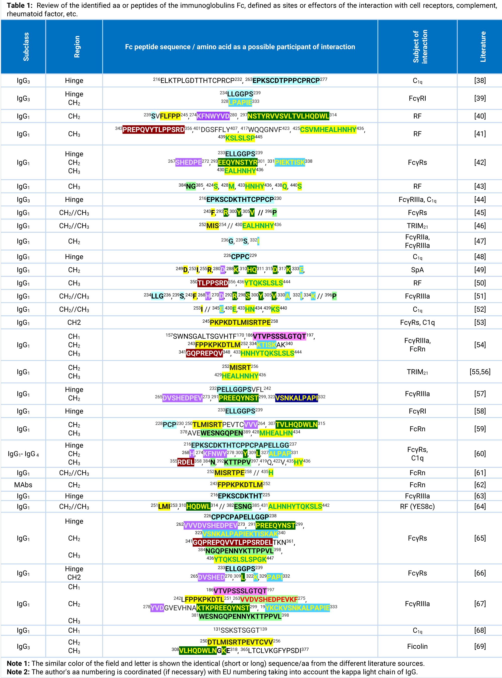
A picky and careful analysis of the literature sources shown that there are several aa sequences in the IgG molecule that are available for interaction with other proteins (SpA, SpG, PpL), various cellular receptors (FcγRs and TRIM21), Rheumatoid Factor (RF), C1q complement. Basically, the sites of the above-mentioned interaction are located in the hinge region and CH2 and CH3 domains of the IgG molecule. Although some authors have shown that in the light chain there are available linear sequences that interact with cellular receptors. The most interesting information in the context of this investigation is summarized in the table 1.
This table also includes possible interaction sites located in other parts of the IgG molecule, which were important in the light of the study objectives. In addition, IgM and IgA1,2 aa sequences were tested for analogous sequences to avoid possible cross-linking of these immunoglobulins during affinity isolation of IgG. The several analogous or similar sites and their parts were detected and discussed below.
The results of studies by a number of authors [38-69] systematized in the table 1 are not exhaustive, but are most relevant for considering possible interaction sites that may be available in the IgG molecule. We have noted that the data on IgG light chain kappa/lambda interaction sites were not considered as these chains present in all immunoglobulin spices and their peptide sequences would guarantee the binding of IgAs, IgD, IgE and IgM by future developed affinity chromatographic gels.
Most studies identify aa sequences or individual aas that have a direct or indirect effect on IgG (in this case IgG1) interaction with cells effectors. All tabulated aa sequence clearly covers the middle (150FPE…SGV167) and lower (193VVT…ICN209) parts of the CH1 domain, top (224EPK…THT233), middle (234CPPCP238) and lower (239APELLGGPS247) parts of the hinge, and smears the whole CH2 domain (249FLF…TPE266, 270VVV…YVD280, 288KTK…NGK317, 319YKC…KTI340) and two short parts of CH3 domain. But if we consider the aas which are significant for binding only the two shorter essential sequences can be distinguished at the CH1 domain: 158FPE…SGA170 and 193VVT…GTQ204, five at the CH2 domain: 241FLFPPKPK248, 252MISRTPEV259, 266VSHEDPEV273, 394EQYNSTYR301, 331PIEKTISK338, and two at the CH3 domain: 380ESNGQPEN388 and 429HEALHNHYTQK439.
The latter sequence of the CH3 domain interacts with the PRYSPRY domain of the homodimeric TRIM21 with very high affinity 0.6 nM [70]. Identical (except the IgG3 Y/F mutation) sequences were found in the IgG2-4 molecules with Kd = 37 ÷ 200 nM [71]. Unfortunately, analogues are also present in the domains of IgM and IgA1-2. Although the affinity of IgM and IgA for monomeric TRIM21 PRYSPRY is much weaker, 17 and 50 μM, respectively [70,72], which is due to large differences in the core aa compositions (IgM: HEALPNRVTER and IgA: HEALPLAFTQK), the ability to interact remains quite high. Therefore, this sequence 429H-K439 of the CH3 domain was excluded from the following experiments, but was taken into account for the development of an affinity peptide for the total isolation of immunoglobulins A, G1-4 and M classes. As for other selected sites, their analogues were not found in IgM/A1-2/D/E.
Anti-sense sequences to selected sites on the IgG1 molecule in 8-aas overlapping peptides were by our proposed level of 50% desorption of immunoglobulin from membrane-synthesized peptide (pH50%) [15] and presented in the table 2. Fragments of sense peptides, which according to our analysis of physicochemical properties may represent the interaction sites of the Fc domain (CH1-3) of IgG1 and Fc receptors (D1,2 domains FcγRn, FcγRI/II/III), highlighted in bold.
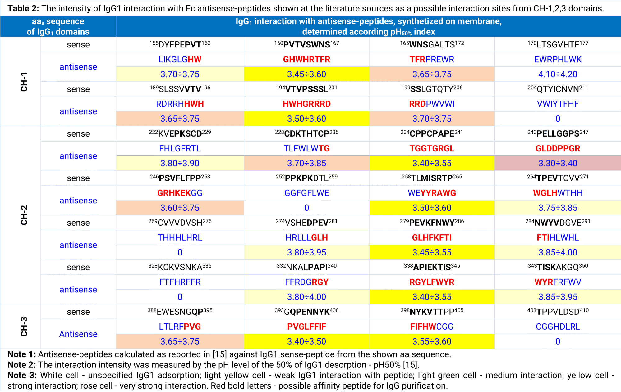
Incubation of anti-sense peptides synthesized on the membrane with hIgG1 showed varying degrees of peptide-protein interaction in all cases membrane-synthesized. At the previous study it was shown that a nonspecific interaction carries on at pH50%>4.00, specific weak interaction - at pH50% = 3.75 ÷ 4.00, specific average - at pH50% = 3.60 ÷ 3.75, specific strong - at pH50% = 3.40 ÷ 3.60, specific powerful - at pH50% = 3.20 ÷ 3.40, specific superpower - at pH50% < 3.20. Specific superpower interaction is, of course, a subjunctive term, but complete protein desorption from the peptide in this case requires lowering pH to 2.0 and below, which always leads to marked denaturation of the protein.
The analysis of the results, presented in the table 2, determined a number of peptides synthesized on the membrane, which forcefully captured IgG1 during the 5 min incubation: GHWHRTFR (No.1), HWHGRRRD (No.2), GRGLDDPPGRHKEK (No.3), YYRAWGHWGFKH (No.4), GLHFKFTI (No.5), RGYLFWYR (No.6), and PVGLFFIF (No.7). It should be noted that all the affinity peptides calculated by our method closely coincide with the affinity for IgG peptides developed by SPR (surface plasmon resonance) [73], several chemical modifications with mass spectrometric analysis [74], biomimetic design strategy [75], combinatorial phage-display library screening [40,76,77].
The aas physic-chemical properties of potential IgG1 interaction sites and identified peptides carried out the change-points of aas according to date of table 3 to improve the effectiveness of the IgG1 capture. In addition, the reduction of up to 8 aas long peptides was carried out in order to standardize their synthesis.
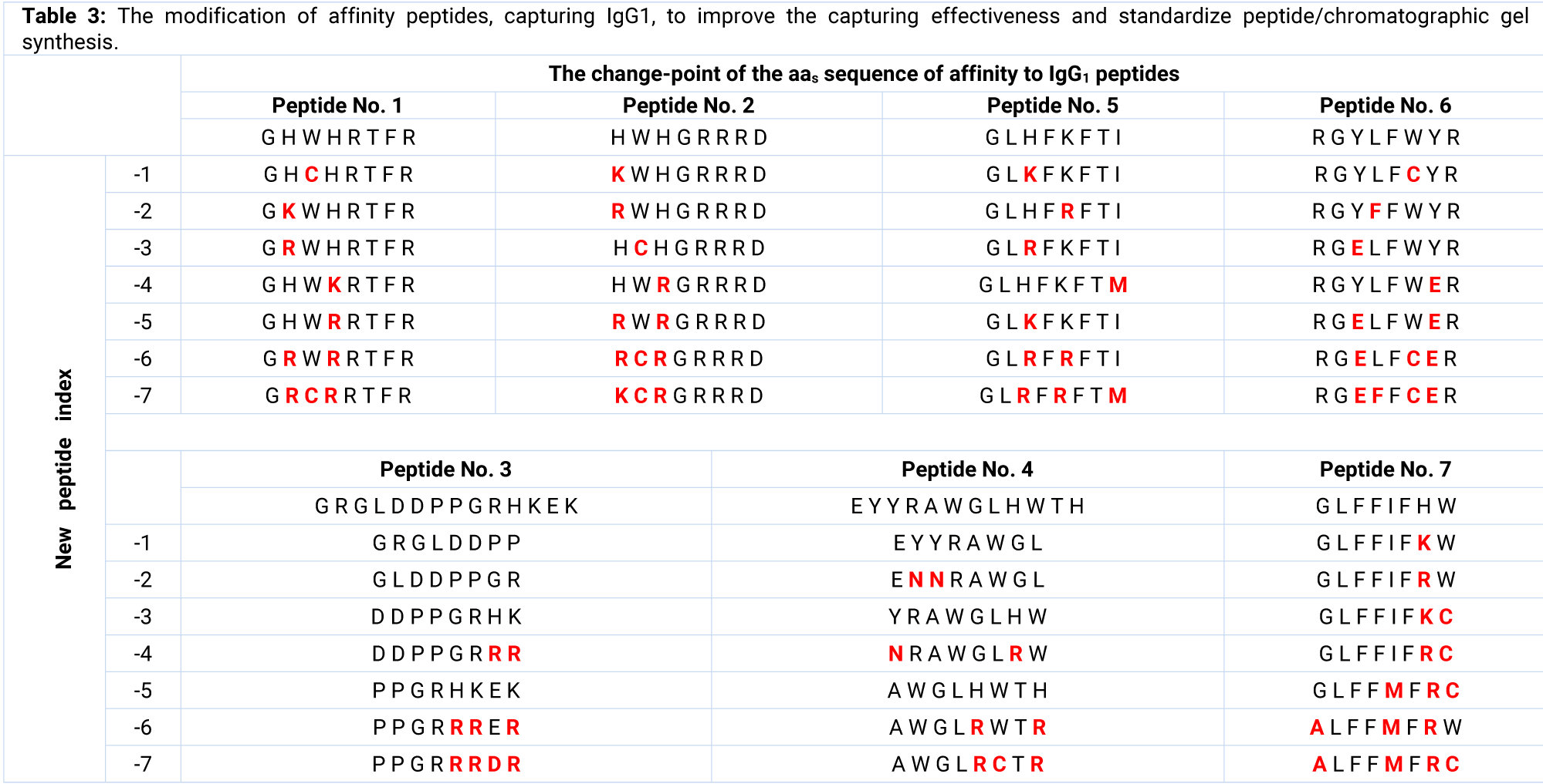
From the proposed (Table 3) modified peptides, the best results of IgG1 CH1 binding showed peptides no. 1-7 GRCRRTFR (pH50% = 3.30, Kd = 32 nM) and no. 2-6 RCRGRRRD (pH50% = 3.35, Kd = 51 nM), IgG1 CH2 binding - No. 3-4 DDPPGRRR (pH50% = 3.20, Kd = 14 nM), no. 4-4 NRAWGLRW (pH50% = 3.40, Kd = 67 nM), no. 5-6 GLRFRFTI (pH50% = 3.45, Kd = 72 nM) and no. RGELFWER (pH50% = 3.40, Kd = 61 nM), IgG1 CH3 binding - no. 7-6 ALFFMFRW (pH50% = 3.50, Kd = 82 nM). The most effective peptide was DDPPGRRR, but each of the seven listed can be used for chromatographic purification of IgG/antibodies as an affinity ligand. Their chromatographic properties will be determined in a separate study. So far, we have obtained seven high affinity peptides that interact with the immunoglobulin molecule at seven spaced sites, which, as previously planned, will provide a multipoint interaction of the IgG with chromatographic gel.
Multipoint peptide-affinity chromatographic gel properties, determination DBC and TDC
After peptides “sewing” the following chromatographic gel was obtained: DDPPGRRR- and 7-PEPs-WorkBeads 40/1000 with a total peptide density 21.8 mg × mL-1 (~22.3 μmol × mL-1), each peptide was sewed in approximately equal proportion.
It was shown that target chromatographic fraction eluted with citric acid-Na2HPO4 buffer, pH 2.6, contained IgG1,2,3,4 in proportion 67.3:20.8:8.5:3.4 approximately as at the total IgG re-purified sample for both gels. The IgA and IgM in the eluate were not found. The purity of the total IgG was 98.6% and 98.0, corresponding.
The DBC with enough residence time 10 min for both gels was around 62-65 and 50-54 mg × mL-1 of total IgG, corresponding (Figure 1).
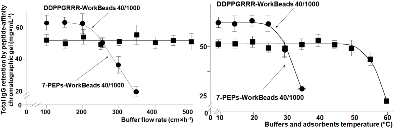
The DBC and TDC of a monopeptide affinity gel, as expected, dropped sharply with increasing speed (>200 cm × h-1) or buffer temperature (>30°C), while the same parameters of a multi-peptide affinity gel practically didn’t change at a linear buffer flow rate at 500 cm×h-1 or increasing its temperature to 45°C at a stable speed of 300 cm × h-1 (Figure 1). The data obtained indicate that the IgG molecule bounded at several sites on the chromatographic gel is sharply limited in mobility, which in turn preserves its native state.
The other conclusion following from the results of the 7-PEPs-WorkBeads 40/1000 gel TDC determination were following: the direct process of IgG virus inactivation and elimination was possible to carry on under the temperature 45°C and buffer flow rate maximum 250 cm × h-1. The temperature around 50°C is already in the zone of the critical retention of immunoglobulin by chromatographic gel.
Direct virus inactivation by S/D treatment during IgG purification process by peptide-affinity chromatography method
S/D treatment of IgG was performed according to scheme shown earlier [1,2]. Our published dates and previous studies on virus inactivation of immunoglobulins dates (in full will be published shortly) have shown that proper virus removal during chromatographic purification depends on the nature of the Solvent/Detergent (S/D) and their concentration, time and temperature of action on the IgG adsorbed by the chromatographic gel. The most effective virus inactivation/elimination conditions were selected for the presented work, namely: TnBP/Triton X100 concentrations 1.0% and 2.5%, respectively; temperature 45°C and exposure to high T° for 5 hours (linear buffer gradient, which T° increases from 20 to 45°C during 1 hour; exposure to constant buffer T° 45°C during 3 hours; linear buffer gradient, which T° decreases from 45 to 20°C during 1 hour).
Results of the S/D virus inactivation of IgG directly in a chromatographic column depending on process temperature are presented in table 4.
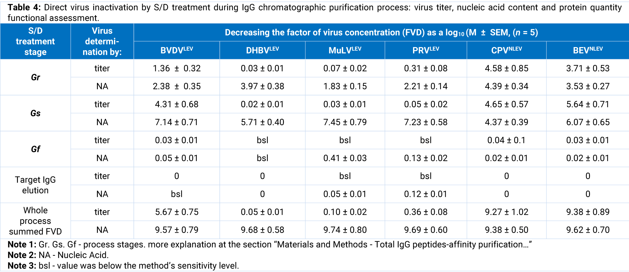
For further comparison we continue to evaluate the virus inactivation by three or two mentioned methods in the present study. In fact, we received full repetition of previously detected pattern: the method of the virus titer determination gave an adequate result according to target protein infectivity in the case of LEV and NLEV models [1,2]. The LEV titer determination didn’t leave the hope to calculate process kinetics since the loss in virus infectivity due to the virus particles destruction and protein denaturation. The virus titration was more acceptable for NLEV infectivity mass balance determination - maximum 18% of the total infectivity was lost against 4-6% determined by RT-PCR. Unfortunately, the titration accuracy was very poor again (variants deviation more than 15-20% in the best cases) and didn’t allow definite conclusions.
Determination of viral proteins by IFA approximately simulated the virus titer and nucleic acids measurement but could in no way to confirm the presence or absence of infectivity. Quantification of viral nucleic acids by RT-PCR in target proteins and buffer fractions obtained during inactivation allowed to calculate the process mass balance within 95.4 ± 4.9%. It means that stability of the virus nucleic acid (in most experiments not more than 3-4% of losses, rarely up to 6.5%) allows to calculate the process kinetic to assess what happens to model virus in the process of S/D treatment with small variant’s diversity not higher than 8-10%. In contrast, diversity of variants of virus titer of target proteins was more than 25-30%.
On the other hand, the virus titer determination compared with the determination of its nucleic acid in the eluate samples is very indicative in terms of resistance of the virus to the S/D mixture. For example, if the titer of most LEVs at the eluate (phase Gr) was almost on the limit of detection, and nucleic acids amount was 2-4 Log, it can mean only one thing: S/D mixture destroyed the virus envelope with loss of virulence and nucleic acids were released into solution. If we found equal amount of virus in the eluate by titer and nucleic acid, it means that LEVs almost were not damaged by S/D, but S/D caused dissociation of virus and IgG and virus was washed out of the column. Thus, the process of virus elimination occurs with CPVNLEV and BEVNLEV (Table 4, stage Gr, Gf). Thus, it’s well known that LEVs destruction by S/D is 3-4 times more effective than that for NLEVs [78] that also supported by our data shown before [1,2] and in the table 4. BVDVLEV conduct itself is a bit asymmetric, namely: partially destroyed and partially undamaged washed out, which confirms its previously determined medium resistance to S/D treatment [79].
In the present investigation we demonstrated again that virus inactivation by S/D treatment of peptide/protein preparation directly in the chromatographic column cooperates two processes, namely: 1) destruction of a virus particles by solvent and/or detergent (mainly for LEVs with minor effect for NLEVs) and 2) dissociation and washing away of virus material which was associated with protein where the outcome that was successful both for LEVs and NLEVs. The IgG yield and purity were not measurably changed when the virus-inactivation/elimination was introduced in the chromatographic purification process.
High affinity peptides to the human aRIgG calculation and development
The level of vaccination in Ukraine against RV is about 78% and among the younger generation (up to 25 years) - more than 82% [23]. The quantity of the specific anti-rubella IgG (aRIgG ) was determined in the donor blood near 29 IU×mL-1, that left the possibility of a noticeable purification of specific IgG during the experiment. A total 4 g primary purified IgG (93.4% purity or 3736 mg pure IgG) from 1 L plasma was processing over 5 chromatographic cycles through the ECOPLUS Glass column (TAC25/125SLPE0-AB-2) packed with 20 ml of 7-PEPs-WorkBeads 40/1000 and received 3.734 g of target fraction with amount of IgG1-4 - 3660 mg, that means a purity 98,0%. The total anti-RV activity was 24308 IU, thus specific activity was calculated according to pure IgG as 6.64 IU×mg-1.
Of course, we hoped that RV has a sufficiently specific receptor on the cell surface for the primary invasion to calculate on this basis a possible high-affinity peptide against aRIgG . In addition, a sufficient number of publications offers enough options for the interaction of RV capsid proteins with cells, thus exposing potential sites of interaction [26,80-100]. The capsid protein C is the second contender for interaction with the cell receptor [26]. So, these three capsid proteins would be the ideal choice for a determination of peptide epitopes that interact with cell surface, i.e., capsid proteins. In the table 5 the most important data concerning the determination of possible sites of interaction on the RV capsid proteins were systematized. The table dates shown that at least 10 epitopes of RV proteins E1, E2 and C are available for interaction and several of them are possible and are specific for interaction with the cellular receptor Myelin-Oligodendrocyte Glycoprotein (MOG) [32].
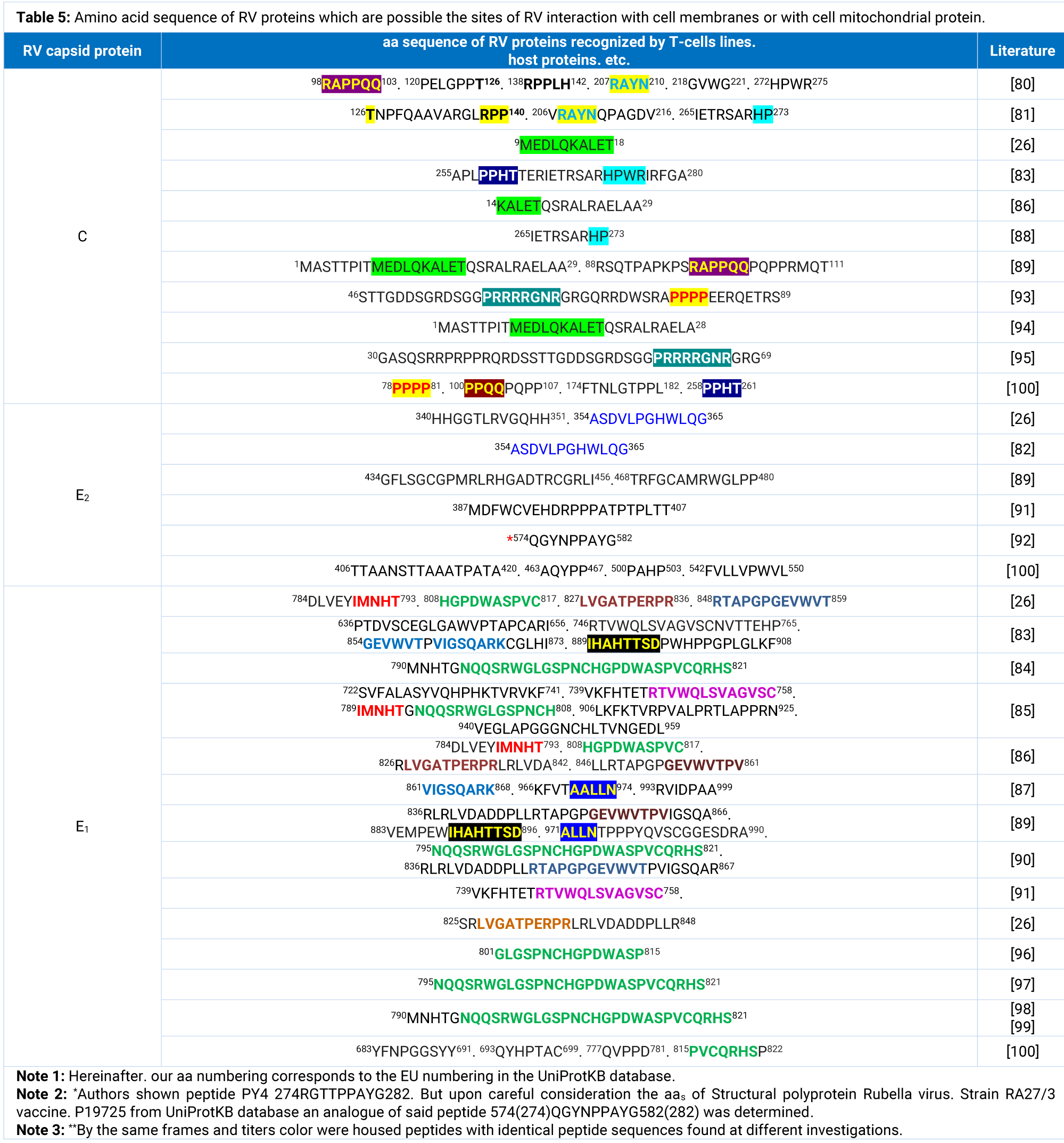
The analysis of the dates at the table 5 gives the chance to concentrate attention at the 4 capsid C sites with confident interaction (no. 1-C - 10EDLQKALET18; no. 2-C - 59PRRRRGNR66; no. 3-C - 78PPPPEERQ85; no. 4-C - 98RAPPQQPQ105), on the 3 protein E2 sites (no. 5-E2 - 354VLPGHWLQ365, no. 6-E2 - 543VLLVPWVL550, no. 7-E2 - 574QGYNPPAY582) and on the 5 protein E1 sites (no. 8-E1 - 742HTETRTVW749, no. 9-E1 - 829GATPERPR836, no. 10-E1 - 861VIGSQARK868, no. 11-E1 - 889IHAHTTSD896, no. 12-E1 - 918RTLAPPRN925). We do not rule out that other sites listed in the table interact with different cellular effectors, but we choose for experimental study those that are confidently identified by first researchers and confirmed by other.
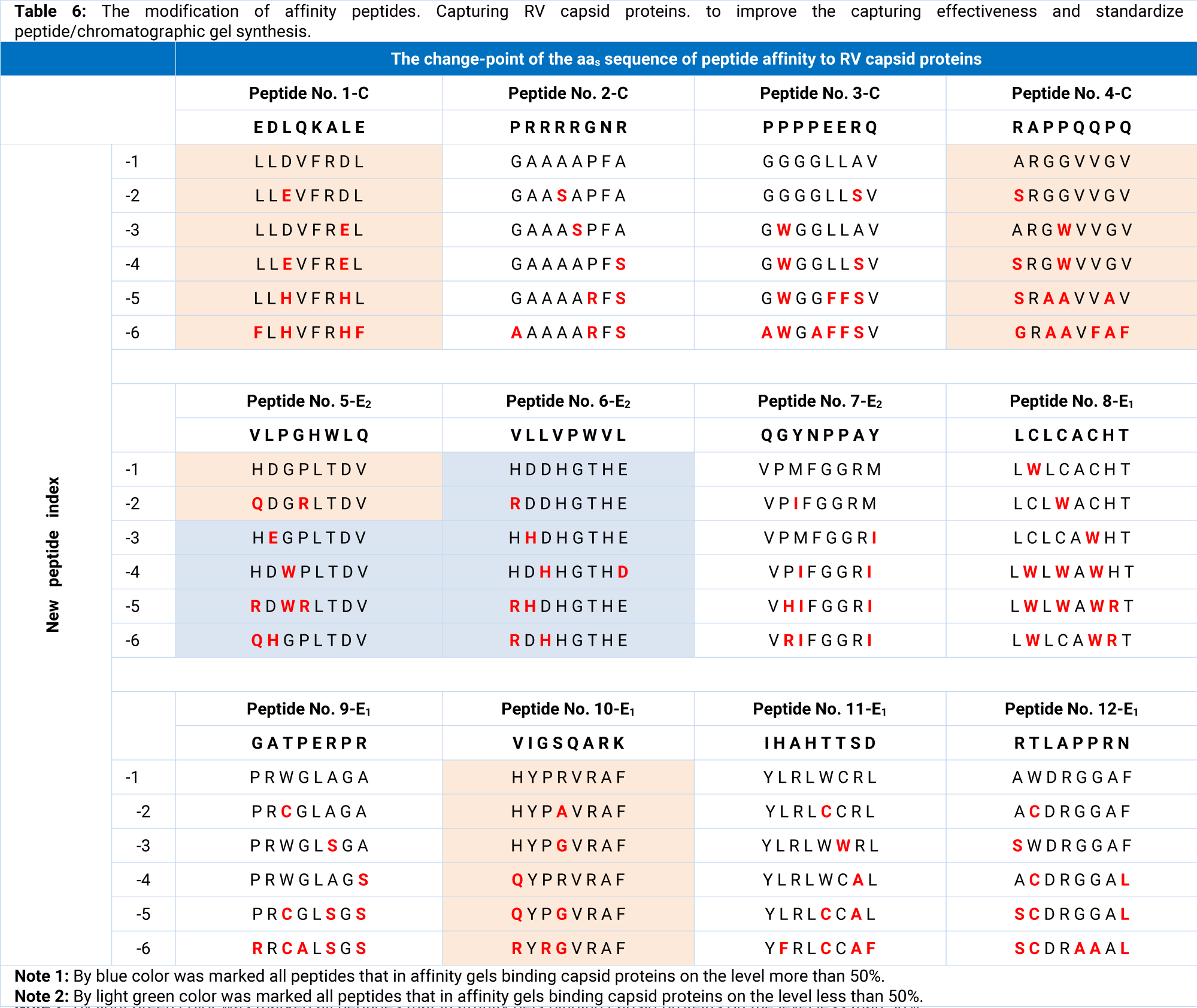
Against selected peptides we developed anti-sense peptides [15] shown in the table 6 and tested their affinity by incubation with corresponding capsid proteins. Peptides LLDVFRDL (no. 1-1-C), GAAASPFА (no. 2-3-C), HDDHGTHD (no. 6-4-E2), HYPGVRAF (no. 10-3-E1) and AWDRGGAF (no. 12-1-E1) showed effective interaction during incubation on the level pH50% = 3.45 ÷ 3.65. The following aas replacement Ll,2/F1,2 and D3/H3 in peptide no. 1-1-C; A2/S2 and P6/R6 in peptide 2-3-C; P3/R3 in peptide no. 10-3-E2; W2D3/C2К3 in peptide 12-1-E1 raised the level of interaction to a strong one (pH50% = 3.40 ÷ 3.50) for last peptides: FFHVFRDL, GSAASRFA, HDDHGTHD, HYRGVRAF and ACKRGGAF. Kd for each peptide interaction with corresponding КМ capsid protein were 29 ÷ 41 nM.
aRIgG separation from total IgG by peptide-affinity chromatography
As described in section “Material and methods” five peptide affinity gels were synthetized and their chromatographic properties were examined. The peptide density of each gel was 20.3 ÷ 22.6 mg × mL-1 (~21.9 ÷ 23.2 μmol × mL-1).
It was shown that target chromatographic fraction was eluted from these chromatographic gels with citric acid-Na2HPO4 buffer, pH 2.6, contained from 41 to 81% of total anti-rubella activity. The best result was received with RDHHGTHE-WorkBeads 40/1000 gel 81%), a little worse – RHDHGTHE-gel (78%, the difference was statistically unsignificant compare with gel no. 6-6-E2, p > 0.1) and 5-6-4/2/3/1-, and 5-6/5/4/3-E2-gels (66-52%, corresponding; the difference was statistically significant compare with gel no. 6-6-E2, p < 0.001 ÷ 0.02). All gels binding capsid proteins on the level more than 50% marked by blue color, less than 50% - by light green color in the table 6. Given that DBC of peptide affinity column was around 50 mg × mL-1 on the XK 16/20 column with 25 ml RDHHGTHE-WorkBeads 40/1000 gel was uploaded 20 ml of 5% solution of virus-inactivated/eliminated IgG1-4, that means 1000 mg IgG with ~6640 IU of aRIgG was uploaded. 959.3 mg IgG/1135 IU aRIgG were removed from column with first washing by equilibration buffer and second washing by equilibration buffer including 200 mM NaCl, pH 7.4. The target fraction eluted by 20 mM citric acid-Na2HPO4 buffer, pH 2.6, included 6.8 mg IgG and 5509 IU of aRIgG . The last 35.9 mg IgG and 127 IU of aRIgG were removed from column during sanitization process by 0.1 M glycine-HCl, pH 2.5, and 0.05 M NaOH. The table 7 showed the process mass-ballans.
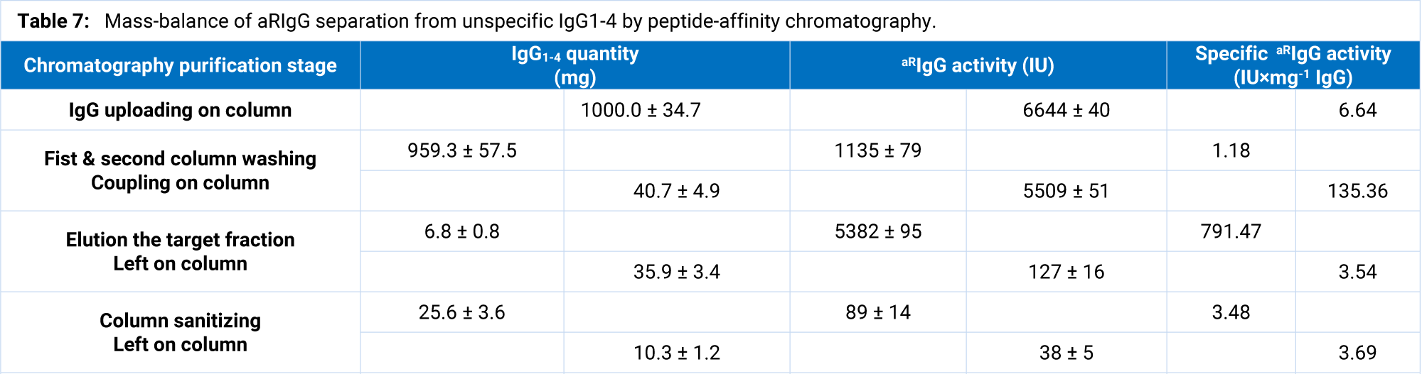
A 10 min of residence time was enough for coupling the ~83% of target activity aRIgG activity by peptide-affinity gel (the difference between total and coupling activity on the uploading and washing stages). The residence time increasing did not lead to significant changes in the binding of target activity. Note that the main part of this activity was associated with 4% of the IgG only, that suggested the coupling in basic the aRIgG . 35.9 mg IgG with specific activity 3.54 IU × mg-1 IgG. We do not refer to significant losses of aRIgG . Most likely this is due to the presence in the human immunoglobulin family the IgGs with higher affinity to the synthesized peptide. But its IgGs do not have high aRIgG activity. The proteins washed at the sanitization stage were the denatured IgGs that quantity around 1% is normal for chromatographic process.
The anti-Rubella IgG separated from the total human IgG1-4 family has a specific activity closed to 800 IU × mg-1. Given that the aRIgG titer in the donor blood was about 29 IU × mL-1, and in 1 mL of plasma was about 4 mg IgG, it means that specific activity was ~7,5 ÷ 8.0 IU × mg-1 and that we achieved an increasing in aRIgG titer by 100 times.
In a total for the 4 chromatographic cycles during 2 hours 27 mg of immunoglobulin with a total anti-rubella activity 21,500 IU was isolated from 1 L of standard donor plasma. Thus, it can be argued that the main goal of the investigation was achieved - the developing a peptide affinity chromatographic gel with satisfactory properties for the isolation of specific anti-rubella immunoglobulin. We do not rule out that on further work on the described approach we will have not only a more effective adsorbent, but also adsorbents for isolation from standard donor plasma of anti-herpes, anti-measles, anti-polio, anti-diphtheria and other IgG, according to which large-scale vaccinations with long-life immunity can be carried out on these diseases.
The high aRIgG titer of separated immunoglobulins show that affinity RDHHGTHE peptide and its family (6-1/2/3/4/5/6-E2) or their closed by physic-chemical properties peptides no. -3-6 from 5-E2 (shown in the table 6) captures exactly this aRIgG . Cong, Jiang and Tien back in 2011 found that the role of MOG, expressed in the central nervous system and in other tissues such as spleen, liver and thymus of mice, as a receptor of RV attachment to cell cannot be excluded [24]. Nobody has denied this fact so far, due to which we suggested that MOG could have a similar analogue of the peptide no. 6-6-E2 for RVs binding on the cell surface. In order to verify, we performed analysis of MOG aas sequence represented at UniProt_KB (Q16653 MOG human, Q63345 MOG rat, Q61885 MOG mouse, P55803 MOG bovine, Q9BGS7 MOG monkey macaca, and Q29ZQ1 MOG monkey marmoset). Particular attention was paid to the sites of possible interaction MOG with RV capsid proteins (131DHSYQEEAAMELK143 and 236AGQFLEELR244) indicated by the authors early publication [24]. The site 196TFDPHFLRV204 [24] was rejected as inadequate to interact with the virus because it was topologically located inside the cell, not even transmembrane. On the base both analyzed sites were determined following peptides at human MOG: 130RDHSYQEE137 and 232HRRLAGQF239.
A comparison of the developed RDHHGTHE and the found peptide 130RDHSYQEE137 shows that they are identical by 50% «1R2D3H…6E» and other almost does not differ in physic-chemical properties. Thus, at 4H/S replacement that should interact with V at 543VLLVPWVL550 of E2 the histidine looks more acceptable amino acid than serine, due to the fact that it will interact with valine not only by hydrophilicity but also by charge, while serine interacts with valine only due to pronounced hydrophilicity. The charge of serine and valine, which is close to zero, has no special effect on the interaction (HPIH = −1.7, pIH = 7.6; HPIS = −1.1, pIS=5.7; HPIV = 2.3, pIV = 6.0). The same reasoning applies to another pair of amino acids 7H/E, where histidine will also be preferable to glutamine in interaction with valine (HPIH = −1.7, pIH = 7.6; HPIE = −2.6, pIE = 3.2; HPIV = 2.3, pIV = 6.0). At the 5G/Y replacement that should interact with P the glycine due to high hydrophilicity more preferable for interaction (HPIg = 0.7, pIG = 6.0; HPIY = 0.1, pIY = 5.7; HPIP = −0.3, pIP=6.5). In the 6T/Q pair, tyrosine is the only one antisense aa for tryptophan [14,15], so it will be preferred in any case (HPIT = −0.8, pIT = 5.9; HPIQ = −2.9, pIQ = 5.7; HPIW = 1.5, pIW = 6.0).
The peptide RDHHGTHE as analog of the MOG 232HRRLAGQF239 has a greater potential to interact with another site of RV E2 – 354VLPGHWLQ36 [26,82], namely: a powerful positive charged core with high hydrophilicity (RDHH) against the low negative charge and medium hydrophobicity (VLPG) and three couples of the sense-antisense aas (2D/2L, 6T/6W, 7H/7L). The level of interaction is lower than with 543VLLVPWVL550 due to the presence of antagonistic by the physical sense aas (the same charge and hydrophobicity) at peptides positions 3H/3P and 8E/8Q.
Another developed peptide QHGPLTDV (shown 66% aRIgG activity coupling as an affinity ligand) corresponds to the possibility of interaction with 543VLLVPWVL550, but showed low efficiency. This may be caused both by the only partial conformational availability of 543VLLVPWVL550 and by the weak interaction due to the absence of amino acids with a strong charge and sufficient hydropathy properties.
The same potential RV-receptor core peptides were found at the MOG aas sequences of other animals with a short offset of 1-2 aa positions. The presence of T-cell epitopes on E2 protein of RV with same or closed aas sequence [82] and immune epitope with anti-sense sequence on the developed against RV’ E2 protein [100] add confidence that human MOG peptides 130RDHSYQEE197 and 232HRRLAGQF239 both or one of them are/is the key site(s) of cell receptor and RV interaction. Which one peptide is including into RV-receptor, will be possible to recognize in the next study.
Conclusion
With each new study, we are increasingly finding that biochemical process of protein-protein interaction occurs due to hydropathic and electrostatic contact between linear sequences built from amino acid partner couples. This emphasizes once again the non-randomness and the logical genetic program (sense and antisense sequences on each of the interaction partner proteins) of recognition, such as receptor recognition by the effector. Conversely, the interaction due to conformational convergence of partner amino acids seems random and very difficult for genetic programming. From the same point of view, it no longer seems speculative and fantastic that the interaction amino acids partner couples (or by J. Biro sense-antisense) are the same genetic code for proteins as the nucleotide pairs for nucleic acids.
Based on the above the definition at the MOG receptor RV the linear peptides of effective interaction with virus antisense peptides and more than 66-81% extraction of aRIgG from a mixture of IgG1-4 plasma donors suggests that MOG peptides 130RDHSYQEE137 or 232HRRLAGQF239 may be a key receptor’s sequence for RV. On the other hand, we hope that peptides QHGPLTDV/RDHHGTHE, which we developed in this study, could work no less effectively in the living organism as blockers of the virus landing site on the cellular receptor. If the assumptions made in the following experiments are proven, it will pave the way for the development of reliable natural peptide safeguards pharmaceuticals not only against viruses and bacteria.
Acknowledgement
Authors express profound gratitude to the staff of pilot plant of scientific and manufacturing firm Shijir International LCC, Sukhbaatar sq., Bodi Tower building, Ulaanbaatar, Mongolia, for the opportunity to intervene in the real manufacturing processes and to test the represented method on the pilot plant in the Raining (Boroo) Valley of Mongolia.
Funding information
This study was funded by Neutromics Ukraine TOV (Ukraine) and Shijir International LCC (Mongolia) in equal shares. The scientific data and practical results obtained by authors belong to Neutromics Ukraine TOV (Ukraine) and Shijir International LCC (Mongolia) and fixed by the agreement and the priority certificate for the granted patent of Ukraine. Both companies have allowed publication in the open press with the transfer of rights on the article to the appropriate publisher.
Conflict of Interest Statement
Authors declare no conflict of interest. The study was performed at a time when authors were employees of Neutromics Ukraine TOV and working together at the biotechnology pilot plant in Raining (Boroo) Valley, Mongolia.
All authors contributed to the study conception and design. Material preparation, data collection and analysis were performed by all authors too. The first draft of the manuscript was written by Serhiy P. Havryliuk and Heorgii L. Volkov and all authors commented on previous versions of the manuscript. All authors read and approved the final manuscript.
Ethical Approval
This article does not contain any studies with human participation or animal performed by any of the authors.
References
- Volkov GL, Havryliuk SP, Krasnobryzha IM, Havryliuk OS. The protein/peptide direct virus inactivation during chromatographic process: developing approaches. Appl Biochem Biotechnol. 2017;181(1):233-249. doi: 10.1007/s12010-016-2209-2.
- Havryliuk, SP, Krasnobryzha IM, Havryliuk OS, Volkov GL. The simultaneous human FVIII/vWF purification and virus inactivation combined in chromatographic column. J Biomol Res Ther. 2017;6(2):1000157. doi: 10.4172/2167-7956.1000157.
- Yoshidome T, Kinoshita M. Physical origin of hydrophobicity studied in terms of cold denaturation of proteins: comparison between water and simple fluids. Phys Chem Chem Phys. 2012;14(42):14554-14566. doi: 10.1039/C2CP41738C.
- Risso PH, Borraccetti, DM, Araujo C, Hidalgo ME, Gatti CA. Effect of temperature and pH on the aggregation and the surface hydrophobicity of bovine κ-casein. Colloid Polym Sci. 2008;286(12):1369-1378. doi: 10.1007/s00396-008-1906-y.
- Huang DM, Chandler D. Temperature and length scale dependence of hydrophobic effects and their possible implications for protein folding. PNAS. 2000;97(15):8324-8327. doi: 10.1073/pnas.120176397.
- Van der Vegt, Nayar, D. The hydrophobic effect and the role of cosolvents. J Phys Chem B. 2017;121(43):9986-9998. doi: 10.1021/acs.jpcb.7b06453.
- Mouratou B, Béhar G, Pecorari F. Artificial affinity proteins as ligands of immunoglobulins. Biomolecules. 2015;5:60-75. doi: 10.3390/biom5010060.
- Krepper W, Satzer P, Beyer BM, Jungbauer A. Temperature dependence of antibody adsorption in protein affinity chromatography. J Chromatogr A. 2018;1551:59-68. doi: 10.1016/j.chroma.2018.03.059.
- Alexander P, Fahnestock S, Lee T, Orban J, Bryan P. Thermodynamic analysis of the folding of the streptococcal protein G IgG-binding domains B1 and B2: why small proteins tend to have high denaturation temperatures. Biochemistry. 1992;31(14):3597-3603. doi: 10.1021/bi00129a007.
- Akerström B, Lindahl D, Björck L, Lindqvist A. Protein Arp and protein H from group A streptococci. Ig binding and dimerization are regulated by temperature. J Immunol. 1992;148(10):3238-3243. https://tinyurl.com/hyd2kf8m.
- Franklin JN, Victa C, Donald P, Fahrner R. Fragments of protein an eluted during protein an affinity chromatography. J Chromatogr A. 2007;1163(1-2):105-111. doi: 10.1016/j.chroma.2007.06.012.
- Shimoni J, Gunawan F, Thomas A, Vanderlaan M, Stults J. Trace level analysis of leached protein in bioprocess samples without interference from the large excess of rhMAb IgG. J Immunol Methods. 2009;28;341(1-2):59-67. doi: 10.1016/j.jim.2008.10.015.
- Basle YL, Chennell P, Tokhadze N, Astier A, Sautou V. Physicochemical stability of monoclonal antibodies: A review. J Phar Sci. 2020;109(1):169-190. doi: 10.1016/j.xphs.2019.08.009.
- Biro J. System and method to obtain oligo-peptides with specific high affinity to query proteins. US 8145437 B2. 2012. https://tinyurl.com/yc297svx
- Havryliuk SP, Krasnobryzha M, Havryliuk OS, Volkov GL. A simple, cheap and fast method for calculation of peptides to be used as ligands in affinity chromatography or in other applications in biotech and pharma industry. Am J Pharmacol Ther. 2020;4(1):007-018. doi: 10.3787/ajpt.id20.
- Detmer F, Griesbach J, Sleijpen K, Hazenoot J, Hermans P. Rapid implementation of novel affinity purification. Manufacture of commercial-scale next-generation antibody therapies. Bio Process International. Featured report “Speed to market”. 2019;17(10):12-17. https://tinyurl.com/2dsrc7re
- Muguruma K, Fujita K, Fukuda A, Kishimoto S, Sakamoto S, Arima R. Kinetics-based structural requirements of human immunoglobulin G binding peptides. ACS Omega. 2019;4(11);14390-14397. doi: 10.1021/acsomega.9b01104.
- Toughiri R, Wu X, Ruiz D, Huang F, Crissman JW, Dickey M, Froning K, Conner EM, Cujec TP, Demarest SJ. Comparing domain interactions within antibody Fabs with kappa and lambda light chains. MAbs. 2016;8(7):1276-1285. doi: 10.1080/19420862.2016.1214785.
- Biro JC. Genetic code completed the proteomic code and nucleic acid assisted protein folding. Bio International Convention/Boston Convention and Exhibition Center, USA. 2012.
- Chen X, Li R, Pan Z, Qian C, Yang Y. Human monoclonal antibodies block the binding of SARS-CoV-2 spike protein to angiotensin converting enzyme 2 receptor. Cell Mol Immunol. 2020;17(6):647–649. doi: 10.1038/s41423-020-0426-7.
- Huang RY, Lin MS, Kuo TC, Chen CL, Lin CC, Chou YC. Humanized cOVID‐19 decoy antibody effectively blocks viral entry and prevents SARS‐CoV‐2 infection. EMBO Mol Med. 2021;13(1):e12828. doi: 10.15252/emmm.202012828.
- Dimech W, Arachchi N, Cai J, Sahin T, Wilson K. Investigation into low-level anti-rubella virus IgG results reported by commercial immunoassays. Clin Vaccine Immunol. 2013;20(2):255-261. doi: 10.1128/CVI.00603-12.
- Lyashko: The level of immunization against measles by the end of the year will be 78%. 2020. https://tinyurl.com/2p9evnm6
- Cong H, Jiang Y, Tien P. Identification of the myelin oligodendrocyte glycoprotein as a cellular receptor for rubella virus. J Virol. 2011;85(21):11038-11047. doi: 10.1128/JVI.05398-11.
- Trinh QD, Pham NTK, Takada K, Komine S, Hayakawa S. Myelin oligodendrocyte glycoprotein-independent rubella infection of keratinocytes and resistance of first-trimester trophoblast cells to rubella virus in vitro. Viruses. 2018;10(1):23. doi: 10.3390/v10010023.
- McCarthy M, Lovett A, Kerman RH, Overstreet A, Wolinsky JS. Immunodominant T-cell epitopes of rubella virus structural proteins defined by synthetic peptides. J Virol. 1993;67(2):673–681. doi: 10.1128/jvi.67.2.673-681.1993.
- Volkov G, Andrianov, S., Slominskiy A, Havrylyuk O, Goroshnikova T. Advantages of the EBA method for the first stage chromatography plasma proteins re-purification. 2005.
- Volkov GL, Andrianov SI, Slominskiy AY, Havrylyuk OS, Goroshnikova TV. Advantages of the EBA method for the plasma proteins re-purification obtained by alcohol Kohn method. IX Ukrainian Biochem. 2006;2:142.
- Bradford MM. A rapid and sensitive method for the quantitation of microgram quantities of protein utilizing the principle of protein-dye binding. Anal Biochem. 1976;72:248-254. doi: 10.1006/abio.1976.9999.
- Stoscheck CM. Quantitation of protein. Method Enzymol. 1990;182;50-69. doi: 10.1016/0076-6879(90)82008-p.
- SP Bajaj, AE Schmidt, AM Mathur, K Padmanabhan, D Zhong, M Mastri, PJ Fay, Factor IXa: Factor VIIIa interaction. Helix 330-338 of factor IXa interacts with residues 558-565 and spatially adjacent regions of the A2 subunit of Factor VIIIa. JBC. 2001;276(19):16302-16309. doi: 10.1074/jbc.M011680200.
- Bio-Works user instruction for Work Beads 40 ACT, IN40400010. https://tinyurl.com/str7bxne.
- Gavara PR, Bibi NS, Sanchez ML, Grasselli M, Lahore M. Chromatographic characterization and process performance of column-packed anion exchange fibrous adsorbents for high throughput and high capacity bioseparations. Processes. 2015;3:204-221. doi: 10.3390/pr3010204.
- Volkov GL, Havryliuk SP, Krasnobryzha IM, Zhukova AI, Havryliuk OS. Method for isolation complex of Factor VIII and von Willebrand Factor from blood plasma using gel-exclusion column chromatography, UA 110920, 2016.
- Kaerber G. Contribution to the collective treatment of pharmacological series experiments. Naunyn Schmiedberg's Arch. Exp Pathol Pharmakol. 1931;162:480-483. https://tinyurl.com/3dhe4b5p
- Spearman C. The method of “right and wrong cases” (“constant stimuli”) without Gauss’s formulae. Br J Psyhol. 1908;2:277-282. https://tinyurl.com/32p7rk9p
- Zhang J, Storey KB. Bioplot: An easy-to-use R pipeline for automated statistical analysis and data visualization in molecular biology and biochemistry. Peer J. 2016;4:e2436. doi: 10.7717/peerj.2436.
- Tan LK, Shopes RJ, Oi VT, Morrison SL. Influence of the hinge region on complement activation, C1q binding, and segmental flexibility in chimeric human immunoglobulins. PNAS. 1990;87(1):162-166. doi: 10.1073/pnas.87.1.162.
- Canfield SM, Morrison SL. The binding affinity of human IgG for its high affinity Fc receptor is determined by multiple amino acids in the CH2 domain and is modulated by the hinge region. J Exp Med. 1991;173(6):1483-1491. Doi: 10.1084/jem.173.6.1483.
- Williams RC, Malone CC. Rheumatoid-factor-reactive sites on CH2 established by analysis of overlapping peptides of primary sequence. Scand J Immunol. 1994;40(4):443-456. Ddoi: 10.1111/j.1365-3083.1994.tb03487.x.
- Peterson C, Malone CC, Williams RC. Rheumatoid-factor-reactive sites on CH3 established by overlapping 7-mer peptide epitope analysis. Mol Immunol. 1995;32(1):57-75. doi: 10.1016/0161-5890(94)00122-h.
- Shields RL, Namenuk AK, Hong K, Meng YG, Rae J, Briggs J. High resolution mapping of the binding site on human IgG1 for FcγRI, FcγRII, FcγRIII, and FcRn and design of IgG1 variants with improved binding to the FcγR. JBC. 2001;276(9):6591-6604. doi: 10.1074/jbc.M009483200.
- Yuriev E, Ramsland PA, Edmundson AB. Recognition of IgG-derived peptides by a human IgM with an unusual combining site. Scand J Immunol. 2002;55(3):242-255. doi: 10.1046/j.0300-9475.2002.01032.x.
- Dall'Acqua WF, Cook KE, Damschroder MM, Woods RM, Wu H. Modulation of the effector functions of a human IgG1 through engineering of its hinge region. J Immunol. 2006;177(2):1129-1138. doi: 10.4049/jimmunol.177.2.1129.
- Stavenhagen JB, Gorlatov S, Tuaillon N, Rankin CT, Li H. Fc optimization of therapeutic antibodies enhances their ability to kill tumor cells in vitro and controls tumor expansion in vivo via low-affinity activating Fcγ receptors. Cancer Res. 2007;67(18):8882-8890. doi: 10.1158/0008-5472.CAN-07-0696.
- Keeble AH, Zahra K, Forster A, James LC. RIM21 is an IgG receptor that is structurally, thermodynamically, and kinetically conserved. PNAS. 2008;105(16):6045-6050. doi: 10.1073/pnas.0800159105.
- Richards JO, Karki S, Lazar GA, Chen H, Dang W, Desjarlais JR. Optimization of antibody binding to Fc;RIIa enhances macrophage phagocytosis of tumor cells. Mol Cancer Ther. 2008;7(8):2517-2527. doi: 10.1158/1535-7163.MCT-08-0201.
- Jefferis R. The antibody paradigm: Present and future development as a scaffold for biopharmaceutical drugs, Biotechnol. Genet Eng Rev. 2010;26:1-42. doi: 10.5661/bger-26-1.
- Huang B, Liu FF, Dong XY, Sun Y. Molecular mechanism of the affinity interactions between protein A and human immunoglobulin G1 revealed by molecular simulations. J Phys Chem B. 2011;115(14):4168-4176. doi: 10.1021/jp111216g.
- Lee YC, Tsai KC, Leu S, Wang TJ, Liu CY, Yang Y. Isolation, characterization, and molecular modeling of a rheumatoid factor from a Hepatitis C virus infected patient with Sjögren's syndrome. Scientific World J. 2013;516. doi: 10.1155/2013/516516.
- Mimoto F, Igawa T, Kuramochi T, Katada H, Kadono S, Kamikawa T. Novel asymmetrically engineered antibody Fc variant with superior FcγR binding affinity and specificity compared with afucosylated Fc variant. 2013;5(2):229-236. doi: 10.4161/mabs.23452.
- Diebolder CA, Beurskens FJ, Jong RN, Koning RI, Strumane K, Lindorfer MA. Complement is activated by IgG hexamers assembled at the cell surface. Science. 2014;343(6176):1260–1263. doi: 10.1126/science.1248943.
- Kelton W, Mehta N, Charab W, Lee J, Lee CH, Kojima T. IgGA: A “cross-isotype” engineered human Fc antibody domain that displays both IgG-like and IgA-like effector functions. Chem Biol. 2014;21(12):1603-1609. doi: 10.1016/j.chembiol.2014.10.017.
- Jensen PF, Larraillet V, Schlothauer T, Kettenberger H, Hilger M. Investigating the interaction between the neonatal Fc receptor and monoclonal antibody variants by hydrogen/deuterium exchange mass spectrometry. Mol Cell Proteomics. 2015;14(1):148-161. doi: 10.1074/mcp.M114.042044.
- Foss S, Bottermann M, Jonsson A, Sandlie I, James LC, Andersen JT. TRIM21 – from intracellular immunity to therapy. Front Immunol. 2019;10:2049. doi: 10.3389/fimmu.2019.02049.
- Foss S, Watkinson R, Sandlie I, James LC, Andersen JT. TRIM21: A cytosolic Fc receptor with broad antibody isotype specificity. Immunol Rev. 2015;268(1):328-339. doi: 10.1111/imr.12363.
- Kiyoshi M, Caaveiro JMM, Kawai T, Tashiro S, Ide, T, Asaoka Y. Structural basis for binding of human IgG1 to its high-affinity human receptor FcγRI. Nat Commun. 2015;6:6866. doi: 10.1038/ncomms7866.
- Oganesyan V, Mazor Y, Yang C, Cook KE, Woods RM, Ferguson A. Structural insights into the interaction of human IgG1 with FcγRI: No direct role of glycans in binding. Acta Cryst. 2015;71(Pt 11):2354-2361. doi: 10.1107/S1399004715018015.
- Monnet C, Jorieux S, Urbain R, Fournier N, Bouayadi K, De Romeuf. Selection of IgG variants with increased FcRn binding using random and directed mutagenesis: impact on effector functions. Front Immunol. 2015;6:39. doi: 10.3389/fimmu.2015.00039.
- Irani V, Guy AJ, Andrewa D, Beeson JG, Ramsland PA, Richards JS. Molecular properties of human IgG subclasses and their implications for designing therapeutic monoclonal antibodies against infectious diseases. Mol Immunol. 2015;67(2 Pt A):171-182. doi: 10.1016/j.molimm.2015.03.255.
- Walters, Jensen PF, Larailet V, Lin K, Patapoff T, Schlothauer T. Conformational destabilization of immunoglobulin G increases the low pH binding affinity with the neonatal Fc receptor. JBC. 2016;291(4):1817-1825. doi: 10.1074/jbc.M115.691568.
- Jensen PF, Schoch A, Larraillet V, Hilger M, Schlothauer T, Emrich T, Rand KD. A two-pronged binding mechanism of IgG to the neonatal Fc receptor controls complex stability and IgG serum half-life. Molecular & Cellular Proteomics. 2016;16(3):451-456. doi: 10.1074/mcp.M116.064675.
- Ashoor DN, Ben Khalaf, Hachemi S, Marzouq MH, Fathallah MD. Engineering of the upper hinge region of human IgG1 Fc enhances the binding affinity to FcγIIIa (CD16a) receptor isoform. Protein Engineering Design & Selection. 2018;31(6):205-212. doi: 10.1093/protein/gzy019.
- Shiroishi M, Ito Y, Shimokawa K, Lee JM, Kusakabe T, Ueda T. Structure–function analyses of a stereotypic rheumatoid factor unravel the structural basis for germline-encoded antibody autoreactivity. JBC. 2018;293(18):7008-7016. doi: 10.1074/jbc.M117.814475.
- Wang Х, Mathieu M, Brezski RJ. IgG Fc engineering to modulate antibody effector functions. Protein Cell 2018;9(1):63–73. Doi: 10.1007/s13238-017-0473-8.
- Saunders KO. Conceptual approaches to modulating antibody effector functions and circulation half-life. Front Immunol. 2019;10:1296. doi: 10.3389/fimmu.2019.01296.
- Yogo R, Yamaguchi Y, Watanabe H, Yagi H, Satoh T, Nakanishi M. The Fab portion of immunoglobulin G contributes to its binding to Fcγ receptor III. Sci Rep. 2019;9(1):11957. doi: 10.1038/s41598-019-48323-w.
- Vidarte L, Pastor C, Mas S, Blázquez AB, Rios V, Guerrero R, Vivanco F. Serine 132 is the C3 covalent attachment point on the CH1 domain of human IgG1. JBC. 2001;276(41):38217-38223. doi: 10.1074/jbc.M104870200.
- Panda S, Zhang J, Yang L, Anand GS, Ding JL. Molecular interaction between natural IgG and ficolin - mechanistic insights on adaptive-innate immune crosstalk. Sci Rep. 2014;4:3675. doi: 10.1038/srep03675.
- Mallery DL, McEwan WA, Bidgood SR, Towers G, Johnson CM, James LC. Antibodies mediate intracellular immunity through tripartite motif containing 21 (TRIM21). PNAS. 2010;107(46):19985-19990. doi: 10.1073/pnas.1014074107.
- Foss S, Watkinson RE, Grevys A, McAdam MB, Bern M, Hoydahl LS. TRIM21 immune signaling is more sensitive to antibody affinity than its neutralization activity. J Immunol. 2016;196(8):3452-3459. doi: 10.4049/jimmunol.1502601.
- Bidgood SR, Tam JC, McEwan WA, Mallery DL, James LC. Translocalized IgA mediates neutralization and stimulates innate immunity inside infected cells. PNAS. 2014;111(37):13463-13468. doi: 10.1073/pnas.1410980111.
- Islam N, Shen F, Gurgel PV, Rojas OJ, Carbonell R.G. Dynamic and equilibrium performance of sensors based on short peptide ligands for affinity adsorption of human IgG using surface plasmon resonance. Biosens Bioelectron. 2014;58:380-387. doi: 10.1016/j.bios.2014.02.069.
- Yang H, Gurgel PV, Williams DK, Bobay BG, Cavanagh J, Muddiman DC, Carbonell RG. Binding site on human immunoglobulin G for the affinity ligand HWRGWV. J Mol Recognit. 2010;23(3):271-282. doi: 10.1002/jmr.967.
- Zhao WW, Liu FF, Shi QH, Sun Y. Octapeptide-based affinity chromatography of human immunoglobulin G: Comparisons of three different ligands. J Chromatogr A. 2014;1359:100-111. doi: 10.1016/j.chroma.2014.07.023.
- Kruljec N, Molek P, Hodnik V, Anderluh G, Bratkovič T. Development and characterization of peptide ligands of immunoglobulin G Fc region. Bioconjug Chem. 2018;29(8):2763-2775. doi: 10.1021/acs.bioconjchem.8b00395.
- Williams RC Jr, Malone CC. Rheumatoid-factor-reactive sites on CH2 established by analysis of overlapping peptides of primary sequence. Scand J Immunol. 1994;40(4):443-456. doi: 10.1111/j.1365-3083.1994.tb03487.x.
- Remington KM. Fundamental strategies for viral clearance. Part 2: Technical approaches. BioProcess International. 2015;13(5):10-17. https://tinyurl.com/3hpchs6x
- Alonso WR, Trukawinski S, Savage M, Tenold RA, Hammond DJ. Viral inactivation of intramuscular immune serum globulins. Biologicals. 2000;28(1):5-15. doi: 10.1006/biol.1999.0234.
- Ou D, Chong P, McVeish P, Jefferies WA, Gillam S. Characterization of the specificity and genetic restriction of human CD4+ cytotoxic T cell clones reactive to capsid antigen of rubella virus. Virology. 1992;191(2):680-686. doi: 10.1016/0042-6822(92)90243-i.
- Ou D, Chong P, Tripet B, Gillam S. Analysis of T- and B-cell epitopes of capsid protein of rubella virus by using synthetic peptides. J Virol. 1992;66(3):1674-1681. doi: 10.1128/JVI.66.3.1674-1681.1992.
- Ou D, Chong P, Choi Y, McVeigh, Jefferies WA, Koloitis G, Tingle AJ, Gillam S. Identification of T-cell epitopes on E2 protein of rubella virus, as recognized by human T-cell lines and clones. J Virol. 1992;66(11):6788-6793. doi: 10.1128/JVI.66.11.6788-6793.1992.
- Ou D, Chong P, Tingle AJ, Gillam S. Mapping T‐cell epitopes of rubella virus structural proteins E1, E2, and C recognized by T‐cell lines and clones derived from infected and immunized populations. J Med Virol. 1993;40(3):175-183. doi: 10.1002/JMV.1890400302.
- Wolinsky JS, Sukholutsky E, Moore WT, Lovett A, McCarthy M, Adame B. An antibody- and synthetic peptide-defined rubella virus E1 glycoprotein neutralization domain. J Virol. 1993;67(2):961-968. doi: 10.1128/JVI.67.2.961-968.1993.
- Chaye H, Ou D, Chong P, Gillam S. Human T- and B-cell epitopes of E1 glycoprotein of rubella virus. J Clin Immunol. 1993;13(2):93-100. doi: 10.1007/BF00919265.
- Lovett E, McCarthy M, Wolinsky JS. Mapping cell-mediated immunodominant domains of the rubella virus structural proteins using recombinant proteins and synthetic peptides. J Gen Virol. 1993;74(Pt 3):445-452. doi: 10.1099/0022-1317-74-3-445.
- Marttila J, Ilonen J, Lehtinen M, Parkkonen P, Salmi A. Definition of three minimal T helper cell epitopes of rubella virus E1 glycoprotein. Clin Exp Immunol. 1996;104(3):394-397. doi: 10.1046/j.1365-2249.1996.54762.x.
- Ou D, Mitchell LA, Décarie D, Tingle AJ, Nepom GT. Promiscuous T-cell recognition of a rubella capsid protein epitope restricted by DRB1*0403 and DRB1*0901 molecules sharing an HLA DR supertype. Human Immunol. 1998;59(3):149-157. doi: 10.1016/s0198-8859(98)00006-8.
- Mitchell LA, Tingle AJ, Décarie D, Shukin R. Identification of rubella virus T-cell epitopes recognized in anamnestic response to RA27/3 vaccine: associations with boost in neutralizing antibody titer. Vaccine. 199914;17(19):2356-2365. doi: 10.1016/s0264-410x(99)00040-7.
- Mitchell LA. Sex differences in antibody- and cell-mediated immune response to rubella re-immunisation. J Med Microbiol. 1999;48(12):1075-1080. doi: 10.1099/00222615-48-12-1075.
- Ou D, Mitchell LA, Metzger DL, Gillam S, Tingle AJ. Cross-reactive rubella virus and glutamic acid decarboxylase (65 and 67) protein determinants recognised by T cells of patients with type I diabetes mellitus. Diabetologia. 2000;43(6):750-762. doi: 10.1007/s001250051373.
- Robinson K, Mostratos A, Grencis RK. Generation of rubella virus-neutralizing antibodies by vaccination with synthetic peptides. FEMS Immunol Med Microbiol. 1995;10(3-4):191-198. doi: 10.1111/j.1574-695X.1995.tb00033.x.
- Beatch MD, Hobman TC. Rubella virus capsid associates with host cell protein p32 and localizes to mitochondria. J Virol. 2000;74(12):5569-5576. doi: 10.1128/JVI.74.12.5569-5576.2000.
- Ghebrehiwet B, Atreya, CD. The N-terminal conserved domain of rubella virus capsid interacts with the C-terminal region of cellular p32 and overexpression of p32 enhances the viral infectivity. Virus Res. 2002;85(2):151-161. doi: 10.1016/s0168-1702(02)00030-8.
- Everitt JC, Law LJ, Hobman TC. Interactions between rubella virus capsid and host protein p32 are important for virus replication. J Virol. 2005;79(16):10807-10820. doi: 10.1128/jvi.79.16.10807-10820.2005.
- Chaye H, Chong P, Tripet B, Brush B, Gillam S. Localization of the virus neutralizing and hemagglutinin epitopes of E1 glycoprotein of rubella virus. Virology. 1992;189(2):483-492. doi: 10.1016/0042-6822(92)90572-7.
- Mitchell LA, Zhang T, Ho M, Décarie D, Tingle AJ, Zrein M, Lacroix M. Characterization of rubella virus-specific antibody responses by using a new synthetic peptide-based enzyme-linked immunosorbent assay. J Clin Microbiol. 1992;30(7):1841-1847. doi: 10.1128/jcm.30.7.1841-1847.1992.
- Cordoba P, Grutadauria SL, Cuffini C, Zapata MT. Presence of a neutralizing domain in isolates of rubella virus in Cordoba, Argentina. Clin Diagn Lab Immunol. 1997;4(4):493-495. doi: 10.1128/cdli.4.4.493-495.1997.
- Cordoba P, Lanoel A, Grutadauria S, Zapata M. Evaluation of antibodies against a rubella virus neutralizing domain for determination of immune status. Clin Diagn Lab Immunol. 2000;7(6):964-966. doi: 10.1128/CDLI.7.6.964-966.2000.
- Elgenaid SN, Hajj EM, Ibrahim AM, Essa ME, Hassan MA. Prediction of multiple peptide based vaccine from e1, e2 and capsid proteins of rubella virus: An in-silico approach. Immunome Res. 2018;14(1):1-13. doi: 10.4172/1745-7580.1000146.





Content Alerts
SignUp to our
Content alerts.
 This work is licensed under a Creative Commons Attribution 4.0 International License.
