
2026 January 20;7(1):001-011. doi: 10.37871/jbres2251.
-
Subject area(s):

 |
|
 |
|
 |
|

 Review Article
Anthrax Pathogenesis as Overcoming Lymph Node Macrophage Barrier
Noskov Anatoly N*
Introduction
Anthrax is especially dangerous infectious disease caused by the gram-positive spore-forming microorganism Bacillus anthracis, and with expressed toxic syndrome and high mortality of infected animals or humans [1,2]. Bacillus anthracis belongs in the Bacillus cereus group according to biotaxonomic characteristics [3,4]. The of these two microorganisms is more than 90%, and from the evolution B. cereus is considered as a precursor of the anthrax pathogen [5].
A single point mutation in the gene of the global transcription regulator plcR induced the “silence” of a large gene cluster responsible for the synthesis of more than 20 proteins produced by B. cereus and the Bacillus anthracis forming [6,7]. Two high-molecular plasmids pX01 and pX02 were necessary for formation as new bacilli species [8-10]. These plasmids determined the new phenotype and the ecological niche of Bacillus anthracis - a highly virulent spore-forming bacilli with a powerful pathogenicity complex for animals and human [11,12].
The pX01 plasmid contains the structural genes of the exotoxin pag, lef, and cya, and gene of new global trans-activator atxA that regulates the transcription of the toxin and capsule formation genes, as well as the additional operon gerX encoding three germination genes A, B, and C [13-16]. These genes are located on a fragment of 44.8 thousand base pairs flanked by inverted IS-elements and called the "pathogenicity island " [17].
pX02 plasmid contains the cap operon that includes the capB, capC, capA, and capD genes responsible for the synthesis of D-glutamyl polypeptide and capsule assembly [18], as well as the dep gene encoding the capsule degradation process [19], and the regulatory genes acpA, acpB, and abrB [20,21] pX02 plasmid contains also the amiA gene that controls the peptidoglycan hydrolysis as important and needing stage at spore germination when envelope is destroying [22].
The global transcription regulator atxA is strongly dependent from CO2 (5-10%), and this played a crucial role in the in vivo selection of new bacilli [23-26].
In nature the anthrax pathogen infecting occurs only by spores [1,2,28] and therefore transmission of infection is possible only after the death of the host and the spore formation in it.
Some pathways of spore entry into the host body are described: subcutaneous, oral-intestinal, intranasal and aerogenic.
Peroral-intestinal infection occurs with the help of water by washing out the spores from remains of anthrax died animals and eating the meat of these animals. The lethal dose ranges from several hundred spores and the mortality rate reaches 40% [27,28]. This infection pathway is the most significant in the spread of anthrax from an epidemiological point of view.
As for subcutaneous pathway, the possibility of such infection for animals is minimal for animals [26,28]. People are infected most often subcutaneously when are working with infected animals at enterprises engaged in the processing of animal hair and leather raw materials. Domestic infection is usually associated with butchering the meat of animals that have died from anthrax. The anthrax cutaneous form is expressed in inflammation of closest regional lymph nodes and is often accompanied by the formation of suppurations and ulcers. Human subcutaneous infection is characterized by a low mortality rate, since in this case the disease does not reach the stage of generalized infection [1,26,28]. The mouse model of infection with subcutaneous injection is the most effective for determination of the degree of pathogenicity of various Bacillus anthracis strains. Death of animals occurs usually on 2-3 days and lethal doses range from 1-10 spores. This pathway is used in laboratory researches as it makes it possible to control the conditions of the experiment to the greatest extent. Significant destruction of the skin occurs during skin infection in humans and this leads to a powerful inflammatory reaction. As a result, while the phagocytosed by macrophages pathogen is localized in the lymph node, an inflammatory zone is formed due to vasoconstriction of blood vessels, blocking the pathogen's progress into the blood. Therefore, the pathogen that has multiplied in the lymph node still needs to overcome the inflammatory zone. If the pathogen fails to do this inflammatory barrier, then skin cells lysis as ulcers occurs. The infectious disease prognosis is favorable in this case.
Aerogenic infecting is used only in experimental conditions. Lethal doses for aerogenic infecting range from 7000 to 50,000 spores according to various authors [28,29]. This parameter varies depending on the type of infected animals used in experiments, as well as on the volume of aerosol and other numerous conditions. However, the lethal dose in this case does not fall below several thousand spores [1,28,29]. Such high lethal doses are associated with most die of the germinating spores in alveolar macrophages, as was found on similar mouse adenocarcinoma cells J774.A [30,31].
B. anthracis spores, regardless of the method of entry into the host body, are phagocytosed by macrophages and transported to the lymph stream to the regional lymph node [32-36]. Spores germinate only in macrophages [37-42]. The components of the surrounding nutrient medium penetrate through the swollen shell into the spore and the GPR germination protease begins to hydrolyze SASP proteins that compactly pack DNA [43]. Degradation of protective proteins leads to the release of DNA regions carrying ger operon genes responsible for germination, as well as toxin formation genes [2,40].
The germinating spores are sensitive to aggressive phagosome conditions: rapid acidification and action of proteolytic and oxidative systems [44,45]. The rapid lysis of the phagosome membrane is most effective pathway to survival of germinating spores. The bacteria escape from phagosome to the macrophage cytoplasm during phagocytosis was shown firstly for listeria producing the pore-forming protein Listeriolysin O [46,47]. This protein lysed phagosomal membrane and thereby accelerated the bacterial escape from the phagosome to the cytoplasm.
Bacillus anthracis produces two pore-forming proteins: anthrolysin O [48,49] and protective antigen [50,51]. Anthrolysin synthesis is constitutive while bacterial survival on germination stage connects early expression of toxin formation genes [11]. It has been established also that the activity of the atxA (inducing toxin synthesis) that is necessary for the survival of germinating spores inside macrophages [2,15,17]. The atxA coordinates the germinating spore's responses to external signals, primarily to elevated CO2 levels [14,25]. Toxin production is regulated in vitro and in vivo through the positive control of the pag, lef, and cya by atxA [14,24]. This indicates the direct involvement of the toxin in the survival of germinating spores during phagocytosis. However, for the conversion of PA83 into the pore-forming protein PA63 capable to lysing the cell membrane is necessary also hydrolysis it by proteases [52].
From our point of view, immediately after the start of synthesis and secretion, the molecules of the protective antigen PA83 diffuse through the peptidoglycan layer, in that they undergo hydrolysis by bacterial proteases to PA63. So, it was demonstrated that immune inhibitor A1 (InhA1) is a secreted metalloprotease that is unique to pathogenic members of the Bacillus genus and has been associated with cleavage of host and bacterial proteins during infection [53].
Acidic phagosome conditions in the surface layer of peptidoglycan create to rapid oligomerization of PA63 molecules [50]. These oligomers are bound with the phagosome membrane and form pores in it without interacting with specific receptors [51]. The first oligomer sharply reduces the rate of acidification as protons escape from the phagosome through forming PA63 pores. Next PA63 oligomers bind with the phagosome membrane and destroy it. Thus, the survival of germinating spores during phagocytosis depends on the rate of synthesis of the protective antigen PA83 and its hydrolysis by bacterial proteases to PA63. Furthermore, swelling of germinating spores occurs unevenly and only in the part that is most likely adjacent to the phagosome membrane [41]. The remaining part of the spore shell protects the germinating spore that not yet covered a full-scale bacterial wall (Figure 1).
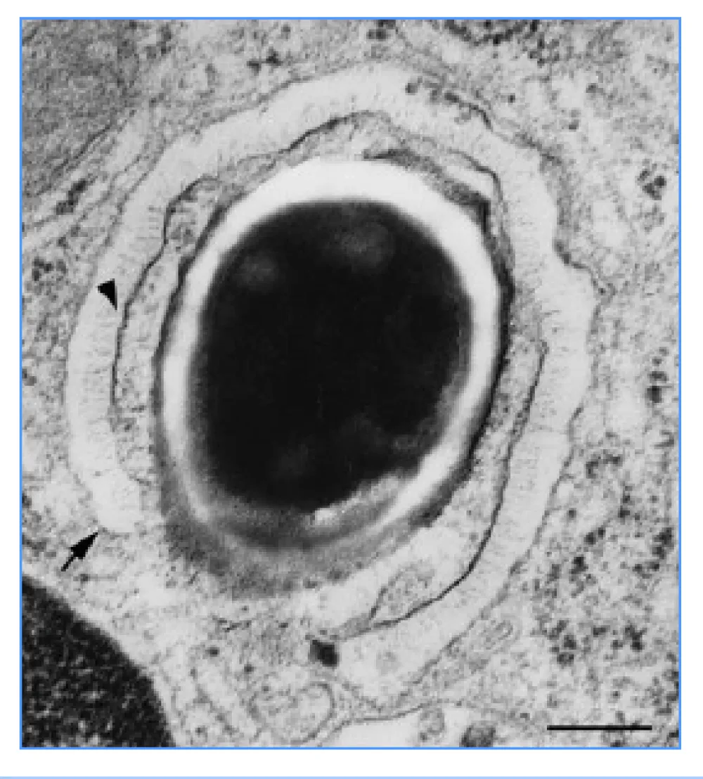
PA63-oligomer destroys phagosome membrane that connected with swelling part of germinating spore and it escape from phagosome.
Unusual and extremely interesting data was obtained at the study of spore survival with different genotypes during phagocytosis by macrophage-like J774A cells (Table 1).
| Table 1: Spore survival of different strain during phagocytosis by macrophage-like cell J774.1.[31]. | |||||
| N/N | STI-Rif pXO1-pXO2- | ∆Ames pXO1-pXO2+ | 81/1 pXO1+pXO2+ | 71/12 pXO1+pXO2-/+ | STI-1 pXO1+pXO2- |
| *LD50 | >109 | >107 | 1-10 | 102 | 105 |
| PA83/ml, 3 h | < 5 ng | < 5 ng | 380 ng | 310 ng | 520 ng |
| PA83/ml, 24 h | < 5 ng | < 5 ng | 19.3 mkg | 15.4 mkg | 24.6 mkg |
| Time, h | Seeding from macrophages, log CFU | ||||
| 0,5 (Control) | 5,99 | 5,98 | 6,03 | 5,99 | 6,01 |
| 2 | 0,68 | 1,25 | 2,87 | 3,65 | 5,12 |
| 4 | 0,16 | 0,42 | 1,6 | 2,29 | 5,76 |
| 6 | 0,15 | 0,24 | 0,42 | 1,58 | 5,43 |
| 12 | 0,11 | 0,24 | 0,29 | 1,26 | 5,34 |
| *LD50 for white inbred mice by subcutaneous injection. | |||||
Germinated spores of vaccine strain STI-1 (pXO1+pXO2-) survived better then spores high virulent strain 81/1 (pXO1+pXO2+): 5,12 KOE against 2,87 KOE according data at 2 hour. The analysis of PA synthesis data put everything in place. STI-1 synthesized PA significantly more than 81/1, accordingly 520 ng/ml and 380 ng/ml for 3 hours. But to 24 hours of cultivation, the difference decreased, accordingly 24.6 mkg/ml and 19.3 mkg/ml.
Thus, the effective survival of germinating spores in macrophages at an early stage of phagocytosis depends from the rate of PA83 synthesis (Figure 2).
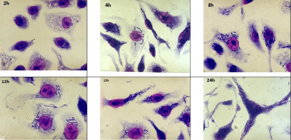
The investigation of the pathogen multiplication within macrophages showed the STI-1 vaccine strain vegetative cells multiplied in macrophages more 24 hours of observation. Macrophages not lysed although bacterial cells synthesized LF. Usually, the macrophages are lysed within one hour at lethal toxin (PA+LF) addition [30]. Therefore, the macrophages should have lysed within 1-2 hours after infection by spores of toxin-producing strains.
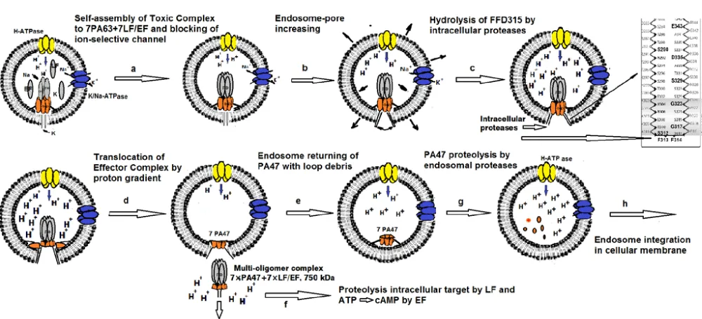
The macrophage lysis absence directly indicates that monomeric LF molecules, synthesized by vegetative cells, are not effective. Previously us was proposed translocation model according to which only the multi-oligomeric complex 7×PA17+7×LF/EF penetrates into the target-cell [55].
This oligomeric complex induces high and rapid cytotoxicity and not monomeric LF molecules. These results cast doubt on the proposed "unfolding-folding" as mechanism for the penetration of LF individual molecules into target cells [56]. Also, it is also questionable the supposed intracellular target for LF, although it was demonstrated LF hydrolyzes MAPK-kinases in solution and in macrophages [57,58] but not in Bacillus anthracis infected macrophages (Figure 3) [31,53,59,60].
Further, it was demonstrated the virulent strain 81/1 vegetative cells escape macrophages to 4-6 hours observation and the macrophages maintained their integrity at the same time [31,53,60] (Figure 4).
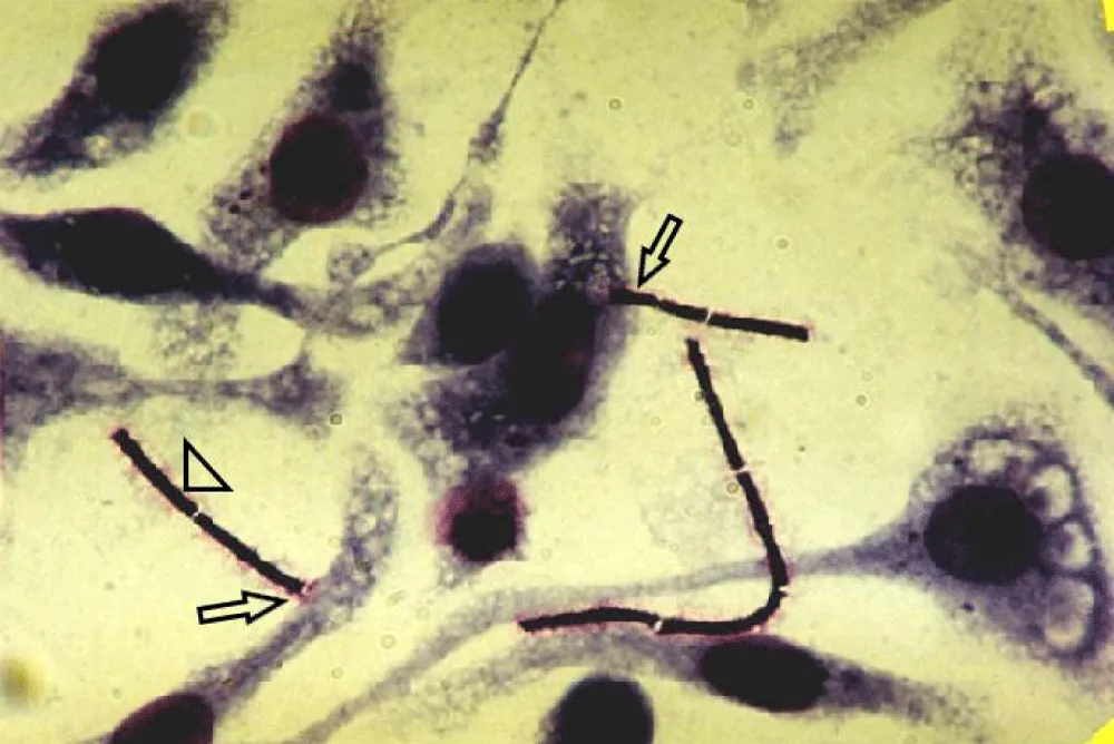
This moment should be discussed in more detail as it is fundamental for bacterial escape from macrophages. This phenomenon is the exocytosis of bacteria using pore-forming proteins. Numerous pores on the side of the inner membrane induce exocytosis and bacteria escape macrophages. The membrane closes behind the bacterial cells and the eukaryotic cell remains intact.
Bacterial exocytosis is a common and effective mechanism bacterial escape macrophages and other cells using pore-forming proteins (Bacillus, Listeria, Staphylococci, Streptococci etc.).
Concerning the Bacillus anthracis, only capsule-coated and toxin-synthesizing bacteria escape the macrophages. After synthesis and secretion, the molecules PA83 are hydrolyzed by bacterial proteases to PA63, that are oligomerized to 7×PA63 diffusing in the acidic capsule space (poly-D-glutamic acid) [26,61]. Therefore, pore-forming proteins 7×PA63 come out of the capsule and interact with the membrane inducing exocytosis. At this stage of infection, the capsule plays not a protective but a functional role as a space in that PA83 is activated.
Concerning the Bacillus anthracis, only capsule-coated and toxin-synthesizing bacteria escape quickly the macrophages by means exocytosis. With our point of view synthesized the PA83 molecules are hydrolyzed by bacterial proteases to PA63 that are oligomerized to 7×PA63 at diffusing in the acidic capsule space (poly-D-glutamic acid) [26,61]. Therefore, 7×PA63 pore-forming proteins diffuse from capsule and interact with the membrane inducing exocytosis. At this stage of infection, the capsule plays not a protective but a functional role as a space in that PA83 is activated.
Thus, vegetative cells of virulent strains escape quickly macrophages, while vaccine strain cells (pXO1+pXO2-) remain in macrophages for a long time.
Concerning non-toxigenic capsule strains (pXO1-pXO2+), they escape more slowly macrophages with exocytosis induced by pore-forming anthrolysin O. For the oligomer formation is required acidic conditions that appear at diffusing in the acidic capsule space. Certainly, antrolysin accelerates the escape of capsule-coated and toxin-synthesizing cells (pXO1+pXO2+).
These processes take place in the lymph nodes during infection. Therefore, vegetative cells of virulent strains, protected by capsule from rephagocytosis [62], escape quickly macrophages, multiply within lymph node and synthesize exotoxin. This exotoxin attacks and lyses macrophages and cells of the epithelial-endothelial barrier (EEB) [63]. As a result, the EEB is destroyed and bacterial cells penetrate into bloodstream. Using methods in vivo imaging with bioluminescent, it was found that during 30-35 hours after phagocytosis of spores, bacteria are detected only in one or more lymph nodes [64,65].
But why do bacteria multiply only in the lymph nodes during more 30 hours, and not disseminate to the lymphatic system with the lymph flow?
From our point of view, lymph node blockade is one of the key moments in anthrax pathogenesis (and other infections as plague, tularemia, and caused viruses etc.) and is induced by cytokines in response to the appearance of infected macrophages. During the reproduction of bacterial cells in macrophages, infection markers appear on their surface in the form of MHCI with peptides from secreted pathogen proteins. In the lymph node, these structures interact with T-cell receptors of T-cells. This interaction induces IL-1, TNFa, IL-2 and INFg synthesis by macrophages and T-cells. Synthesized cytokines stimulate the phagocytosis, maturation and proliferation of cytotoxic lymphocytes (CTL) to destroy the infectious agent. The cytokines induce also vasoconstriction of lymphatic vessels to lymph node blockade. Key moment, the immune system blocks the infected macrophages for pathogen elimination, so that it does not escape beyond the lymph node. IL-1 is a pro-inflammatory cytokine and its action is associated with endothelial cells. IL-2 responsible for cell proliferation.
Probably, TNFa or INFg (TNFa + INFg) can act as vasoconstriction inductors of lymphatic vessels. In addition, the synthesized cytokines penetrate to the bloodstream at toxin epithelial-endothelial barrier destruction and cause a prodromal state (first non-specific symptom) as a predecessor of cytokine storm.
Essential note, anthrax pathogenesis proceeds asymptomatic during 30-35 hours up to pathogen penetration into bloodstream and yet more 6-8 hours to first detected symptoms as temperature, frequent pulse and breathing.
Bioluminescence was used to reveal how bacterial cells multiply in mice immunized with a protective antigen. In immunized mice, a small bacterial growth observed at first, which quickly decayed. [64,65]. This allows us to conclude that infection of the lymph node occurs regardless of the presence of antibodies to PA that blocked the cytotoxic effect on all sensitive cells: macrophages and cells of the epithelial-endothelial barrier [64,65]. As a result, the pathogen is destroyed cooperatively in the lymph node by immune system cells and cannot enter the bloodstream although anti-PA antibodies do not have a direct antibacterial effect. Therefore, all used today vaccines induce PA-antibodies [66-68]. An important moment - phagocytosis and spore germination, and the penetration into the macrophage cytoplasm, do not depend on the presence of anti-PA antibodies.
Penetrated into bloodstream bacterial cells of virulent strains, and covered with a capsule and therefore protected from rephagocytosis, multiply rapidly. The infected host is converted in a kind of fermenter, in that a huge pathogen mass produces the exotoxin. The concentration of exotoxin can reach 20 mg/ml at the stage of bacteremia in 109 CFU. This is despite the fact that lethal doses for macrophages are about 1 mkg/ml [30]. The exotoxin attacks macrophages and endothelial cells of micro-vessels that leads to impaired gas exchange in the lungs, disruptive at work of the heart and other vital organs.
Thus, toxemia is ended as a powerful toxic and cardiovascular shock inducing death of infected organism [69,70].
After infected host death and a body temperature decreasing, ideal conditions for spore formation are created. The vegetative cell multiplication is stopped, genes responsible for capsule depolymerization are activated, toxin synthesis is inhibited, cell chains are shortened, etc. [68].
The died from anthrax animals, both in their natural environment and into burial grounds, are sources of long-term preservation of the spore form.
Conclusion
The spores, after entering the host body, are phagocytized by macrophages that migrate to near regional lymph node. Phagocytosed spores germinate in phagosomes and destroy the phagosome membrane by means PA63 oligomers that are formed during proteolysis by bacterial proteases PA83. Germinated spores penetrate to macrophage cytoplasm and transform into vegetative cells covered with a poly-d-glutamine capsule. Synthesized and secreted PA83 molecules diffuse through the acidic capsule space and are undergo proteolysis by bacterial proteases to PA63. The PA63 molecules form oligomers that interact with the macrophage membrane, form pores in it, and induce the exocytosis of vegetative cells. This mechanism of bacterial exit from macrophages is common to intracellular pathogens with pore-forming activity. The covered with a poly-d-glutamine capsule vegetative cells multiply rapidly in the lumen of the lymph node. The exotoxin synthesized by bacterial cells attacks and lyses macrophages and epithelial-endothelial barrier cells. Multiplied bacterial cell after destruction of the barrier between lymph and blood enter into the bloodstream. The rapidly increasing bacterial mass synthesizes a huge amount of exotoxin, which attacks and lyses macrophages and endothelial cells that form blood vessels. Final pathogenic stage is toxic and cardiovascular shock that culminating in the rapid host death.
Thus, the Bacillus anthracis spore form ensures the survival of this biological species in the harshest environmental conditions, and controlled by “quorum system” synthesis of pathogenic factors such as capsule, exotoxin and proteases make this pathogen the most dangerous for human and animal life.
References
- Sangwan N, Gangwal A, Jain P, Langtso C, Srivastava S, Dhawan U, Baweja R, Singh Y. Anthrax: Transmission, Pathogenesis, Prevention and Treatment. Toxins (Basel). 2025 Jan 24;17(2):56. doi: 10.3390/toxins17020056. PMID: 39998073; PMCID: PMC11860322.
- Mock M, Fouet A. Anthrax. Annu Rev Microbiol. 2001;55:647-71. doi: 10.1146/annurev.micro.55.1.647. PMID: 11544370.
- Read TD, Peterson SN, Tourasse N, Baillie LW, Paulsen IT, Nelson KE, Tettelin H, Fouts DE, Eisen JA, Gill SR, Holtzapple EK, Okstad OA, Helgason E, Rilstone J, Wu M, Kolonay JF, Beanan MJ, Dodson RJ, Brinkac LM, Gwinn M, DeBoy RT, Madpu R, Daugherty SC, Durkin AS, Haft DH, Nelson WC, Peterson JD, Pop M, Khouri HM, Radune D, Benton JL, Mahamoud Y, Jiang L, Hance IR, Weidman JF, Berry KJ, Plaut RD, Wolf AM, Watkins KL, Nierman WC, Hazen A, Cline R, Redmond C, Thwaite JE, White O, Salzberg SL, Thomason B, Friedlander AM, Koehler TM, Hanna PC, Kolstø AB, Fraser CM. The genome sequence of Bacillus anthracis Ames and comparison to closely related bacteria. Nature. 2003 May 1;423(6935):81-6. doi: 10.1038/nature01586. PMID: 12721629.
- Helgason E, Okstad OA, Caugant DA, Johansen HA, Fouet A, Mock M, Hegna I, Kolstø AB. Bacillus anthracis, Bacillus cereus, and Bacillus thuringiensis--one species on the basis of genetic evidence. Appl Environ Microbiol. 2000 Jun;66(6):2627-30. doi: 10.1128/AEM.66.6.2627-2630.2000. PMID: 10831447; PMCID: PMC110590.
- Kolstø AB, Tourasse NJ, Økstad OA. What sets Bacillus anthracis apart from other Bacillus species? Annu Rev Microbiol. 2009;63:451-76. doi: 10.1146/annurev.micro.091208.073255. PMID: 19514852.
- Agaisse H, Gominet M, Okstad OA, Kolstø AB, Lereclus D. PlcR is a pleiotropic regulator of extracellular virulence factor gene expression in Bacillus thuringiensis. Mol Microbiol. 1999 Jun;32(5):1043-53. doi: 10.1046/j.1365-2958.1999.01419.x. PMID: 10361306.
- Mignot T, Mock M, Robichon D, Landier A, Lereclus D, Fouet A. The incompatibility between the PlcR- and AtxA-controlled regulons may have selected a nonsense mutation in Bacillus anthracis. Mol Microbiol. 2001 Dec;42(5):1189-98. doi: 10.1046/j.1365-2958.2001.02692.x. PMID: 11886551.
- Green BD, Battisti L, Koehler TM, Thorne CB, Ivins BE. Demonstration of a capsule plasmid in Bacillus anthracis. Infect Immun. 1985 Aug;49(2):291-7. doi: 10.1128/iai.49.2.291-297.1985. PMID: 3926644; PMCID: PMC262013.
- Uchida I, Hashimoto K, Terakado N. Virulence and immunogenicity in experimental animals of Bacillus anthracis strains harbouring or lacking 110 MDa and 60 MDa plasmids. J Gen Microbiol. 1986 Feb;132(2):557-9. doi: 10.1099/00221287-132-2-557. PMID: 3086499.
- Okinaka RT, Cloud K, Hampton O, Hoffmaster AR, Hill KK, Keim P, Koehler TM, Lamke G, Kumano S, Mahillon J, Manter D, Martinez Y, Ricke D, Svensson R, Jackson PJ. Sequence and organization of pXO1, the large Bacillus anthracis plasmid harboring the anthrax toxin genes. J Bacteriol. 1999 Oct;181(20):6509-15. doi: 10.1128/JB.181.20.6509-6515.1999. PMID: 10515943; PMCID: PMC103788.
- Koehler TM. Bacillus anthracis physiology and genetics. Mol Aspects Med. 2009 Dec;30(6):386-96. doi: 10.1016/j.mam.2009.07.004. Epub 2009 Aug 3. PMID: 19654018; PMCID: PMC2784286.
- Brézillon C, Haustant M, Dupke S, Corre JP, Lander A, Franz T, Monot M, Couture-Tosi E, Jouvion G, Leendertz FH, Grunow R, Mock ME, Klee SR, Goossens PL. Capsules, toxins and AtxA as virulence factors of emerging Bacillus cereus biovar anthracis. PLoS Negl Trop Dis. 2015 Apr 1;9(4):e0003455. doi: 10.1371/journal.pntd.0003455. Erratum in: PLoS Negl Trop Dis. 2015 Apr 22;9(4):e0003746. doi: 10.1371/journal.pntd.0003746. PMID: 25830379; PMCID: PMC4382292.
- Bartkus JM, Leppla SH. Transcriptional regulation of the protective antigen gene of Bacillus anthracis. Infect Immun. 1989 Aug;57(8):2295-300. doi: 10.1128/iai.57.8.2295-2300.1989. PMID: 2501216; PMCID: PMC313445.
- Dai Z, Sirard JC, Mock M, Koehler TM. The atxA gene product activates transcription of the anthrax toxin genes and is essential for virulence. Mol Microbiol. 1995 Jun;16(6):1171-81. doi: 10.1111/j.1365-2958.1995.tb02340.x. PMID: 8577251.
- Guidi-Rontani C, Pereira Y, Ruffie S, Sirard JC, Weber-Levy M, Mock M. Identification and characterization of a germination operon on the virulence plasmid pXO1 of Bacillus anthracis. Mol Microbiol. 1999 Jul;33(2):407-14. doi: 10.1046/j.1365-2958.1999.01485.x. PMID: 10411756.
- Guignot J, Mock M, Fouet A. AtxA activates the transcription of genes harbored by both Bacillus anthracis virulence plasmids. FEMS Microbiol Lett. 1997 Feb 15;147(2):203-7. doi: 10.1111/j.1574-6968.1997.tb10242.x. PMID: 9119194.
- Sirard JC, Guidi-Rontani C, Fouet A, Mock M. Characterization of a plasmid region involved in Bacillus anthracis toxin production and pathogenesis. Int J Med Microbiol. 2000 Oct;290(4-5):313-6. doi: 10.1016/S1438-4221(00)80030-2. PMID: 11111904.
- Makino S, Uchida I, Terakado N, Sasakawa C, Yoshikawa M. Molecular characterization and protein analysis of the cap region, which is essential for encapsulation in Bacillus anthracis. J Bacteriol. 1989 Feb;171(2):722-30. doi: 10.1128/jb.171.2.722-730.1989. PMID: 2536679; PMCID: PMC209657.
- Uchida I, Makino S, Sasakawa C, Yoshikawa M, Sugimoto C, Terakado N. Identification of a novel gene, dep, associated with depolymerization of the capsular polymer in Bacillus anthracis. Mol Microbiol. 1993 Aug;9(3):487-96. doi: 10.1111/j.1365-2958.1993.tb01710.x. PMID: 8105361.
- Drysdale M, Bourgogne A, Hilsenbeck SG, Koehler TM. atxA controls Bacillus anthracis capsule synthesis via acpA and a newly discovered regulator, acpB. J Bacteriol. 2004 Jan;186(2):307-15. doi: 10.1128/JB.186.2.307-315.2004. PMID: 14702298; PMCID: PMC305762.
- Saile E, Koehler TM. Control of anthrax toxin gene expression by the transition state regulator abrB. J Bacteriol. 2002 Jan;184(2):370-80. doi: 10.1128/JB.184.2.370-380.2002. PMID: 11751813; PMCID: PMC139583.
- Mesnage S, Fouet A. Plasmid-encoded autolysin in Bacillus anthracis: modular structure and catalytic properties. J Bacteriol. 2002 Jan;184(1):331-4. doi: 10.1128/JB.184.1.331-334.2002. PMID: 11741877; PMCID: PMC134760.
- Uchida I, Hornung JM, Thorne CB, Klimpel KR, Leppla SH. Cloning and characterization of a gene whose product is a trans-activator of anthrax toxin synthesis. J Bacteriol. 1993 Sep;175(17):5329-38. doi: 10.1128/jb.175.17.5329-5338.1993. PMID: 8366021; PMCID: PMC206586.
- Sirard JC, Mock M, Fouet A. The three Bacillus anthracis toxin genes are coordinately regulated by bicarbonate and temperature. J Bacteriol. 1994 Aug;176(16):5188-92. doi: 10.1128/jb.176.16.5188-5192.1994. PMID: 8051039; PMCID: PMC196368.
- Koehler TM, Dai Z, Kaufman-Yarbray M. Regulation of the Bacillus anthracis protective antigen gene: CO2 and a trans-acting element activate transcription from one of two promoters. J Bacteriol. 1994 Feb;176(3):586-95. doi: 10.1128/jb.176.3.586-595.1994. PMID: 8300513; PMCID: PMC205094.
- Doganay M, Metan G, Alp E. A review of cutaneous anthrax and its outcome. J Infect Public Health. 2010;3(3):98-105. doi: 10.1016/j.jiph.2010.07.004. Epub 2010 Aug 24. PMID: 20869669.
- Sirisanthana T, Brown AE. Anthrax of the gastrointestinal tract. Emerg Infect Dis. 2002 Jul;8(7):649-51. doi: 10.3201/eid0807.020062. PMID: 12095428; PMCID: PMC2730335.
- Doganay M, Dinc G, Kutmanova A, Baillie L. Human Anthrax: Update of the Diagnosis and Treatment. Diagnostics (Basel). 2023 Mar 10;13(6):1056. doi: 10.3390/diagnostics13061056. PMID: 36980364; PMCID: PMC10046981.
- Brachman PS. Inhalation anthrax. Ann N Y Acad Sci. 1980;353:83-93. doi: 10.1111/j.1749-6632.1980.tb18910.x. PMID: 7013615.
- Friedlander AM. Macrophages are sensitive to anthrax lethal toxin through an acid-dependent process. J Biol Chem. 1986 Jun 5;261(16):7123-6. PMID: 3711080.
- Bakhteeva IV, Titareva GM, Kravchenko TB, Mironova RI, Noskov AN. [Anthrax: early steps of the intracellular stage of infection development]. Mol Gen Mikrobiol Virusol. 2005;(4):3-9. Russian. PMID: 16334217.
- Wenner KA, Kenner JR. Anthrax. Dermatol Clin. 2004 Jul;22(3):247-56, v. doi: 10.1016/j.det.2004.03.001. PMID: 15207306.
- Glomski IJ, Piris-Gimenez A, Huerre M, Mock M, Goossens PL. Primary involvement of pharynx and peyer's patch in inhalational and intestinal anthrax. PLoS Pathog. 2007 Jun;3(6):e76. doi: 10.1371/journal.ppat.0030076. PMID: 17542645; PMCID: PMC1885272.
- Dietrich CF, Chichakli M, Bargon J, Wehrmann T, Wiewrodt R, Buhl R, Caspary WF. Mediastinal lymph nodes demonstrated by mediastinal sonography: activity marker in patients with cystic fibrosis. J Clin Ultrasound. 1999 Jan;27(1):9-14. doi: 10.1002/(sici)1097-0096(199901)27:1<9::aid-jcu2>3.0.co;2-r. PMID: 9888093.
- Schmidt AF Jr, Rodrigues OR, Matheus RS, Kim Jdu U, Jatene FB. Mediastinal lymph node distribution, size and number: definitions based on an anatomical study. J Bras Pneumol. 2007 Mar-Apr;33(2):134-40. English, Portuguese. doi: 10.1590/s1806-37132007000200006. PMID: 17724531.
- Shetron-Rama LM, Herring-Palmer AC, Huffnagle GB, Hanna P. Transport of Bacillus anthracis from the lungs to the draining lymph nodes is a rapid process facilitated by CD11c+ cells. Microb Pathog. 2010 Jul-Aug;49(1-2):38-46. doi: 10.1016/j.micpath.2010.02.004. Epub 2010 Feb 25. PMID: 20188814.
- Weiner MA, Hanna PC. Macrophage-mediated germination of Bacillus anthracis endospores requires the gerH operon. Infect Immun. 2003 Jul;71(7):3954-9. doi: 10.1128/IAI.71.7.3954-3959.2003. PMID: 12819082; PMCID: PMC161980.
- Hachisuka Y. Germination of B. anthracis spores in the peritoneal cavity of rats and establishment of anthrax. Jpn J Microbiol. 1969 Jun;13(2):199-207. doi: 10.1111/j.1348-0421.1969.tb00454.x. PMID: 4980413.
- Guidi-Rontani C, Weber-Levy M, Labruyère E, Mock M. Germination of Bacillus anthracis spores within alveolar macrophages. Mol Microbiol. 1999 Jan;31(1):9-17. doi: 10.1046/j.1365-2958.1999.01137.x. PMID: 9987105.
- Dixon TC, Fadl AA, Koehler TM, Swanson JA, Hanna PC. Early Bacillus anthracis-macrophage interactions: intracellular survival survival and escape. Cell Microbiol. 2000 Dec;2(6):453-63. doi: 10.1046/j.1462-5822.2000.00067.x. PMID: 11207600.
- Guidi-Rontani C, Levy M, Ohayon H, Mock M. Fate of germinated Bacillus anthracis spores in primary murine macrophages. Mol Microbiol. 2001 Nov;42(4):931-8. doi: 10.1046/j.1365-2958.2001.02695.x. Erratum in: Mol Microbiol 2002 Apr;44(1):297. PMID: 11737637.
- Ruthel G, Ribot WJ, Bavari S, Hoover TA. Time-lapse confocal imaging of development of Bacillus anthracis in macrophages. J Infect Dis. 2004 Apr 1;189(7):1313-6. doi: 10.1086/382656. Epub 2004 Mar 19. PMID: 15031802.
- Lee KS, Bumbaca D, Kosman J, Setlow P, Jedrzejas MJ. Structure of a protein-DNA complex essential for DNA protection in spores of Bacillus species. Proc Natl Acad Sci U S A. 2008 Feb 26;105(8):2806-11. doi: 10.1073/pnas.0708244105. Epub 2008 Feb 19. PMID: 18287075; PMCID: PMC2268541.
- Lukacs GL, Rotstein OD, Grinstein S. Phagosomal acidification is mediated by a vacuolar-type H(+)-ATPase in murine macrophages. J Biol Chem. 1990 Dec 5;265(34):21099-107. PMID: 2147429.
- Tonello F, Zornetta I. Bacillus anthracis factors for phagosomal escape. Toxins (Basel). 2012 Jul;4(7):536-53. doi: 10.3390/toxins4070536. Epub 2012 Jul 10. PMID: 22852067; PMCID: PMC3407891.
- Kuhn M, Kathariou S, Goebel W. Hemolysin supports survival but not entry of the intracellular bacterium Listeria monocytogenes. Infect Immun. 1988 Jan;56(1):79-82. doi: 10.1128/iai.56.1.79-82.1988. PMID: 3121515; PMCID: PMC259237.
- Portnoy DA, Chakraborty T, Goebel W, Cossart P. Molecular determinants of Listeria monocytogenes pathogenesis. Infect Immun. 1992 Apr;60(4):1263-7. doi: 10.1128/iai.60.4.1263-1267.1992. PMID: 1312514; PMCID: PMC256991.
- Shannon JG, Ross CL, Koehler TM, Rest RF. Characterization of anthrolysin O, the Bacillus anthracis cholesterol-dependent cytolysin. Infect Immun. 2003 Jun;71(6):3183-9. doi: 10.1128/IAI.71.6.3183-3189.2003. PMID: 12761097; PMCID: PMC155736.
- Mosser EM, Rest RF. The Bacillus anthracis cholesterol-dependent cytolysin, Anthrolysin O, kills human neutrophils, monocytes and macrophages. BMC Microbiol. 2006 Jun 21;6:56. doi: 10.1186/1471-2180-6-56. PMID: 16790055; PMCID: PMC1550246.
- Milne JC, Furlong D, Hanna PC, Wall JS, Collier RJ. Anthrax protective antigen forms oligomers during intoxication of mammalian cells. J Biol Chem. 1994 Aug 12;269(32):20607-12. PMID: 8051159.
- Blaustein RO, Koehler TM, Collier RJ, Finkelstein A. Anthrax toxin: channel-forming activity of protective antigen in planar phospholipid bilayers. Proc Natl Acad Sci U S A. 1989 Apr;86(7):2209-13. doi: 10.1073/pnas.86.7.2209. PMID: 2467303; PMCID: PMC286881.
- Klimpel KR, Molloy SS, Thomas G, Leppla SH. Anthrax toxin protective antigen is activated by a cell surface protease with the sequence specificity and catalytic properties of furin. Proc Natl Acad Sci U S A. 1992 Nov 1;89(21):10277-81. doi: 10.1073/pnas.89.21.10277. PMID: 1438214; PMCID: PMC50321.
- Pflughoeft KJ, Swick MC, Engler DA, Yeo HJ, Koehler TM. Modulation of the Bacillus anthracis secretome by the immune inhibitor A1 protease. J Bacteriol. 2014 Jan;196(2):424-35. doi: 10.1128/JB.00690-13. Epub 2013 Nov 8. PMID: 24214942; PMCID: PMC3911237.
- Welkos S, Friedlander A, Weeks S, Little S, Mendelson I. In-vitro characterisation of the phagocytosis and fate of anthrax spores in macrophages and the effects of anti-PA antibody. J Med Microbiol. 2002 Oct;51(10):821-831. doi: 10.1099/0022-1317-51-10-821. PMID: 12435060.
- Noskov AN. [Molecular model of anthrax toxin translocation into target-cells]. Bioorg Khim. 2014 Jul-Aug;40(4):399-404. Russian. doi: 10.1134/s1068162014040098. PMID: 25898749.
- Krantz BA. Anthrax Toxin: Model System for Studying Protein Translocation. J Mol Biol. 2024 Apr 15;436(8):168521. doi: 10.1016/j.jmb.2024.168521. Epub 2024 Mar 7. PMID: 38458604.
- Duesbery NS, Webb CP, Leppla SH, Gordon VM, Klimpel KR, Copeland TD, Ahn NG, Oskarsson MK, Fukasawa K, Paull KD, Vande Woude GF. Proteolytic inactivation of MAP-kinase-kinase by anthrax lethal factor. Science. 1998 May 1;280(5364):734-7. doi: 10.1126/science.280.5364.734. PMID: 9563949.
- Vitale G, Pellizzari R, Recchi C, Napolitani G, Mock M, Montecucco C. Anthrax lethal factor cleaves the N-terminus of MAPKKs and induces tyrosine/threonine phosphorylation of MAPKs in cultured macrophages. Biochem Biophys Res Commun. 1998 Jul 30;248(3):706-11. doi: 10.1006/bbrc.1998.9040. PMID: 9703991.
- Dixon TC, Fadl AA, Koehler TM, Swanson JA, Hanna PC. Early Bacillus anthracis-macrophage interactions: intracellular survival survival and escape. Cell Microbiol. 2000 Dec;2(6):453-63. doi: 10.1046/j.1462-5822.2000.00067.x. PMID: 11207600.
- Noskov AN. [Molecular aspects of anthrax pathogenesis]. Zh Mikrobiol Epidemiol Immunobiol. 2014 Jul-Aug;(4):92-101. Russian. PMID: 25286538.
- ZWARTOUW HT, SMITH H. Polyglutamic acid from Bacillus anthracis grown in vivo; structure and aggressin activity. Biochem J. 1956 Jul;63(3):437-42. doi: 10.1042/bj0630437. PMID: 13341899; PMCID: PMC1216191.
- Ezzell JW, Welkos SL. The capsule of bacillus anthracis, a review. J Appl Microbiol. 1999 Aug;87(2):250. doi: 10.1046/j.1365-2672.1999.00881.x. PMID: 10475959.
- Weiner ZP, Glomski IJ. Updating perspectives on the initiation of Bacillus anthracis growth and dissemination through its host. Infect Immun. 2012 May;80(5):1626-33. doi: 10.1128/IAI.06061-11. Epub 2012 Feb 21. PMID: 22354031; PMCID: PMC3347428.
- Glomski IJ, Piris-Gimenez A, Huerre M, Mock M, Goossens PL. Primary involvement of pharynx and peyer's patch in inhalational and intestinal anthrax. PLoS Pathog. 2007 Jun;3(6):e76. doi: 10.1371/journal.ppat.0030076. PMID: 17542645; PMCID: PMC1885272.
- Glomski IJ, Dumetz F, Jouvion G, Huerre MR, Mock M, Goossens PL. Inhaled non-capsulated Bacillus anthracis in A/J mice: nasopharynx and alveolar space as dual portals of entry, delayed dissemination, and specific organ targeting. Microbes Infect. 2008 Oct;10(12-13):1398-404. doi: 10.1016/j.micinf.2008.07.042. Epub 2008 Aug 13. PMID: 18762267.
- Brey RN. Molecular basis for improved anthrax vaccines. Adv Drug Deliv Rev. 2005 Jun 17;57(9):1266-92. doi: 10.1016/j.addr.2005.01.028. Epub 2005 Apr 21. PMID: 15935874.
- Cybulski RJ Jr, Sanz P, O'Brien AD. Anthrax vaccination strategies. Mol Aspects Med. 2009 Dec;30(6):490-502. doi: 10.1016/j.mam.2009.08.006. Epub 2009 Sep 1. PMID: 19729034; PMCID: PMC2783700.
- Sangwan N, Gangwal A, Jain P, Langtso C, Srivastava S, Dhawan U, Baweja R, Singh Y. Anthrax: Transmission, Pathogenesis, Prevention and Treatment. Toxins (Basel). 2025 Jan 24;17(2):56. doi: 10.3390/toxins17020056. PMID: 39998073; PMCID: PMC11860322.
- Watson LE, Kuo SR, Katki K, Dang T, Park SK, Dostal DE, Tang WJ, Leppla SH, Frankel AE. Anthrax toxins induce shock in rats by depressed cardiac ventricular function. PLoS One. 2007 May 23;2(5):e466. doi: 10.1371/journal.pone.0000466. PMID: 17520025; PMCID: PMC1867860.
- Kuo SR, Willingham MC, Bour SH, Andreas EA, Park SK, Jackson C, Duesbery NS, Leppla SH, Tang WJ, Frankel AE. Anthrax toxin-induced shock in rats is associated with pulmonary edema and hemorrhage. Microb Pathog. 2008 Jun;44(6):467-72. doi: 10.1016/j.micpath.2007.12.001. Epub 2007 Dec 23. PMID: 18222626.





Content Alerts
SignUp to our
Content alerts.
 This work is licensed under a Creative Commons Attribution 4.0 International License.
