
Medicine Group . 2022 January 18;3(1):065-073. doi: 10.37871/jbres1401.





 Case Series
Combined Sinus Grafting and Lateral Augmentation by a Hyaluronic Acid-Facilitated Guided Bone Regeneration Protocol – Case Series Supported by Human Histologic Analysis
Anton Friedmann1* and Werner Goetz2
2Department for Orthodontics, Friedrich-Wilhelm-University Bonn, Germany
- Guided Bone Regeneration (GBR)
- Guided Tissue Regeneration (GTR)
- Hyaluronic Acid (HA)
- Wound healing
- Wound complication
- Sinus Floor Elevation (SFE)
- Bone substitute
- Crosslinked collagen membrane
- Biologics
- Human histology
- Sticky bone
Abstract
Guided Bone Regeneration (GBR) is a well-established procedure for the regeneration of alveolar bone defects. In the case of highly complex defect situations, however, inconsistent treatment results are often achieved. Hyaluronic Acid (HA) fulfills several relevant preliminary success criteria for improved regenerative treatment outcomes in complex defects: HA supports the creation of a toxin-free wound area, HA improves wound space stabilization, accelerates wound healing and supports regenerative processes crucial for bone regeneration. The novelty in the reported cases is the use of porcine derived bone substitute particulate hydrated with a cross-linked hyaluronic acid for simultaneous sinus grafting and lateral/vertical ridge augmentation in combination with a ribose cross-linked collagen membrane. The approval of the feasibility is provided with the clinical and histological observations. Three consecutive cases received the abovementioned material combination for staged sinus floor elevation and additional augmentation procedure by one operator according to a standardized protocol. All three sites constantly demonstrate superior clinical outcomes in terms of radiographically impressive tissue enhancement and implant function. The clinical outcomes are supported by qualitative histological analysis reflecting great similarity between samples regarding the observed process of new bone formation and bone substitute behavior. The specific staining allows for detection of osteoclastic activity and indicates the tendency of the particulate bone substitute to degrade over time once integrated.
Introduction
The lateral window approach introduced by Tatum 1986 allows for accessing the sinus cavity in atrophic maxillae with significantly reduced subantral amount of alveolar bone [1]. The bone formation within the sinus cavity occurs once the Schneiderian membrane separated from the bone surface is sufficiently retained in a position caudally to the eye socket creating room for blood clot formation [2]. Numerous xenogenic or synthetic bone substitutes, autogenous bone or allografts are considered suitable for grafting the sinus, sufficient cranial fixation of the Schneiderian membrane and support of new bone formation within the cavity [2-5]. With the Guided Bone Regeneration (GBR) an established regenerative procedure is available for the long-term function of osseointegrated dental implants [6-10]. The use of bone substitutes is also considered effective in creating and maintaining space within the defect providing the membrane with the mechanical support and stabilizing the blood clot in the wound area. As in the GBR, the use of membrane for cover the lateral window is shown to mechanically stabilize the bone graft within the sinus cavity [11]. Thus, the combination of a slowly resorbing Deproteinized Bovine Bone Mineral (DBBM) with a Native Collagen Membrane (NCM), disclosed good clinical success within small standard defects [7]. In rather complex situations, patient outcomes are inconsistent [12]. The amount of newly formed bone obtained after the external sinus lift required for sufficient implant retention and later function remains a matter of debates. An extended healing period is required, when bone substitute materials are in use alone for grafting [5]. Tarnow, et al. [13] histologically monitored the bone formation within sinus cavities grafted with or without a membrane cover in a case series. Furthermore, the protease enzyme activity may harm the regenerative process, since tissues may not be completely mature in the beginning of healing if a soft tissue complication occurs [14].
The adjunct use of so-called “Biologics” recently has aroused increasing interest in attempt to increase the predictability of augmentation procedures. One meta-analysis figures out that the combination of bovine particulate xenograft with a blood centrifugate applied for sinus grafting results in constantly higher amount of newly generated bone compared to other grafting alternatives (Trimmel, et al. [15]). The current hyaluronic acid derivatives feature regenerative properties that can improve the augmentative outcome and may represent a new biologic option for bone regeneration.
Hyaluronic Acid (HA) is an important natural component of the extracellular matrix and is detected almost ubiquitously in the tissues of a mammalian organisms, e.g. in the skin, joints, eyes and in most other organs and tissues including periodontium [15].
Thanks to its biocompatibility and completely absent immunogenicity, as well as its biodegradability and its involvement in numerous biological processes such as tissue healing, HA has been recognized and extensively investigated as a potent biomaterial for various clinical applications in recent decades [17,18]. Moreover, several studies showed bacteriostatic [19,20] fungostatic [21] anti-inflammatory [22] anti-edematous [23] osteoinductive [20,24-26] and proangiogenetic [27] activities of HA.
Natural HA features the highest regenerative potential but shows a fast degradation profile in vivo. Crosslinked HA is manufactured from natural HA following well-established technologies. With an increasing crosslinking rate, the degradation pattern extends for up to several months or even years. Concomitantly HA becomes increasingly inert by losing its physiological properties. Slightly crosslinked derivatives featuring longer resorption profiles of several weeks are adjunctively used in bone augmentation or periodontal regeneration (HYADENT BG, REGEDENT GmbH, Germany).
Several factors are crucial for successful regeneration of alveolar ridge defects at the local level [28]:
- Creation of a toxin-free wound area
- Wound stabilization / undisturbed coagulum formation
- Uneventful primary wound healing
- Space maintenance to ensure bone regeneration
Effects in angiogenesis & wound healing
The results of a recently published meta-analysis clearly showed that unexposed areas resulted in a significantly higher bone gain than areas with and after wound dehiscence, both with one-step and with two-step augmentation procedures [29]. Thus, there is a need for techniques and materials to improve wound stability and accelerate the healing.
In animal studies, HA also showed promising results for the healing of connective tissue [30,31] and facilitated re-epithelialization, ensured good elasticity of the connective tissue and increased microvascular density when used surgically in skin wounds. The additional application of HA accelerates the early neoangiogenesis and significantly reduces the healing time [32]. This property has some clinical relevance. The use of HA on human skin wounds and skin ulcers led to faster wound healing compared to controls, both for intraoral [33,34] and extraoral wounds, respectively [35,36].
Wound area protection
Due to strong hygroscopic properties (1 g HA absorbs up to 6 l water) [37] HA binds immediately the blood flooding the wound, which accelerates the coagulum formation and immediately stabilizes the wound area. The abovementioned bacteriostatic effect on pathogens [19,20], means that intraoperative use of HA can reduce bacterial contamination of the surgical wound space and reduce the risk of post-operative infections.
Bone graft stabilization
Particularly during wound closure and during the early healing phase, compressive pressure at the augmented site can lead to a membrane collapse and to a partial displacement of graft material [38,39]. Therefore, the risk of connective tissue ingrowth into the grafted area instead of successful bone regeneration is increased [40,41].
Mixing particulate bone graft with APCs results in a stable bone putty (“sticky bone”). This increases the initial mechanical stability of the bone graft and facilitates the application procedure. Thanks to pasty consistency, HA also significantly improves the stability of particulate bone substitute material by forming “sticky bone”, comparable to that obtained with APCs.
Support of bone regeneration by HA
HA favors cell attachment & proliferation of various relevant cell types (fibroblasts, osteoblasts, etc.) [42,43]. Based on the promising in vitro data for HA towards the stimulation of osteoblasts [17,44,45] several in vivo studies have been carried out to evaluate the regenerative potential of HA on bone regeneration.
Several animal experiments impressively showed that HA significantly accelerated the bone healing process after tooth extraction, particularly during the critical initial phase [26,46]. A clinical study corroborated this result. Additional application of HA into extraction sockets resulted in a significantly higher percentage of newly formed bone than the blood clot did alone [47].
In an external sinus lift study, Synthetic Tricalcium Phosphate (TCP) applied with HA showed significantly more new bone formation, less residual bone graft material and a higher volume stability of the augmented area than the TCP use alone [48].
Materials and Methods
The three patients are periodontitis patients regularly visiting the SPT program at the Department of Periodontology, Witten/Herdecke University, Germany. The history of progredient Furcation Involvement (FI) ranging for the maxillary molars from grade 2 to grade 3 and subclass B to C was the rationale behind the tooth loss, occurred either in the past or during the maintenance phase. All edentulous posterior maxillae characterized by significantly diminished height of the bone ridge with residual 1 to 3 mm of bone in subantral dimension. The treatment strategy followed the concept of staged approach using lateral window technique for the Sinus Floor Elevation (SFE) and biomaterials for grafting the sinuses. The choice of grafting materials was identical for the cases reported. All three patients received the TL implants (SP TL Straumann implant, Straumann group, Germany) at the second stage 6 to 8 months after SFE. This strategy allowed for core biopsy retrieval at osteotomy, all participating patients donated the core biopsies for laboratory processing. The Ethic Committee of Witten/Herdecke University approved the tissue retrieval (188/2015) and signed consents were obtained.
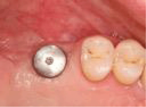
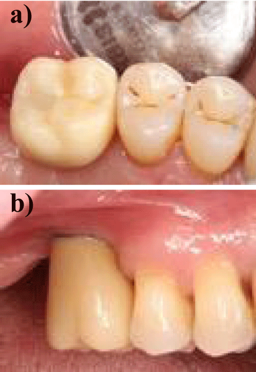
All transmucosally placed implants integrated without complications and were loaded by screw retained zirconia crowns. Meanwhile all implants in the reported cases are functioning for ≥ 12 months without any signs for biological or technical complications.
The bone substitute was a deproteinized porcine bone mineral (DPBM, SMARTGRAFT, REGEDENT, GERMANY) mixed with xHyA, the cross-linked hyaluronic acid (HYADENT BG, REGEDENT, GERMANY). Applied as “sticky bone” it served for sinus grafting and additionally for lateral and/or vertical augmentation. For achieving appropriate barrier function, a Ribose cross-linked collagen membrane (OSSIX® PLUS, REGEDENT, GERMANY) covered on top of the grafted area to seclude the newly created volume from non-ossifying cells of the soft tissue.
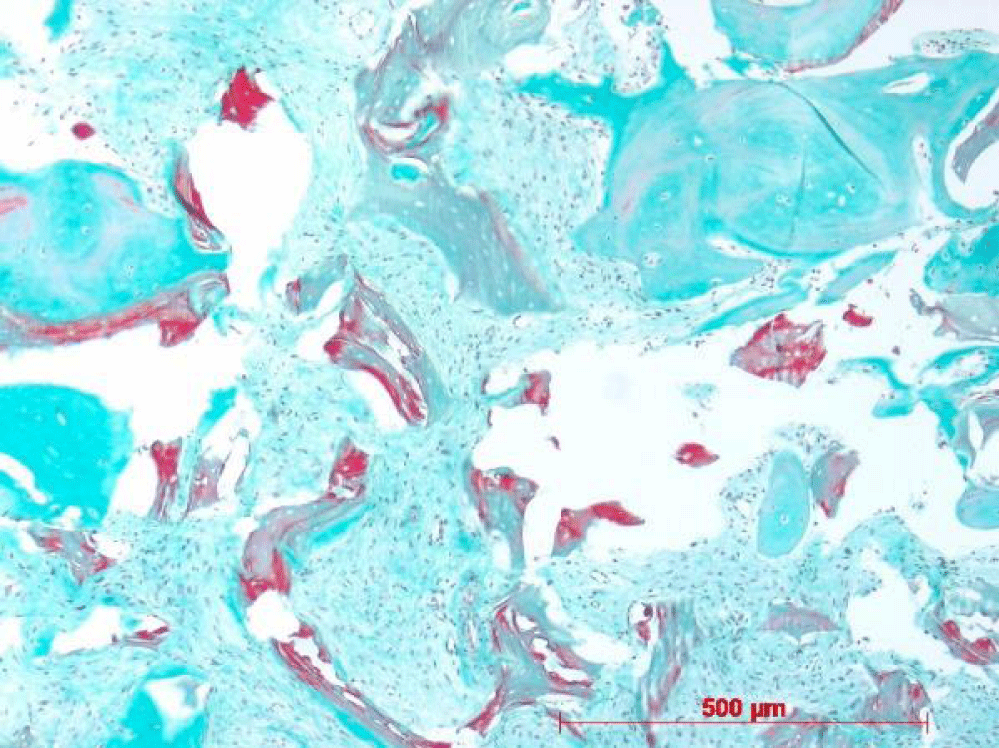
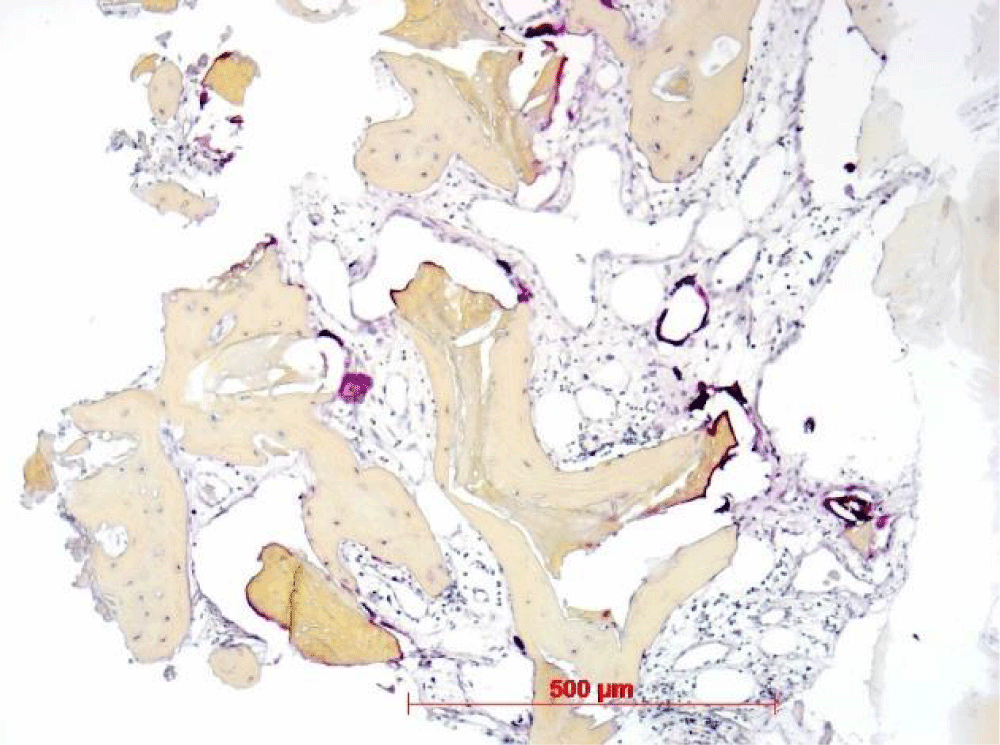
The osteotomies followed the full-thickness flap reflection exposing the newly formed crest of the alveolar ridge and macroscopic considering the amount of mineralized tissue as suitable for implant placement. The retrieved samples were fixated with formalin and processed in paraffin after decalcification. Serial sections were generally stained with H.E., trichrome staining; furthermore, the osteoclast activity was evaluated by TRAP reaction and PAS staining disclosed the process of ossification.
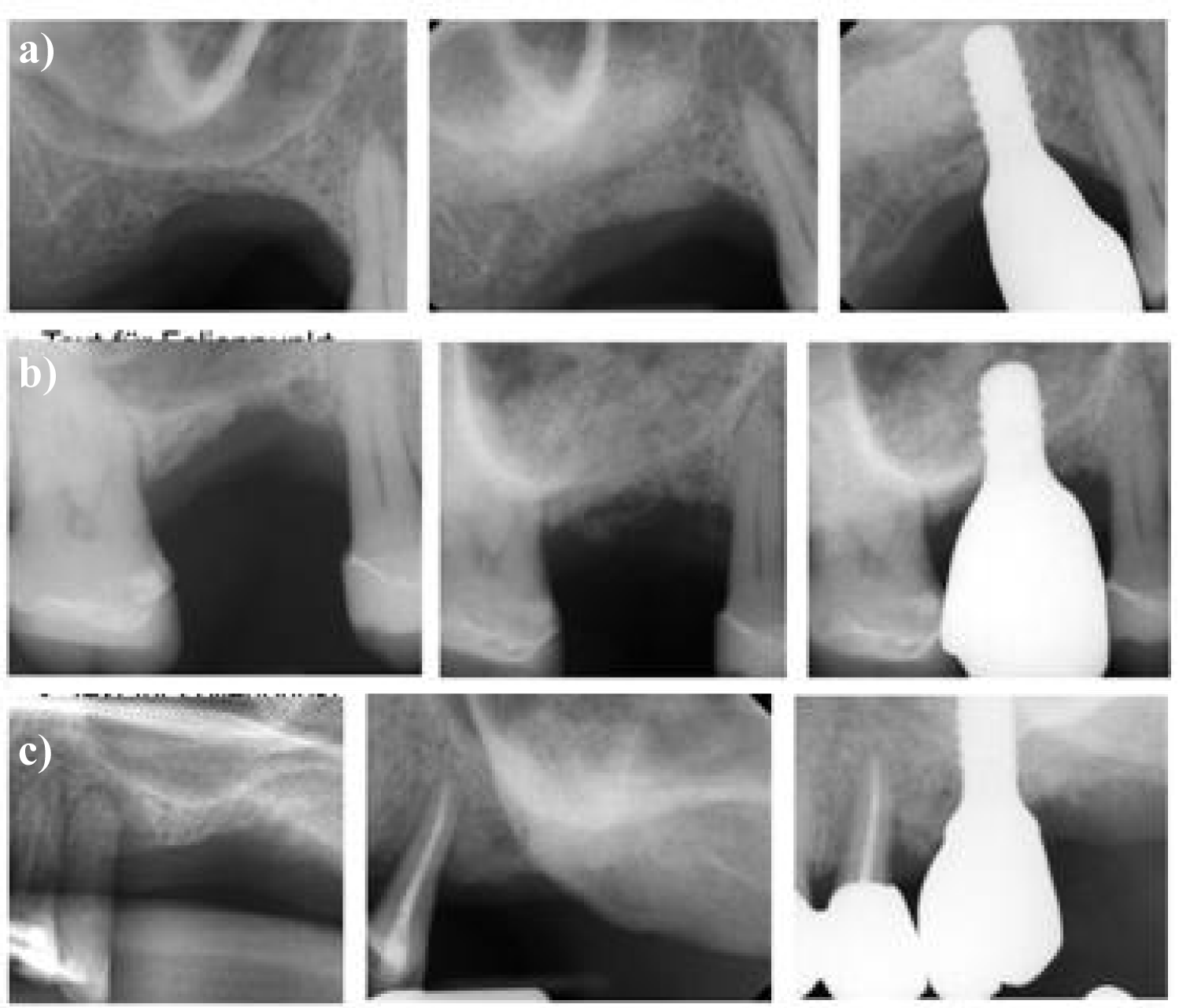
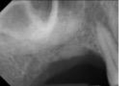
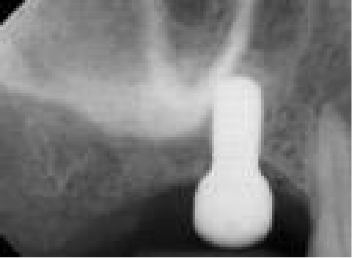
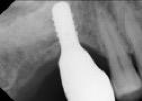
Three triplicates of radiographs document the baseline level of the residual subantral bone in comparison to the level achieved by GBR/sinus lifting and to the new crestal bone level displayed at loaded implants for all three cases (Figure 1).
The formalin fixated tissue samples were forwarded to the lab facility at University of Bonn, Germany. The decalcifying and paraffin embedding followed the sequential dehydration and serial sections were obtained for each sample. A recognized oral biologist released the qualitative microscopic analysis (W.G.).
The microscopic observations revealed newly appositionally formed trabecular bone at the residues of bone substitute in all specimens without exception. The trichrome staining disclosed osteoid formation and the TRAP reaction clearly highlighted the activity of osteoclasts in intimate contact with the residual particulate graft material. All microscopic images are apparently free of inflammatory infiltrate or unspecific multinuclear giant cells.
Case Description
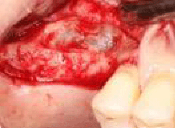
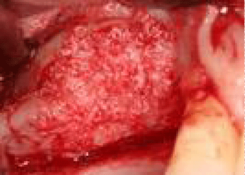
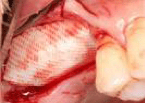
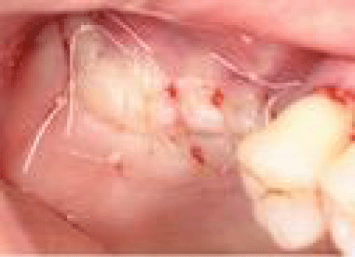
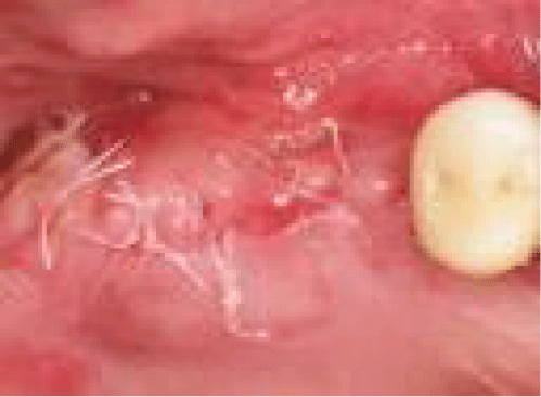
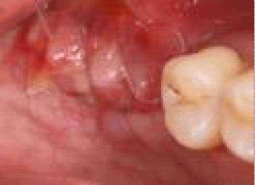
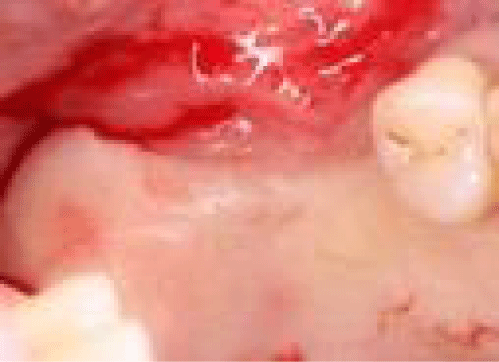
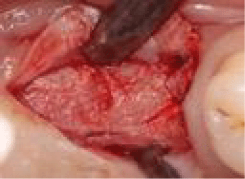
The illustrated case depicts the universal utility of the HA in supporting hard tissue formation. The challenge in this case was the severe loss in crestal bone height after extraction of the two molars, which caused reduction in subantral bone but also a vertical ridge deficiency (Figure 2). Part of the treatment plan was to perform vertical augmentation parallel to lateral window sinus grafting and lateral augmentation using one and the same material combination (Figures 3,4). The sticky bone was prepared on the tray by hydrating the DPBM particulate with xHyA (SMARTGRAFT and HYADENT BG, REGEDENT, GERMANY). Once grafting in both areas completed, the ribose cross-linked collagen membrane was trimmed and positioned for cover the total area across the ridge (OSSIX® PLUS, REGEDENT, GERMANY, Figure 5). The membrane edges overlapped the defect extension for 2-3 mm thus achieving the stabilization effect without any additional fixation. Following the CAF principle, the tensionless suture closely adapted the flap tissue margins and achieved complete closure of the site (Figure 6). The healing was uneventful, the sutures were removed after 7 days (Figures 7,8), and the implant placement was scheduled 7 months later.

Results
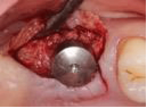
The control x-ray obtained before implant placement displays two vertically augmented areas (Figure 9 arrows), one beneath and one crestal to the residual subantral native bone (Figure 9). The newly formed alveolar crest (Figure 10) exposed by a midcrestal incision impressed with the new dimension in width and displayed residual portion of the CLCM material well attached to the underlying mineralized tissue layer (Figure 11). The core biopsy (Figure 12) retrieved by a hollow cylinder bur revealed clinically suitable grade of mineralization for placement of an WNI TL implant (WNI, SP TL, 10 mm; Straumann Group, Germany) which was inserted supplied with the gingiva former for transmucosal integration (Figure 13). The amount of keratinized mucosa was sufficient to keep up with the rule of a 2 mm zone of keratinized tissue around the implant neck circumferentially. The implant integrated within 8 weeks and a screw retained full zirconia crown was inserted.
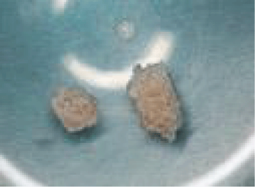
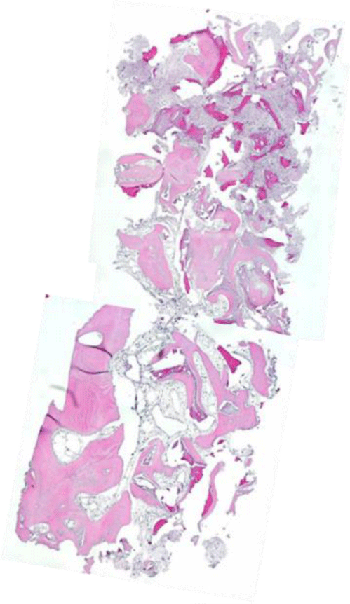
The core biopsy broke apart into two pieces at removing it from the trephine, apparently separating the supracrestal from the subcrestal core portion (Figures 14-17). Both portions, however, displayed newly formed woven bone intimately embedding the residues of particulate graft material (Figure 18). The appositional bone formation revealed by the PAS staining of osteoid activity appeared ubiquitously in all regions of the specimen (Figure 19). Although disrupted into two parts, both display residual subantral bone retained almost in the middle of the core. Besides, the fragments of native bone detected on one side in total length of the core represent the mesial border of the augmented zone. The subsequent degradation of the particulate residues is indicated by the positive TRAP reaction, which specifically highlights the osteoclast activity at the DPBM remnants (Figure 20).
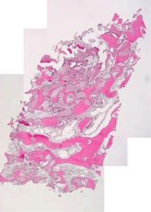
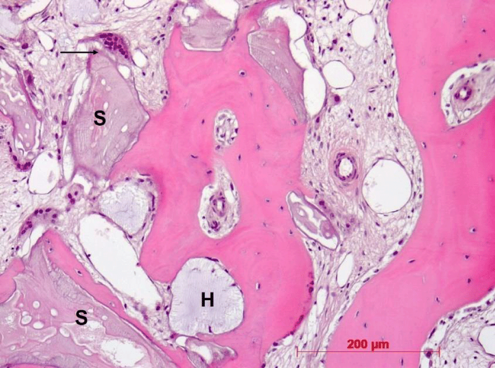
The second displayed core biopsy was obtained from another case 7 months after a sinus grafting procedure, which strictly followed the same treatment protocol and used exactly same biomaterials (Figure 21). The histological findings in this case are closely repeating already reported observations. Surprisingly, besides the similar structures these specimens reveal the presence of residual xHyA encapsulated by appositionally grown bone and appear randomly distributed over the total area of the sample (Figure 22 - “H”). The TRAP reaction does not identify any osteoclasts spatially related to the xHyA-filled areas. The TRAP reaction reveals, however, numerous osteoclasts at the residual particles of the bone substitute.
Discussion
This short case series presents the clinical and histological outcome of sinus floor elevation concomitantly combined either with a lateral or vertical augmentation. While several biomaterials are considered suitable for grafting the sinus cavity [49], the use of particulate bone substitute especially for vertical augmentation of edentulous alveolar ridge without reinforced membrane is critical due to lack of graft stability [50].
To overcome this limitation, we used “sticky bone” which was prepared by hydrating DPBM with xHyA. Thus, implementing bidirectional function, the improved stability of the graft itself and facilitated adhesion at the treated site was achieved. Recent publications confirm the stimulating effect of xHyA molecules on cells of mesenchymal origin promoting osteoblast differentiation and accelerating the matrix formation for mineralization process [45]. Moreover, xHyA exerts numerous positive factors on the soft tissues [32-35]. Therefore, the combination of xHyA as an active biologic applied together with a DPBM represents a novel approach in reconstructive treatment of bone defects. The human histological analysis supports the positive clinical experience by disclosing new appositional bone formation as at the residual bone substitute as at residual xHyA areas which appear vacuole-like embedded in the new tissues. The specific TRAP reaction identifies the osteoclastic activities at the residues of the bone substitute indicating the degradability of the porcine derived particulate. A recent clinical study performs volumetric and qualitative analyses of grafted sinus cavities using the CBCT based approach [51]. The bone substitute used in this study was a bone-albumin impregnated allograft. The authors compare the outcome from the µCT analysis of the core biopsies retrieved at implant placement to the CBCT data. According to the data from an animal study [52] the radiographically based analysis alone should be treated with caution once xenograft particulate material is the substitute of choice.
Besides the sophisticated bone graft combination, one more factor accounts for the treatment success of our modified GBR protocol in lateral and vertical augmentation. The use of a slow-resorbing ribose crosslinked collagen membrane instead of a fast-resorbing conventional native collagen membrane may additionally promote the integration of the substitute into newly formed bone. A recent animal study confirms the impact of membrane longevity towards new bone formation in lateral augmentation of chronic bone defects. The ribose cross-linked collagen membrane served as a sufficient barrier between the augmented site and the soft tissue, characterized by promoting significantly more new bone formation and less residual graft particles compared to a native collagen membrane, even without additional fixation at surgery [52]. A higher number of treated patients is required to substantiate these observations.
Conclusion
Within the limits of this short case series, we conclude that the adjunct use of Hyaluronic Acid (xHyA) in GBR and sinus grafting protocols support the creation of a pathogen-free wound area, improves wound space stabilization, accelerates wound healing and supports regenerative processes for bone regeneration. This potentially results in an accelerated process for new bone/tissue formation accompanied by a higher turnover-rate of substitute material. Therefore, the proposed biomaterial combination presents apparently symbiotic qualities and fulfills several pre-success criteria crucial for facilitating superior regenerative outcome particularly in complex defects.
Acknowledgment
The authors are grateful to Mrs. I. Bay-Müller (Univ. of Bonn) for the technical support in processing the specimens.
Author’s contribution
A.F. case handling, writing the manuscript; W.G. handling of the samples, qualitative microscopy, proofreading the manuscript
References
- Tatum H Jr. Maxillary and sinus implant reconstructions. Dent Clin North Am. 1986 Apr;30(2):207-29. PMID: 3516738.
- Schweikert M, Botticelli D, de Oliveira JA, Scala A, Salata LA, Lang NP. Use of a titanium device in lateral sinus floor elevation: an experimental study in monkeys. Clin Oral Implants Res. 2012 Jan;23(1):100-5. doi: 10.1111/j.1600-0501.2011.02200.x. Epub 2011 Apr 25. PMID: 21518009.
- Chiapasco M, Zaniboni M. Methods to treat the edentulous posterior maxilla: implants with sinus grafting. J Oral Maxillofac Surg. 2009 Apr;67(4):867-71. doi: 10.1016/j.joms.2008.11.023. PMID: 19304048.
- Handschel J, Simonowska M, Naujoks C, Depprich RA, Ommerborn MA, Meyer U, Kübler NR. A histomorphometric meta-analysis of sinus elevation with various grafting materials. Head Face Med. 2009 Jun 11;5:12. doi: 10.1186/1746-160X-5-12. PMID: 19519903; PMCID: PMC2700082.
- Jensen SS, Terheyden H. Bone augmentation procedures in localized defects in the alveolar ridge: clinical results with different bone grafts and bone-substitute materials. Int J Oral Maxillofac Implants. 2009;24 Suppl:218-36. PMID: 19885447.
- Aghaloo TL, Moy PK. Which hard tissue augmentation techniques are the most successful in furnishing bony support for implant placement? Int J Oral Maxillofac Implants. 2007;22 Suppl:49-70. Erratum in: Int J Oral Maxillofac Implants. 2008 Jan-Feb;23(1):56. PMID: 18437791.
- Benic GI, Hämmerle CH. Horizontal bone augmentation by means of guided bone regeneration. Periodontol 2000. 2014 Oct;66(1):13-40. doi: 10.1111/prd.12039. PMID: 25123759.
- Dahlin C, Andersson L, Linde A. Bone augmentation at fenestrated implants by an osteopromotive membrane technique. A controlled clinical study. Clin Oral Implants Res. 1991 Oct-Dec;2(4):159-65. doi: 10.1034/j.1600-0501.1991.020401.x. PMID: 8597617.
- Esposito M, Grusovin MG, Coulthard P, Worthington HV. The efficacy of various bone augmentation procedures for dental implants: a Cochrane systematic review of randomized controlled clinical trials. Int J Oral Maxillofac Implants. 2006 Sep-Oct;21(5):696-710. PMID: 17066630.
- Hämmerle CH, Jung RE, Feloutzis A. A systematic review of the survival of implants in bone sites augmented with barrier membranes (guided bone regeneration) in partially edentulous patients. J Clin Periodontol. 2002;29 Suppl 3:226-31; discussion 232-3. doi: 10.1034/j.1600-051x.29.s3.14.x. PMID: 12787222.
- Ohayon L, Taschieri S, Friedmann A, Del Fabbro M. Bone Graft Displacement After Maxillary Sinus Floor Augmentation With or Without Covering Barrier Membrane: A Retrospective Computed Tomographic Image Evaluation. Int J Oral Maxillofac Implants. 2019 May/June;34(3):681–691. doi: 10.11607/jomi.6940. Epub 2018 Dec 5. PMID: 30521657.
- Bashutski JD, Wang HL. Biologic Agents to Promote Periodontal Regeneration and Bone Augmentation. Clin Adv Periodontics. 2011 Aug;1(2):80-87. doi: 10.1902/cap.2011.110044. PMID: 32698555.
- Tarnow DP, Wallace SS, Froum SJ, Rohrer MD, Cho SC. Histologic and clinical comparison of bilateral sinus floor elevations with and without barrier membrane placement in 12 patients: Part 3 of an ongoing prospective study. Int J Periodontics Restorative Dent. 2000 Apr;20(2):117-25. PMID: 11203554.
- Grosso LT, Iha DK, Niu J, Wakabayashi RC, Johnson PW. Protease profiles of cells isolated from regenerative membranes are associated with clinical outcomes. J Periodontol. 1997 Sep;68(9):809-18. doi: 10.1902/jop.1997.68.9.809. PMID: 9379323.
- Trimmel B, Gede N, Hegyi P, Szakács Z, Mezey GA, Varga E, Kivovics M, Hanák L, Rumbus Z, Szabó G. Relative performance of various biomaterials used for maxillary sinus augmentation: A Bayesian network meta-analysis. Clin Oral Implants Res. 2021 Feb;32(2):135-153. doi: 10.1111/clr.13690. Epub 2021 Jan 6. PMID: 33230862; PMCID: PMC8247032.
- Eliezer M, Imber JC, Sculean A, Pandis N, Teich S. Hyaluronic acid as adjunctive to non-surgical and surgical periodontal therapy: a systematic review and meta-analysis. Clin Oral Investig. 2019 Sep;23(9):3423-3435. doi: 10.1007/s00784-019-03012-w. Epub 2019 Jul 23. PMID: 31338632.
- Chen F, Ni Y, Liu B, Zhou T, Yu C, Su Y, Zhu X, Yu X, Zhou Y. Self-crosslinking and injectable hyaluronic acid/RGD-functionalized pectin hydrogel for cartilage tissue engineering. Carbohydr Polym. 2017 Jun 15;166:31-44. doi: 10.1016/j.carbpol.2017.02.059. Epub 2017 Feb 20. PMID: 28385238.
- Zhao N, Wang X, Qin L, Zhai M, Yuan J, Chen J, Li D. Effect of hyaluronic acid in bone formation and its applications in dentistry. J Biomed Mater Res A. 2016 Jun;104(6):1560-9. doi: 10.1002/jbm.a.35681. Epub 2016 Apr 9. PMID: 27007721.
- Carlson GA, Dragoo JL, Samimi B, Bruckner DA, Bernard GW, Hedrick M, Benhaim P. Bacteriostatic properties of biomatrices against common orthopaedic pathogens. Biochem Biophys Res Commun. 2004 Aug 20;321(2):472-8. doi: 10.1016/j.bbrc.2004.06.165. PMID: 15358200.
- Pirnazar P, Wolinsky L, Nachnani S, Haake S, Pilloni A, Bernard GW. Bacteriostatic effects of hyaluronic acid. J Periodontol. 1999 Apr;70(4):370-4. doi: 10.1902/jop.1999.70.4.370. PMID: 10328647.
- Kang JH, Kim YY, Chang JY, Kho HS. Influences of hyaluronic acid on the anticandidal activities of lysozyme and the peroxidase system. Oral Dis. 2011 Sep;17(6):577-83. doi: 10.1111/j.1601-0825.2011.01807.x. Epub 2011 Apr 8. PMID: 21477181.
- Sasaki T, Watanabe C. Stimulation of osteoinduction in bone wound healing by high-molecular hyaluronic acid. Bone. 1995 Jan;16(1):9-15. doi: 10.1016/s8756-3282(94)00001-8. PMID: 7742090.
- Dahiya P, Kamal R. Hyaluronic Acid: a boon in periodontal therapy. N Am J Med Sci. 2013 May;5(5):309-15. doi: 10.4103/1947-2714.112473. PMID: 23814761; PMCID: PMC3690787.
- de Brito Bezerra B, Mendes Brazão MA, de Campos ML, Casati MZ, Sallum EA, Sallum AW. Association of hyaluronic acid with a collagen scaffold may improve bone healing in critical-size bone defects. Clin Oral Implants Res. 2012 Aug;23(8):938-42. doi: 10.1111/j.1600-0501.2011.02234.x. Epub 2011 Jun 21. PMID: 21689163.
- Kawano M, Ariyoshi W, Iwanaga K, Okinaga T, Habu M, Yoshioka I, Tominaga K, Nishihara T. Mechanism involved in enhancement of osteoblast differentiation by hyaluronic acid. Biochem Biophys Res Commun. 2011 Feb 25;405(4):575-80. doi: 10.1016/j.bbrc.2011.01.071. Epub 2011 Jan 23. PMID: 21266161.
- Mendes RM, Silva GA, Lima MF, Calliari MV, Almeida AP, Alves JB, Ferreira AJ. Sodium hyaluronate accelerates the healing process in tooth sockets of rats. Arch Oral Biol. 2008 Dec;53(12):1155-62. doi: 10.1016/j.archoralbio.2008.07.001. Epub 2008 Aug 9. PMID: 18692778.
- Deed R, Rooney P, Kumar P, Norton JD, Smith J, Freemont AJ, Kumar S. Early-response gene signalling is induced by angiogenic oligosaccharides of hyaluronan in endothelial cells. Inhibition by non-angiogenic, high-molecular-weight hyaluronan. Int J Cancer. 1997 Apr 10;71(2):251-6. doi: 10.1002/(sici)1097-0215(19970410)71:2<251::aid-ijc21>3.0.co;2-j. PMID: 9139851.
- Schwarz F, Sager M, Rothamel D, Herten M, Sculean A, Becker J. Einsatz nativer und quervernetzter Kollagenmembranen für die gesteuerte Gewebe- und Knochenregeneration [Use of native and cross-linked collagen membranes for guided tissue and bone regeneration]. Schweiz Monatsschr Zahnmed. 2006;116(11):1112-23. German. PMID: 17144624.
- Sanz-Sánchez I, Ortiz-Vigón A, Sanz-Martín I, Figuero E, Sanz M. Effectiveness of Lateral Bone Augmentation on the Alveolar Crest Dimension: A Systematic Review and Meta-analysis. J Dent Res. 2015 Sep;94(9 Suppl):128S-42S. doi: 10.1177/0022034515594780. Epub 2015 Jul 27. PMID: 26215467.
- Oryan A, Moshiri A, Meimandi Parizi AH, Raayat Jahromi A. Repeated administration of exogenous Sodium-hyaluronate improved tendon healing in an in vivo transection model. J Tissue Viability. 2012 Aug;21(3):88-102. doi: 10.1016/j.jtv.2012.06.002. Epub 2012 Jul 4. PMID: 22766020.
- Tuncay I, Ozbek H, Atik B, Ozen S, Akpinar F. Effects of hyaluronic acid on postoperative adhesion of tendo calcaneus surgery: an experimental study in rats. J Foot Ankle Surg. 2002 Mar-Apr;41(2):104-8. doi: 10.1016/s1067-2516(02)80033-3. PMID: 11995830.
- King SR, Hickerson WL, Proctor KG. Beneficial actions of exogenous hyaluronic acid on wound healing. Surgery. 1991 Jan;109(1):76-84. PMID: 1984639.
- Pilloni A, Schmidlin PR, Sahrmann P, Sculean A, Rojas MA. Correction to: Effectiveness of adjunctive hyaluronic acid application in coronally advanced flap in Miller class I single gingival recession sites: a randomized controlled clinical trial. Clin Oral Investig. 2018 Nov;22(8):2961-2962. doi: 10.1007/s00784-018-2567-y. Erratum for: Clin Oral Investig. 2018 Jun 30;: PMID: 30027355.
- Yıldırım S, Özener HÖ, Doğan B, Kuru B. Effect of topically applied hyaluronic acid on pain and palatal epithelial wound healing: An examiner-masked, randomized, controlled clinical trial. J Periodontol. 2018 Jan;89(1):36-45. doi: 10.1902/jop.2017.170105. PMID: 28914592.
- Humbert P, Mikosinki J, Benchikhi H, Allaert FA. Efficacy and safety of a gauze pad containing hyaluronic acid in treatment of leg ulcers of venous or mixed origin: a double-blind, randomised, controlled trial. Int Wound J. 2013 Apr;10(2):159-66. doi: 10.1111/j.1742-481X.2012.00957.x. Epub 2012 Mar 8. PMID: 22405094; PMCID: PMC7950449.
- Juhász I, Zoltán P, Erdei I. Treatment of partial thickness burns with Zn-hyaluronan: lessons of a clinical pilot study. Ann Burns Fire Disasters. 2012 Jun 30;25(2):82-5. PMID: 23233826; PMCID: PMC3506212.
- Rajan P, Dusanapudi LN, Kumar CS, Nair D. Hyaluronic acid - a simple, unusual polysaccharide: A potential mediator for periodontal regeneration. Universal Research Journal of Dentistry. 2013;3:113.
- Mertens C, Braun S, Krisam J, Hoffmann J. The influence of wound closure on graft stability: An in vitro comparison of different bone grafting techniques for the treatment of one-wall horizontal bone defects. Clin Implant Dent Relat Res. 2019 Apr;21(2):284-291. doi: 10.1111/cid.12728. Epub 2019 Feb 11. PMID: 30741470.
- Mir-Mari J, Wui H, Jung RE, Hämmerle CH, Benic GI. Influence of blinded wound closure on the volume stability of different GBR materials: an in vitro cone-beam computed tomographic examination. Clin Oral Implants Res. 2016 Feb;27(2):258-65. doi: 10.1111/clr.12590. Epub 2015 Apr 9. PMID: 25856209.
- Meijndert L, Raghoebar GM, Meijer HJ, Vissink A. Clinical and radiographic characteristics of single-tooth replacements preceded by local ridge augmentation: a prospective randomized clinical trial. Clin Oral Implants Res. 2008 Dec;19(12):1295-303. doi: 10.1111/j.1600-0501.2008.01523.x. PMID: 19040446.
- Hämmerle CH, Jung RE, Yaman D, Lang NP. Ridge augmentation by applying bioresorbable membranes and deproteinized bovine bone mineral: a report of twelve consecutive cases. Clin Oral Implants Res. 2008 Jan;19(1):19-25. doi: 10.1111/j.1600-0501.2007.01407.x. Epub 2007 Oct 22. PMID: 17956571.
- Asparuhova MB, Kiryak D, Eliezer M, Mihov D, Sculean A. Activity of two hyaluronan preparations on primary human oral fibroblasts. J Periodontal Res. 2019 Feb;54(1):33-45. doi: 10.1111/jre.12602. Epub 2018 Sep 27. PMID: 30264516; PMCID: PMC6586051.
- Mueller A, Fujioka-Kobayashi M, Mueller HD, Lussi A, Sculean A, Schmidlin PR, Miron RJ. Effect of hyaluronic acid on morphological changes to dentin surfaces and subsequent effect on periodontal ligament cell survival, attachment, and spreading. Clin Oral Investig. 2017 May;21(4):1013-1019. doi: 10.1007/s00784-016-1856-6. Epub 2016 May 19. PMID: 27194052.
- Prince CW. Roles of hyaluronan in bone resorption. BMC Musculoskelet Disord. 2004 Apr 29;5:12. doi: 10.1186/1471-2474-5-12. PMID: 15117412; PMCID: PMC411043.
- Asparuhova MB, Chappuis V, Stähli A, Buser D, Sculean A. Role of hyaluronan in regulating self-renewal and osteogenic differentiation of mesenchymal stromal cells and pre-osteoblasts. Clin Oral Investig. 2020 Nov;24(11):3923-3937. doi: 10.1007/s00784-020-03259-8. Epub 2020 Mar 31. PMID: 32236725; PMCID: PMC7544712.
- Kim JJ, Song HY, Ben Amara H, Kyung-Rim K, Koo KT. Hyaluronic Acid Improves Bone Formation in Extraction Sockets With Chronic Pathology: A Pilot Study in Dogs. J Periodontol. 2016 Jul;87(7):790-5. doi: 10.1902/jop.2016.150707. Epub 2016 Mar 18. PMID: 26991484.
- Alcântara CEP, Castro MAA, Noronha MS, Martins-Junior PA, Mendes RM, Caliari MV, Mesquita RA, Ferreira AJ. Hyaluronic acid accelerates bone repair in human dental sockets: a randomized triple-blind clinical trial. Braz Oral Res. 2018;32:e84. doi: 10.1590/1807-3107bor-2018.vol32.0084. Epub 2018 Sep 13. PMID: 30231173.
- Stiller M, Kluk E, Bohner M, Lopez-Heredia MA, Müller-Mai C, Knabe C. Performance of β-tricalcium phosphate granules and putty, bone grafting materials after bilateral sinus floor augmentation in humans. Biomaterials. 2014 Mar;35(10):3154-63. doi: 10.1016/j.biomaterials.2013.12.068. Epub 2014 Jan 16. PMID: 24439419.
- Danesh-Sani SA, Engebretson SP, Janal MN. Histomorphometric results of different grafting materials and effect of healing time on bone maturation after sinus floor augmentation: a systematic review and meta-analysis. J Periodontal Res. 2017 Jun;52(3):301-312. doi: 10.1111/jre.12402. Epub 2016 Aug 18. PMID: 27534916.
- Urban IA, Montero E, Monje A, Sanz-Sánchez I. Effectiveness of vertical ridge augmentation interventions: A systematic review and meta-analysis. J Clin Periodontol. 2019 Jun;46 Suppl 21:319-339. doi: 10.1111/jcpe.13061. PMID: 30667522.
- Kivovics M, Szabó BT, Németh O, Iványi D, Trimmel B, Szmirnova I, Orhan K, Mijiritsky E, Szabó G, Dobó-Nagy C. Comparison between Micro-Computed Tomography and Cone-Beam Computed Tomography in the Assessment of Bone Quality and a Long-Term Volumetric Study of the Augmented Sinus Grafted with an Albumin Impregnated Allograft. J Clin Med. 2020 Jan 21;9(2):303. doi: 10.3390/jcm9020303. PMID: 31973237; PMCID: PMC7073646.
- Friedmann A, Fickl S, Fischer KR, Dalloul M, Goetz W, Kauffmann F. Horizontal Augmentation of Chronic Mandibular Defects by the Guided Bone Regeneration Approach: A Randomized Study in Dogs. Materials (Basel). 2021 Dec 29;15(1):238. doi: 10.3390/ma15010238. PMID: 35009383; PMCID: PMC8746186.





Content Alerts
SignUp to our
Content alerts.
 This work is licensed under a Creative Commons Attribution 4.0 International License.
