> Medicine Group. 2021 Mar 23;2(3):201-205. doi: 10.37871/jbres1211.
Prevalence and Pattern of Skin Disorders among Human Immuno Deficiency Virus (HIV) Infected Children in Aminu Kano Teaching Hospital (AKTH) Kano, Nigeria
Yahya Aishatu Muhammad*
- Paediatric dermatoses
- HIV infected children
- Kano
- Nigeria
Abstract
Introduction: In HIV infected children, skin disorders are important as they serve as clue to diagnosis of the HIV disease. The Skin is one of the early systems affected by HIV/AIDS, which can affect almost all organs and systems in the body. Prevalence of skin disorders among HIV infected children is up to 90% in some studies.
Objective: To determine the prevalence of skin disorders among HIV infected children attending paediatric infectious disease clinic in Aminu Kano Teaching Hospital Kano, Nigeria.
Materials and Methods: A cross-sectional study was conducted to determine the prevalence of skin manifestations among HIV infected children attending paediatric infectious disease clinic of Aminu Kano Teaching Hospital, Kano, Nigeria. A total of 223 HIV infected participants aged 6weeks to14 years were recruited for this study.
Results: The prevalence of skin disorders among HIV infected children was 78.0%. The leading categories were infections and infestations accounting for 55.1% then inflammatory skin disorders (20.6%) Dermatophytoses were the commonest specific skin disorders observed.
Conclusion: Therefore, the prevalence of skin disorder among HIV infected children in Aminu Kano Teaching Hospital is high (78%). Infections and infestations were the commonest category found followed by inflammatory skin disorders.
Introduction
The Human Immunodeficiency Virus (HIV) is a lentivirus that replicates slowly [1,2]. The HIV virus is divided into two subtypes: HIV type 1 (HIV-1) and HIV type 2 (HIV- 2), each with its own geographical distribution. Human immunodeficiency virus causes progressive damage of the human immune system creating an opportunity for life-threatening opportunistic infections and cancers to thrive [1]. The impact of Human Immunodeficiency Virus (HIV) infection is devastating. It continues to affect the lives of individuals, families and the community, especially in sub Saharan Africa. It reduces the life expectancy, causing loss of man power with significant social and economic impacts [3,4]. In Nigeria, the national HIV prevalence is 1.4% with more than 1.9 million Nigerians living with HIV/AIDS by the beginning of the year 2021, which makes Nigeria the fourth highest country when it comes to HIV burden worldwide [5]. HIV/AIDS is a global health problem, with an estimated 38million (31.6 million -44.5 million) people globally living with HIV at the end of 2019. Among them 1.8 million (1.3million-2.4 million) were children less than 15 years, mostly living in sub- Saharan Africa [6].
Skin disorders, most especially the infections and infestations, are major cause of concern among children as the immunity is not fully developed during childhood [7]. Skin disorders are among common causes of morbidity in developing countries of sub Saharan Africa, together with malaria and diarrhea [8]. They are especially important in HIV infected children and contribute to the risk of other life threatening illnesses like glomerulonephritis, sepsis, heart diseases and cancers [9,10]. In children under five years of age, it accounts for a rise of more than 19% in infant mortality and 36% in under five mortalities [11]. The Skin is one of the early systems affected by HIV/AIDS, which can affect almost all organs and systems in the body [12]. A hospital based prospective study in Rome found the prevalence of skin disorders among HIV-infected children to be 89.0% [13]. In India, Saurabh, et al. [14] found a prevalence rate of skin disorders among HIV infected children to be 72.8% with infections and infestations constituting the largest group Okechukwu, et al. [15] reported prevalence rate of skin disorders of 52.2% among 178 HIV infected children that were receiving care at the special treatment centre of the University of Abuja Teaching Hospital, Nigeria, with Pruritic Papular Eruption, impetigo and cutaneous candidiasis being most common. All these reported prevalence rates point to the magnitude of skin disorders in HIV infected children. There is a knowledge gap about the prevalence and pattern of skin disorders among HIV infected children in Kano. Most of the studies done in Nigeria were conducted in the Southern part of the country. The few that were conducted in the Northern part of Nigeria were mostly among adults. This study therefore aims to determine the prevalence and pattern of skin disorders among HIV infected children in Aminu Kano Teaching Hospital Kano, Nigeria. The findings from this study will be of great public health importance in planning public interventions targeted towards HIV infected children as it will help in planning programs that will help in reducing morbidity and mortality among the children and improve the overall wellbeing of the children and their families which will impact positively on the society at large.
Objectives
To determine the prevalence of skin infection among HIV infected children attending paediatric infectious disease clinic and paediatric outpatient clinic in AKTH.
To determine the pattern of skin disorders among HIV infected children attending paediatric infectious disease clinic and paediatric outpatient clinic in AKTH.
Materials and Methods
The study involved a cross-sectional study to determine the prevalence of skin manifestations among HIV infected children Aged 6 weeks to 14 years attending paediatric infectious disease clinic and paediatric outpatient clinic.
Two hundred and twenty-three HIV infected children were recruited for the study. A systematic sampling technique was employed for recruiting participants into the study. Ethical clearance was obtained from Ethical and Research Committee of Aminu Kano Teaching Hospital (Appendix 1) Informed consent was obtained from the parents and verbal assent from children aged > 7 yrs. The purpose of the study was explained to them and they were free to opt out of the study if they wished to without any consequences. Confidentiality was ensured and they were informed that pictures taken will be displayed in research work with their eyes covered.
This study was conducted between the July 10th and October 20th, 2017. A pre- tested interviewer administered questionnaire was employed for data collection. The socio-economic status of the participants was assessed according to the method suggested by Oyedeji [16] (Appendix 2).
Complete dermatological examination of the hair, scalp, trunk, nails, oral mucosa, genitalia and extremities was carried out on each child in a well-lit room after proper exposure of the patient by the investigator to identify all the possible skin lesions. A chaperon was used for examination of the children. A picture of each lesion was taken. Most of the diagnoses were made clinically. Detailed visualization of the morphology of lesions was enhanced with the use of the common hand lens. Lesions requiring further evaluation and treatment were referred to the dermatology clinic of the Aminu Kano Teaching Hospital while those with readily treatable conditions had drugs prescribed. Furthermore, a complete physical examination of all systems was carried out on the participants.
Rapid HIV antibody testing by determine® method, PCR, CD4+ count, and Full blood count are part of the routine work up for all patients attending the clinic and were retrieved from the participant’s record. The current CD4+ count done within 3 months before patients recruitment was used.
The data was entered into a statistical package for the social sciences SPSS version 25. The data was used to generate frequency tables.
Results
There were 110 (49.3%) males and 113 females (50.7%) recruited into the study with male to female ratio of 1:1.1. The majority of the study participants were in the age group of 5-9 years. Table 1 shows the socio-demographic characteristics of the study population.
Table 1: Socio-demographic characteristics of the study population. |
|
| Characteristic | HIV infected cases n = 223 (%) |
| Gender Male Female |
110 (49.3) 113 (50.7) |
| Age 0-4 years 5-9 years 10-14 years |
77 (34.6) 98 (43.9) 48 (21.5) |
| Socioeconomic class* Lower Middle Upper |
110 (49.3) 80 (35.9) 33 (14.8) |
| Child’s ethnicity Hausa Yoruba Igbo Others |
185 (82.9) 14 (6.3) 12 ( 5.4) 12 (5.4) |
*Social class I & II: upper; social class III: middle and social class; IV & V: lower |
|
Skin disorders were found among 174 (78.0 %) of HIV infected children in this study. Among the 174 HIV infected children with skin disorder, a total of 170 (97.7%) had one skin disorder, 4 (2.3%) had 3 skin disorders.
Infections and infestations were the most common category observed among the children followed by inflammatory skin disorders. This is shown in table 2.
| Table 2: Types of skin disorders among HIV-infected children. | ||
| Category of skin disorder | HIV-infected n = 223 (%) |
|
| Present | Absent | |
| Infections and infestations | 123 (55.2) | 100 (44.8) |
| Viral | 40 (17.9) | 183 (82.1) |
| Fungal | 54 (24.2) | 169 (75.8) |
| Parasitic | 8 (3.6) | 215 (96.4) |
| Bacterial | 21 (9.4) | 202 (90.6) |
| Inflammatory | 46 (20.6) | 177 (79.4) |
| Miscellaneous | 13 (5.8) | 210 (94.2) |
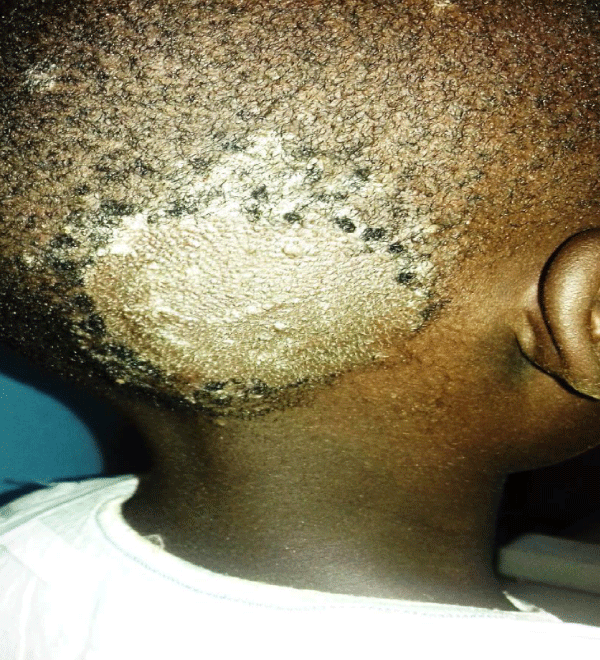
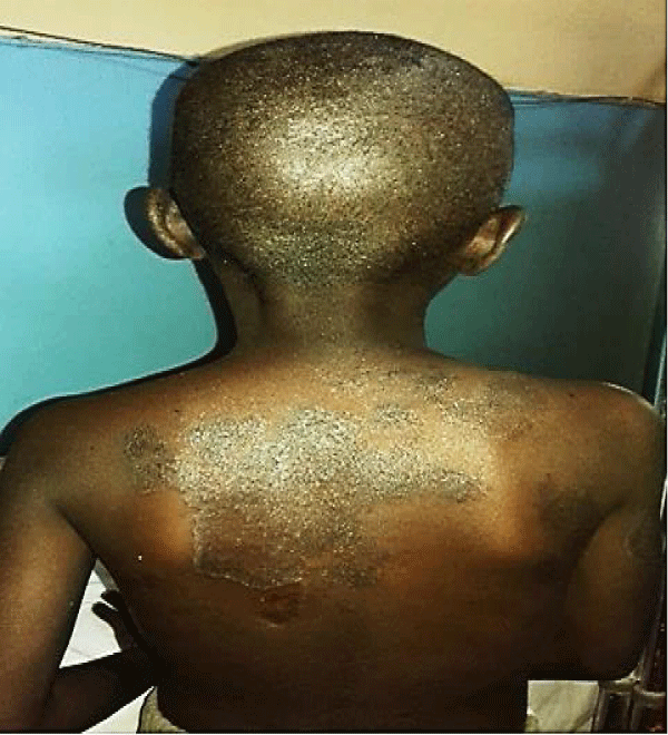
The most frequent lesions observed is dermatophytoses (16.1%), followed by Pruritic Papular Eruption (PPE) (10%) and Oral candidiasis (8.1%). Tinea capitis was the commonest form of dermatophytoses observed among 12.1% of the participants. Tinea capitis and Tinea coporis are represented in clinical photos in figures 1 & 2. These are the most common types of dermatophytoses among the HIV-positive children.
Discussion
The prevalence of skin disorders in this study was high 78.0 %. This is similar to what Umoru, et al. [17] and Katibi, et al. [18] found in Benin and Ibadan, where the prevalence of skin disorders was 64.0% and 12.0%. Another study in Lagos state, Nigeria by Osinaike, et al. [19] also reported high prevalence of skin disorders among HIV infected children (83.0%). This similar trend in prevalence are perhaps because this study also employed a methodology that was similar to that of the aforementioned studies in terms of study site (hospitals) and study design (cross-sectional). Similar higher prevalence of skin disorders among HIV infected children was reported by Lowe, et al. [20] in Zimbabwe (88.0%) and Elhachem, et al. [14] in Rome, Italy (89.5%). The high prevalence of skin disorders among HIV-infected children may be due to impaired immunity as a result of reduced ability of the Langerhans cells in the skin of the HIV infected children to cause T lymphocytes proliferation [21].
Among HIV infected children in this study, infections and infestations (55.2%) was the commonest category of skin disorder. This is similar to findings by other workers [17,18,22,23] Conversely, studies in Lagos [19] and Abuja [16], Nigeria, and that in Zimbabwe [20], reported inflammatory skin disorders as the commonest seen among HIV infected children. Different types of skin disorders observed in dermato-epidemiologic studies underscores the fact that occurrence of skin diseases is often subject to variety of factors such as environmental conditions as well as age of the study population recruited among others, which differ among various studies mentioned above. Some skin disorders have higher prevalence among some age groups and in certain localities which points to the effect of socio-demography in the occurrence of skin disorder [24,25].
Using specific disease conditions commonly found in this study,the four most prevalent types of skin disorders found among the HIV infected children in this study were dermatophytoses, Pruritic papular eruption, viral warts and Oral candidiasis.
Dermatophytosis was the most prevalent diagnosis in this study.This is similar to what was obtained in other studies [18,22].
Pruritic papular eruption was the second commonest skin disorder among HIV infected children found in this study (10.0%). This is similar to finding by umoru, et al. [17] in Benin city, Nigeria who also found PPE to be the second commonest skin disorder among the HIV infected children . The findings in this study differ from findings by Panya, et al. [26] in Tanzania, who reported PPE as the commonest lesion among HIV infected children studied. It is also reported to be the most common skin disorder in HIVinfected children from other African studies [27,28] different from what was found in this study. This difference could be as a result of different environmental conditions of the study sites from that in this study.
PPE is believed to be a form of hypersensitivity reaction of the skin to demodex folliculorum mite which is a normal commensal of the hair follicle. The distortion of the immune system is also responsible for it being frequently encountered among HIV infected people along with other non-infectious dermatoses [29].
Oral candidiasis was the 3rd commonest skin disorder found in this study (8.1%). This differs from finding of Umoru, et al. [17] in Benin city, Nigeria who found oral candidiasis to be the commonest lesion among HIV infected children (25.6%). The duration of ART theraphy among the HIV infected children in this study (48% of the subjects were on ART for six to twelve months) may also be a contributing factor to the differences observed. Oral candidiasis may represent onset of severe immunosuppression among HIV infected subjects and can progress to involve the oesophagus or even become systemic [30].
Viral wart was the fourth common skin disorder found in this study among HIV infected children (6.3%). Plane warts, and mucosal warts were the forms found in this study. Extensive plane warts have previously been described in the context of HIV [31].
Overall, this study has demonstrated that skin disorders are highly prevalent in HIV infected children with infections and infestations being the most common.
Conclusions
This study found that the prevalence of skin disorders among HIV infected children in Aminu Kano Teaching Hospital is high (78%). Infections and infestations were the commonest category found followed by inflammatory skin disorders.
References
- Moir S, Chun TW, Fauci AS. Pathogenic mechanisms of HIV disease. Annu Rev Pathol. 2011;6:223-48. doi: 10.1146/annurev-pathol-011110-130254. PMID: 21034222.
- Letvin NL, Walker BD. Immunopathogenesis and immunotherapy in AIDS virus infections. Nat Med. 2003 Jul;9(7):861-6. doi: 10.1038/nm0703-861. PMID: 12835706.
- WHO. Global update on the health sector response to HIV. WHO Fact sheet, Geneva, switzerland. 2016. https://bit.ly/3c6bXk4
- Saunders R. Equity and access to ART in Ethiopia: Activity report. Washington, DC Futur Group, Heal Policy Initiat Task order 1. 2010. https://bit.ly/2NEHHDv
- Lo J, Nwafor SU, Schwitters AM, Mitchell A, Sebastian V, Stafford KA, Ezirim I, Charurat M, McIntyre AF. Key Population Hotspots in Nigeria for Targeted HIV Program Planning: Mapping, Validation, and Reconciliation. JMIR Public Health Surveill. 2021 Feb 22;7(2):e25623. doi: 10.2196/25623. PMID: 33616537; PMCID: PMC7939933.
- World Health Organization (WHO) . Global HIV and AIDS statistics-2019 fact sheet. 2019. https://bit.ly/393clOo
- Jindal R. The Ages of children and their dermatoses. Clin Paediatr Dermatol. 2016;1:2-4. doi: 10.21767/2472-0143.100003
- Hay RJ, Fuller LC. The assessment of dermatological needs in resource-poor regions. Int J Dermatol. 2011 May;50(5):552-7. doi: 10.1111/j.1365-4632.2011.04953.x. PMID: 21506971.
- Rodgers S, Leslie KS. Skin infections in HIV-infected individuals in the era of HAART. Curr Opin Infect Dis. 2011 Apr;24(2):124-9. doi: 10.1097/QCO.0b013e328342cb31. PMID: 21169832.
- Cedeno-Laurent F, Gómez-Flores M, Mendez N, Ancer-Rodríguez J, Bryant JL, Gaspari AA, Trujillo JR. New insights into HIV-1-primary skin disorders. J Int AIDS Soc. 2011 Jan 24;14:5. doi: 10.1186/1758-2652-14-5. PMID: 21261982; PMCID: PMC3037296.
- UNICEF. State of the World’s Children. NEWYORK USA. 2006. https://uni.cf/3lOkX0B.
- Chu C, Selwyn PA. Complications of HIV infection: a systems-based approach. Am Fam Physician. 2011 Feb 15;83(4):395-406. PMID: 21322514.
- El Hachem M, Bernardi S, Pianosi G, Krzysztofiak A, Livadiotti S, Gattinara GC. Mucocutaneous manifestations in children with HIV infection and AIDS. Pediatr Dermatol. 1998 Nov-Dec;15(6):429-34. doi: 10.1046/j.1525-1470.1998.1998015429.x. PMID: 9875963.
- Mendiratta V, Mittal S, Jain A, Chander R. Mucocutaneous manifestations in children with human immunodeficiency virus infection. Indian J Dermatol Venereol Leprol. 2010 Sep-Oct;76(5):458-66. doi: 10.4103/0378-6323.69041. PMID: 20826983.
- Okechukwu AA, Okechukwu OI, Paul J. Patterns of skin disorder and its relationship with CD4 + cell count in a cohort of HIV-infected children. J Med Med Sci. 2011;2:1131-1138. https://bit.ly/2PdgDf5
- Oyedaji GA. Socioeconomic and cultural background of hospitalized children in Ilesa. Nig J Paediatr. 1985;12:11-17. https://bit.ly/2NCXOkP
- Umoru D, Oviawe O, Ibadin M, Onunu A, Esene H. Mucocutaneous manifestation of pediatric human immunodeficiency virus/acquired immunodeficiency syndrome (HIV/AIDS) in relation to degree of immunosuppression: a study of a West African population. Int J Dermatol. 2012 Mar;51(3):305-12. doi: 10.1111/j.1365-4632.2011.05077.x. PMID: 22348567.
- Katibi OS, Ogunbiyi AO, Oladokun RE, Ernest SK, Osinusi K, Brown BJ, Adedoyin OT, Ojuawo AI. Mucocutaneous Disorders of Pediatric HIV in South West Nigeria: Surrogates for Immunologic and Virologic Indices. J Int Assoc Provid AIDS Care. 2016 Sep;15(5):423-31. doi: 10.1177/2325957413502540. Epub 2013 Sep 20. PMID: 24056797.
- Osinaike BO, Temiye EO, Odusote O, Akinsulie AO, Iroha E. Prevalence of Skin Diseases in Children with Human Immunodeficiency Syndrome Infection in Paediatric HIV Clinic of A Tertiary Hospital in Nigeria. Nig Q J Hosp Med. 2015 Jul-Sep;25(3):164-70. PMID: 27295809.
- Lowe S, Ferrand RA, Morris-Jones R, Salisbury J, Mangeya N, Dimairo M, Miller RF, Corbett EL. Skin disease among human immunodeficiency virus-infected adolescents in Zimbabwe: a strong indicator of underlying HIV infection. Pediatr Infect Dis J. 2010 Apr;29(4):346-51. doi: 10.1097/INF.0b013e3181c15da4. PMID: 19940800; PMCID: PMC3428906.
- Noel GJ. Host defense abnormalities associated with HIV infection. Pediatr Clin North Am. 1991 Feb;38(1):37-43. doi: 10.1016/s0031-3955(16)38041-5. PMID: 1987517.
- Doni SN, Mitchell AL, Bogale Y, Walker SL. Skin disorders affecting human immunodeficiency virus-infected children living in an orphanage in Ethiopia. Clin Exp Dermatol. 2012 Jan;37(1):15-9. doi: 10.1111/j.1365-2230.2011.04202.x. PMID: 22182432.
- Ramesh RP, Raghavendra NV, Bhuvaneshwari CY, Ashok VB. Pattern of muco- cutaneous manifestations in HIV infected children at tertiary care hospital, north Karnataka. Int J Contemp Paediatr. 2015;2:419-423. doi: 10.18203/2349-3291.ijcp20150987
- Bissek AC, Tabah EN, Kouotou E, Sini V, Yepnjio FN, Nditanchou R, Nchufor RN, Defo D, Dema F, Fonsah JY, Njamnshi AK, Muna WF. The spectrum of skin diseases in a rural setting in Cameroon (sub-Saharan Africa). BMC Dermatol. 2012 Jun 21;12:7. doi: 10.1186/1471-5945-12-7. PMID: 22720728; PMCID: PMC3445843.
- Stefanaki C, Stratigos AJ, Stratigos JD. Skin manifestations of HIV-1 infection in children. Clin Dermatol. 2002 Jan-Feb;20(1):74-86. doi: 10.1016/s0738-081x(01)00234-6. PMID: 11849897.
- Panya MF, Mgonda YM, Massawe AW. The pattern of mucocutaneous disorders in HIV-infected children attending care and treatment centres in Dar es Salaam, Tanzania. BMC Public Health. 2009;9:234. https://bit.ly/3ffBUQj
- Goh BK, Chan RK, Sen P, Theng CT, Tan HH, Wu YJ, Paton NI. Spectrum of skin disorders in human immunodeficiency virus-infected patients in Singapore and the relationship to CD4 lymphocyte counts. Int J Dermatol. 2007 Jul;46(7):695-9. doi: 10.1111/j.1365-4632.2007.03164.x. PMID: 17614796.
- Martinez-Rojano H, Morales QC, Torres GF, Gorbea RM. Manifestations of mucocutaneous in HIV-seropositive mothers born children. Pediatr. 2000;67:214-219. https://bit.ly/3vKoy4a
- Prose NS. Mucocutaneous disease in pediatric human immunodeficiency virus infection. Pediatr Clin North Am. 1991 Aug;38(4):977-90. doi: 10.1016/s0031-3955(16)38163-9. PMID: 1870913.
- Don PC, Shen NN, Koestenblatt EK, Sierra MF, Stone RK, Bamji M. Mucocutaneous fungal colonization in HIV-infected children. Acta Derm Venereol. 1995 Jul;75(4):310-11. doi: 10.2340/0001555575310311. PMID: 8578957.
- Carré D, Dompmartin A, Verdon R, Comoz F, Le Brun E, Freymuth F, Leroy D. Epidermodysplasia verruciformis in a patient with HIV infection: no response to highly active antiretroviral therapy. Int J Dermatol. 2003 Apr;42(4):296-300. doi: 10.1046/j.1365-4362.2003.01707_2.x. PMID: 12694498.
Content Alerts
SignUp to our
Content alerts.
 This work is licensed under a Creative Commons Attribution 4.0 International License.


