Medicine Group. 2024 June 24;5(6):649-652. doi: 10.37871/jbres1938.
Study for Diagnosing Knee Osteoarthritis (OA) using Motor Imagery and Deep Neural Networks
Osmar Ferreira Gomes, Oberdan Rocha Pinheiro* and Alex Alísson Bandeira Santos
- Knee osteoarthritis
- Motor imagery
- Electroencephalogram (EEG)
- Deep learning
Abstract
Knee osteoarthritis, also known as osteoarthritis of the knee, is a degenerative condition of the knee joints. It occurs when the cartilage covering the joint surfaces of the knee progressively wears away, resulting in pain, swelling, stiffness and loss of function in the affected knee. Cartilage is a smooth, tough tissue that acts as a shock absorber between bones and allows joints to glide smoothly during movement. Over time, due to aging, overuse, or previous injuries, the cartilage can wear away, which can lead to the development of knee osteoarthritis. Closing the diagnosis of knee Osteoarthritis (OA) is a time-consuming task due to the fact that several different illnesses have very similar signs and symptoms. This research is an attempt to reach a rapid diagnosis of knee OA based on motor imagery through patterns developed in EEG analysis using deep learning.
Introduction
The main risk factors for developing knee Osteoarthritis (OA) include aging, family history of the disease, previous joint injuries, obesity, excessive or inadequate physical activity, and certain medical conditions, such as rheumatoid arthritis. Symptoms of knee osteoarthritis can vary from person to person, but can include pain that worsens with physical activity, swelling, stiffness, crepitus (creaking or popping) when moving the knee, muscle weakness, and limited range of motion. Over time, knee Osteoarthritis (OA) can affect quality of life, making daily activities, such as walking and climbing stairs, more difficult. Diagnosis of knee osteoarthritis is usually made through a combination of clinical examination, detailed medical history, x-rays and, in some cases, other imaging tests such as MRI [1].
The main objective of this work is to study, develop and evaluate a diagnostic tool using motor imagery and deep neural networks (deep learning) to diagnose knee OA. The specific objectives are to digitize, study and organize the database with EEGs of people with and without OA pathology in a convenient format. Then create and simulate the model that represents the system. To conclude: test, evaluate, refine and measure the accuracy of the proposed model to diagnose OA.
With the aim of finding gaps in this field of OA and deep learning, a search was carried out on Google Scholar using the following keywords “Knee Osteoarthritis”, “Deep Learning'', “Convolutional Neural Network” the search resulted in 751 articles, when adding “engine imagery” to the search returned nothing. From the first search, three relevant articles were selected to base the research. In table 1, a summary of the most relevant articles.
| Table 1: Relevant articles. | ||
| Publication | Year | Method |
| Knee osteoarthritis severity prediction using an attentive multi-scale deep convolutional neural network.9 | 2024 | Radiography Analysis through CNN |
| Emergence of Deep Learning in Knee Osteoarthritis Diagnosis.10 | 2021 | Using 2D and 3D MRI images using DL |
| Imaging studies on OA research between January 2019 and April 2020: models of early knee OA, structure modification in established OA, deep learning approaches in image analysis; Eckstein et al. 11 | 2021 | MRI, X-ray (plain radiography) |
Recent research indicates that the electroencephalogram (EEG) is becoming a potential tool for analyzing chronic pain, which is linked to rheumatic diseases such as osteoarthritis, as according to studies carried out by Pinheiro ES, et al. [2], the evaluation of EEG characteristics during a period of time called wakefulness, in which the patient remains completely at rest for a while, demonstrated that chronic neuropathic pain is generally associated with EEG slowness and that the theta and alpha bands have greater density absolute in relation to healthy patients, this shows evidence of patterns that can be used. Another point that has been explored by studies such as Luft C, Andrade A [3]. It is about the imagination of a movement without muscle activation (motor imagery) captured by EEG. The findings indicate that the stimulation of motor imagery causes cortical activation in the pre-motor and motor areas of the brain. In this way, it opens up the possibility of diagnosing patients who have severe pain in certain structures such as skeletal muscles without having to perform movements, which can aggravate the problem and cause severe pain. Based on these studies, we can highlight the following hypothesis: Is the artificial intelligence classification technique capable of identifying patterns in the EEG signals of people with knee osteoarthritis using only the motor imagery of the contraction and relaxation movement of the affected area?
Given the importance of technologies that facilitate the early diagnosis of this disease, as well as the economic and social relevance of studies in this area. The present work aims to enable diagnosis by comparison, in order to determine, through Electroencephalogram (EEG) signal patterns and motor imagery of a group of people with knee Osteoarthritis (OA) and another group of healthy people, a model for estimate the probability of the existence of the pathology in a given person.
Methodology
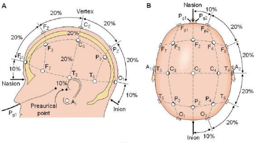
The Electroencephalogram (EEG) records the electrical activities generated by the brain over a period of time. Due to these activities, populations of neurons emit fields with small intensities, these fields are captured and recorded by electrodes fixed to the scalp. To better standardize the capture of these signals, protocols for fixing the electrodes along the skull were developed. The protocol used to acquire EEG signals in this work was the International 10-20 System, which uses 20 points that are marked dividing the skull into proportions of 10% or 20% of the distances between the reference points, Nasion and Inion in the plane medial figure 1(a) in the plane perpendicular to the skull, figure 1(b) the pre-auricular points.
The lobe underneath each electrode is identified by a nomenclature consisting of a maximum of two letters, together with a number or another letter to identify its hemispherical position [4]. A better detail of each point can be seen in table 2.
| Table 2: Points in the 10-20 pattern. | |
| Point | Area Represented |
| Fp | Polar frontal |
| F | Front |
| T | Temporal |
| C | Central |
| P | Parietal |
| O | Occipital |
| z | Medium line |
| Numbers | Odd numbers left side of the midline, even numbers right side |
At this point in the research, the data has already been digitized, processed and placed in spreadsheet format, with each file having already received the appropriate tags to be submitted to the deep neural network (deep learning) CNN now under development. At this stage of research in terms of hardware and software, an I7 processor with 64 GB of RAM and RTX 4070 TI graphics card (GPU) with 12 GB of RAM, 7680 CUDA 192 bit GDDRX type is being used. The software used in this stage is Matlab and the Anaconda platform with Python and its libraries such as Tensorflow and Keras.
Result and Discussion
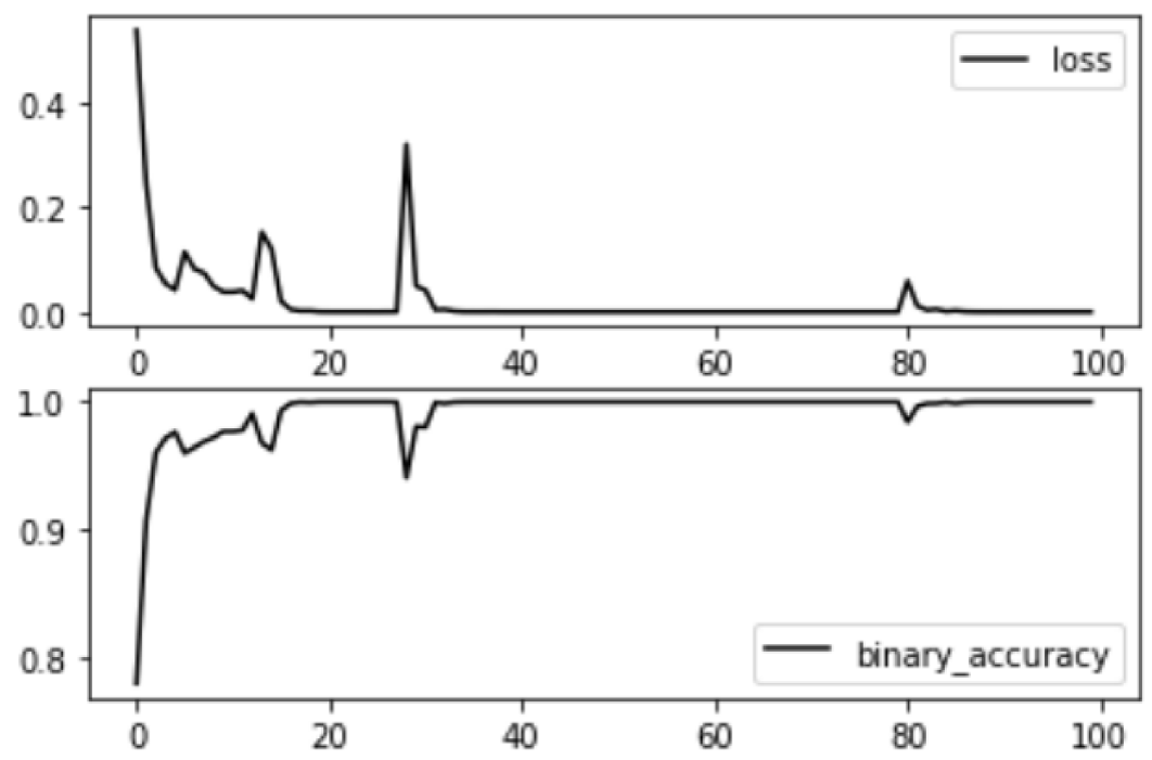
The experiments carried out with the trained Convolutional Neural Network (CNN) showed promising results in terms of accuracy and loss of test data. After the training process, the CNN was evaluated using a set of test data, the results of which indicated an accuracy of 0.96 and a loss of 0.18. The accuracy of 0.96, or 96%, reflects the model's ability to correctly classify the vast majority of examples in the test data set. This high accuracy value suggests that the CNN was able to efficiently learn the relevant features of the data during training, resulting in excellent predictive performance. The loss value of 0.18 is also indicative of the model's efficiency. Loss is a metric that quantifies how well the model is making its predictions, with lower values indicating better performance. A loss of 0.18 is relatively low, suggesting that the CNN not only got most of the predictions right, but also that the incorrect predictions are relatively close to the true classes, minimizing the model's total error. The results can be seen in figure 2.
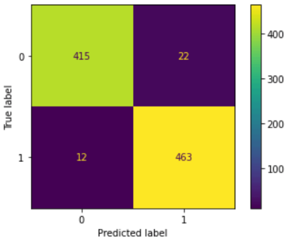
The confusion matrix presented in figure 3 illustrates the performance of the Convolutional Neural Network (CNN) trained on the test data set. The confusion matrix is an essential tool for evaluating the quality of a classification model's predictions, providing a detailed view of correct and incorrect classifications. True Positive (VP): 463 and True Negative (VN): 415.
Conclusion
The results demonstrate that the architecture and hyperparameters chosen for the CNN are effective for the problem in question. The high accuracy and low loss on the test data indicate that the trained model has a good generalization capacity, maintaining good performance not only on the training data, but also on previously unseen data. In summary, the CNN trained in this study proved to be highly effective, achieving an accuracy of 96% and a loss of 0.18 on test data, standing out as a robust solution to the problem addressed. Detailed analysis of the confusion matrix reveals that the trained CNN model performs robustly and efficiently.
Therefore, upon completing this study, it is hoped to be able to present a model that, using a patient's EEG, will be able to diagnose with a good level of probability whether the patient in question has osteoarthritis pathology (OA) [5-8].
Acknowledgement
We would like to thank our dear advisors, Prof. Doctor Alex Alíssson Bandeira and Prof. Doctor Oberdan Pinheiro for the trust placed in this major scientific undertaking. As well as, thank FAPESB for granting these important research grants, as this action is important for the development of scientific research in our state and in Brazil.
References
- Sociedade brasileira de reumatologia. 2023.
- Pinheiro ES, de Queirós FC, Montoya P, Santos CL, do Nascimento MA, Ito CH, Silva M, Nunes Santos DB, Benevides S, Miranda JG, Sá KN, Baptista AF. Electroencephalographic Patterns in Chronic Pain: A Systematic Review of the Literature. PLoS One. 2016 Feb 25;11(2):e0149085. doi: 10.1371/journal.pone.0149085. PMID: 26914356; PMCID: PMC4767709.
- Luft C, Andrade A. Research with EEG applied to the area of motor learning. Portuguese Journal of Sports Science. 2006;6(1):106-115.
- Silva, Rubianes JAI. Co-localization of electrophysiological signals and anatomical MRI signals. 2016.
- Michael FU, Daly JJ, Cavusoglu MC. Assessment of EEG event-related desynchronization in stroke survivors performing shoulder-elbow movements. IEEE Xplore. 2006. doi: 10.1109/ROBOT.2006.1642182.
- Jain Rk, Sharma PK, Gaj S, Sur A, Ghosh P. Knee osteoarthritis severity prediction using an attentive multi-scale deep convolutional neural network. Multimedia Tools and Applications. 2023;83(3):6925-6942. doi: 10.1007/s11042-023-15484.
- Yeoh PSQ, Lai KW, Goh SL, Hasikin K, Hum YC, Tee YK, Dhanalakshmi S. Emergence of Deep Learning in Knee Osteoarthritis Diagnosis. Comput Intell Neurosci. 2021 Nov 10;2021:4931437. doi: 10.1155/2021/4931437. PMID: 34804143; PMCID: PMC8598325.
- Eckstein F, Wirth W, Culvenor AG. Osteoarthritis year in review 2020: imaging. Osteoarthritis Cartilage. 2021 Feb;29(2):170-179. doi: 10.1016/j.joca.2020.12.019. Epub 2021 Jan 5. PMID: 33418028.
Content Alerts
SignUp to our
Content alerts.
 This work is licensed under a Creative Commons Attribution 4.0 International License.


