Medicine Group . 2023 March 29;4(3):515-518. doi: 10.37871/jbres1704.
Dural Tentorial Fistula of Bernasconi-Cassinari Artery, Two Case Reports: Supratentorial and Infratentorial
Patricia Alejandra Garrido Ruiz*, Álvaro Otero, Jesús María Gonçalves, Daniel Pascual, Laura Ruiz, Juan Carlos Roa, Luis Torres and Marta Román Garrido
- Tentorial fistula
- Bernasconi-cassinari artery
- Guillian barré syndrome
Abstract
We report two rare and infrequent cases, one of a patient with a Cognard type V tentorial dural fistula diagnosed at the beginning as a trunk glioma in a patient with gait disturbance. And another with a Guillian Barré syndrome with tetraparesis secondary to a tentorial dural arteriovenous fistula.
Introduction
The Bernasconi-Cassinari artery is a branch of the meningo-pituitary trunk that is not usually visible under normal conditions. However, the artery and its pathological expression can be visualized by arteriography under pathological conditions. Dural fistulas are vascular anomalies with a "shunt" between dural layers. Those that affect said artery are classified in the subgroup of tentorial fistulas. Although the preferred treatment for dural fistulas is endovascular, surgery continues to play an important role in ethmoidal and tentorial fistulas.
Intracranial vascular malformations are well recognized entities that can present with a broad spectrum of neurologic signs and symptoms. Dural Arteriovenous Fistula (DAVF) constitutes 5% to 20% of these malformations. Tentorial DAVFs account for 8.4% of intracranial DAVFs. These are known to be aggressive vascular lesions, as the shunt often results in obliteration of the tentorial sinus with associated cortical venous reflux or involvement of the perimedullary venous plexus.
Clinical Cases Description
Patient 1
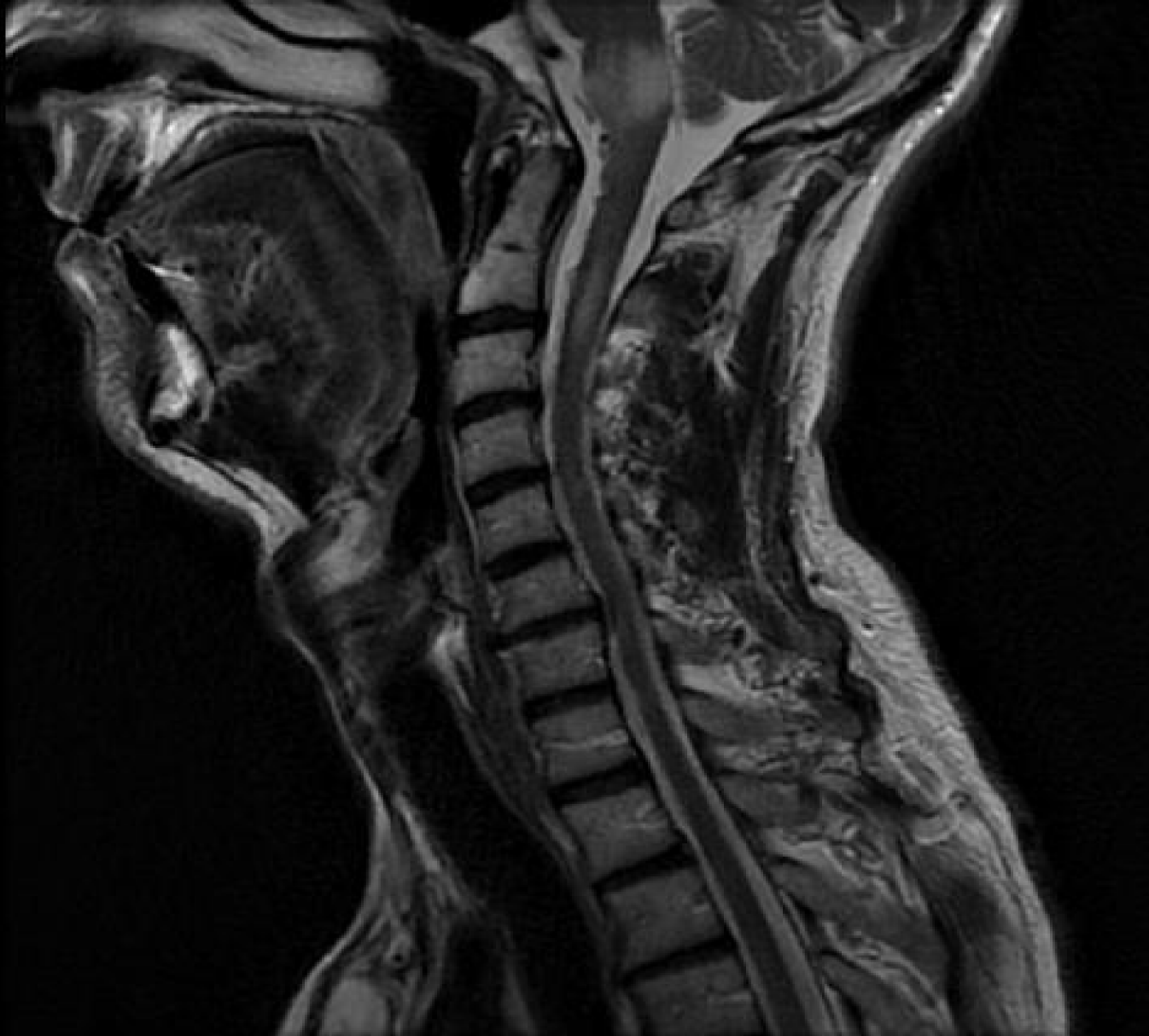
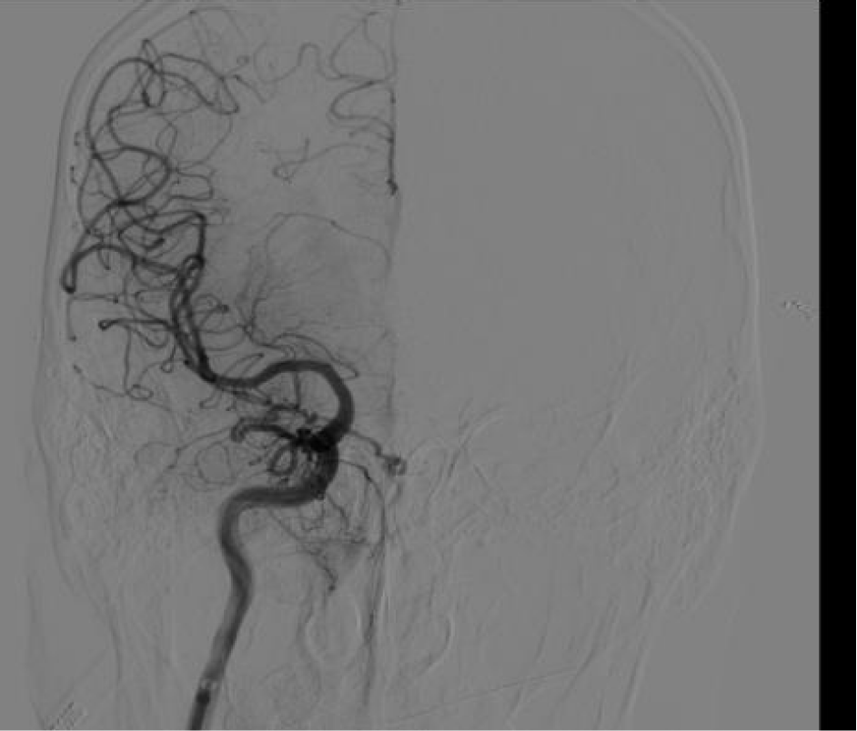
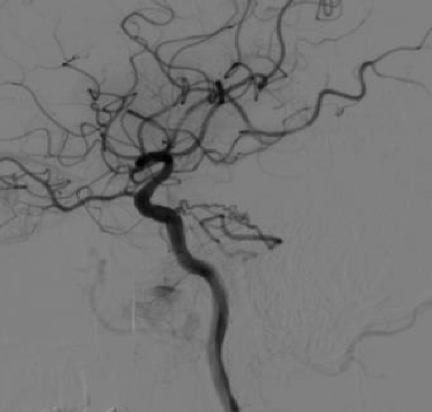
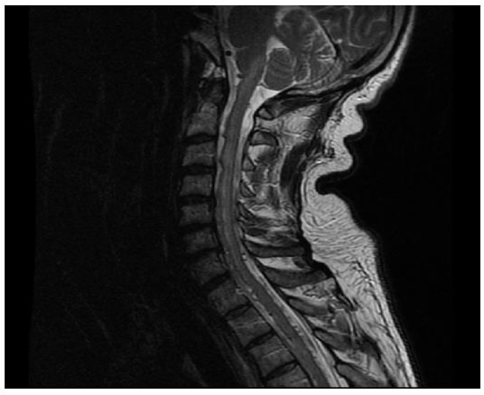
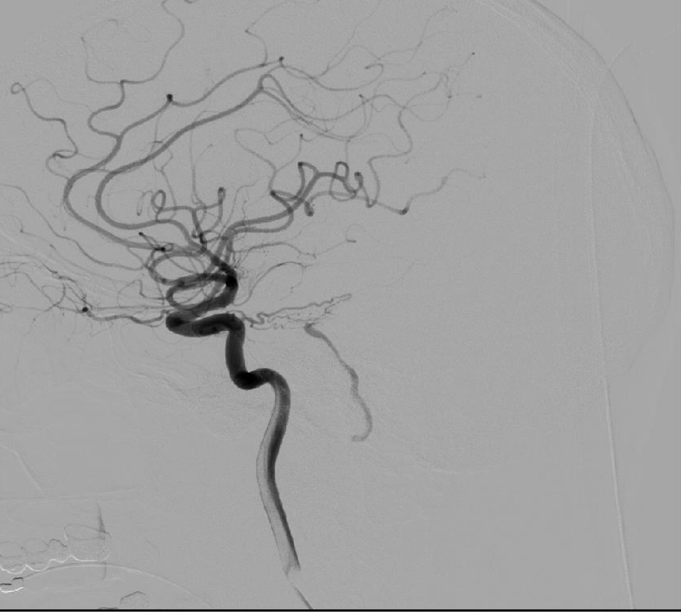
Male, 48 years old, insidious presentation of fluctuating gait disturbance. In MRI, alteration of the bulbopontine signal in T2 interpreted as trunk glioma by Neurology (Figure 1). However, in the radiological control there is spontaneous improvement, so the differential diagnosis is widened. Finally, an arteriography was performed in which a Cognard type V tentorial dural fistula was identified involving the Bernasconi-Cassinari artery [1-3] (Figures 2,3).
Treatment was decided at the Neurovascular Committee. Interventional Neuroradiology confirms difficulty in access, so neurosurgical treatment is decided [4]. Tentorial fistulas are more aggressive, and treatment is mandatory. Endovascular embolization with Onyx or coils is the treatment of election except for ethmoidal and tentorial fistulas. Treatment was planned in a joint session with Neuroradiology and Interventional Neuroradiology, which allows the exact location of the vein foot and the choice of approach.
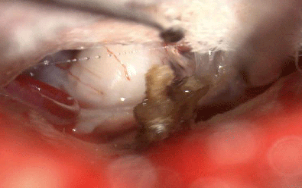
A retrosigmoid suboccipital craniectomy was performed and the vein was identified with the help of video angiography of the foot. After coagulation and cutting (Figures 4,5), disappearance of the flow in affected vessels, including perimedullary veins, is confirmed. The patient evolves without complications and with improvement in neurological function to almost total normality.
Patient 2
53 y/o man with a sudden and rapid onset of acute tetraparesis in less than 12 hours, finally he presented respiratory distress for which he had to be intubated and hospitalized in the ICU with ventilatory support was firstly suspected and treated as a Guillian-Barré syndrome with corticosteroids. CSF leak analysis revealed no abnormalities. MRI: edema could be appreciated in T2 sequences involving the low part of the medulla of the brain stem and the cervical and first segment of the thoracic spinal cord (Figure 6). Angiography: in the cerebral angiography was identified an arteriovenous fistula between the left tentorial artery of Bernasconi and Cassinari (BCA) and its branches that drained in the superior petrous vein (Dandy´s vein) [5] and associated engorgement of the anterior medullary vein (Figure 7).
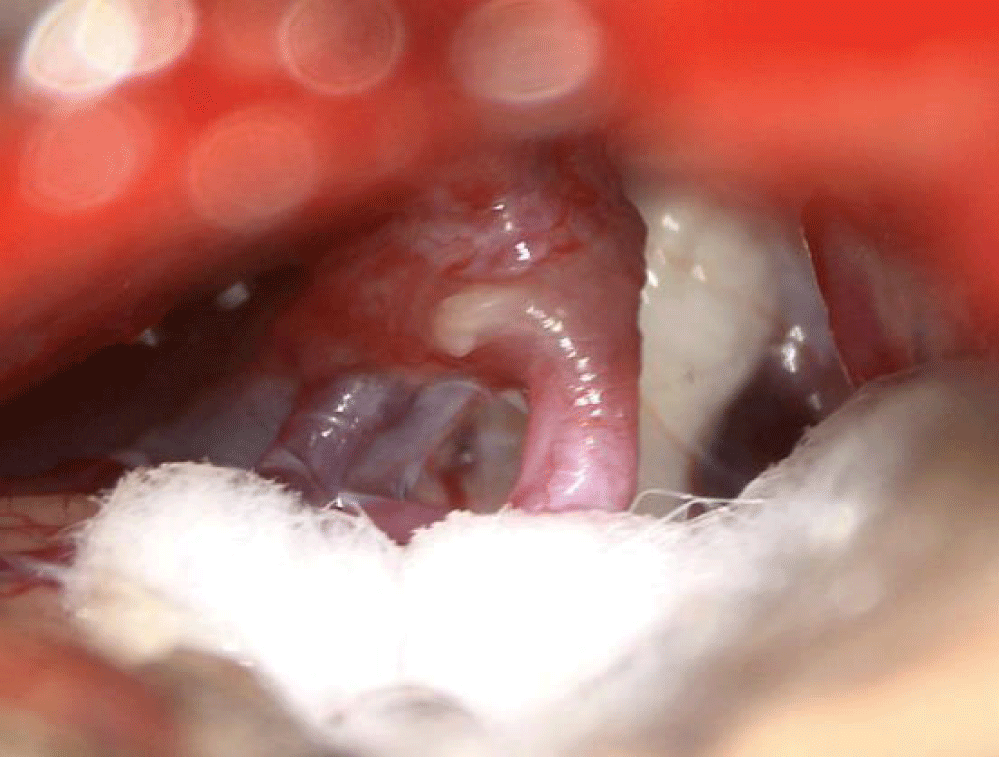
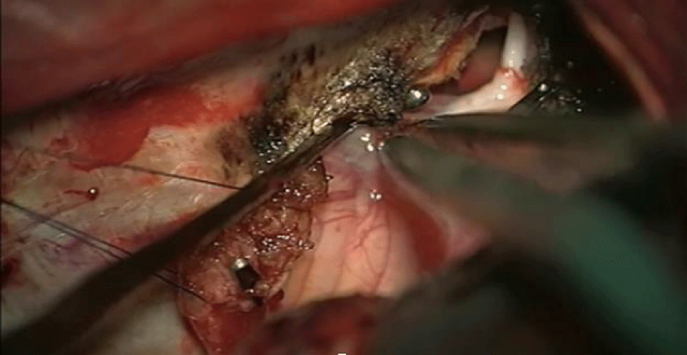
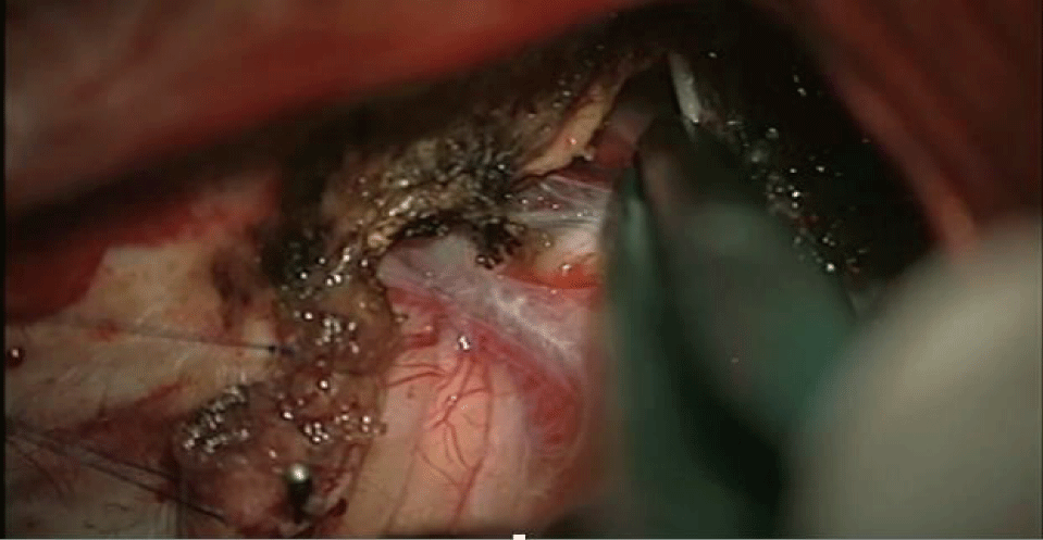
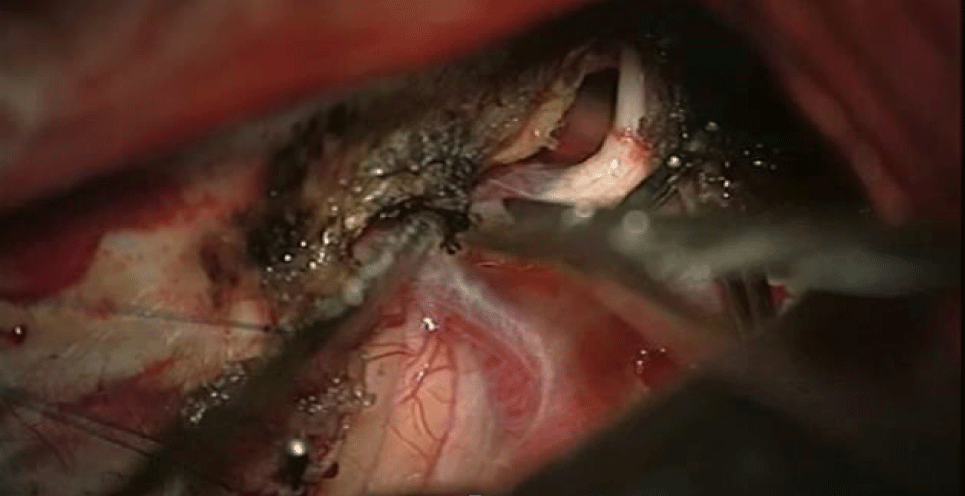
After 72 hours of the onset, a left temporal craniotomy was performed, after dural opening, the left temporal lobe was gently retracted upwards to have access to the whole medial aspect of the After 72 hours of the onset a left temporal craniotomy was performed, after dural opening the left temporal lobe was gently retracted upwards to have access to the whole medial aspect of the tentorium. Afterward was performed an extensive coagulation of the tentorial medial aspect and its arteries, we found the main trunk of the BCA bridging with Dandy´s vein which was arterialized. The BCA was coagulated (Figures 8,9). Afterward, an excision of the tentorial dural leaflet was made (Figure 10). After 8 months patient recovered normal motor function of upper limbs and partial recovery of inferior limbs (4-/5) walking with crutches.
Results and Conclusion
DAVFs are rare but aggressive and potentially fatal vascular malformations. The clinical presentation depends on the type of venous drainage and can cause serious complications frequently (bleeding, myelopathy, etc.). The clinical manifestations and neuroimaging findings can mimic other more common neurologic disorders. Early diagnosis is key, as timely intervention (endovascular or surgery) can reverse the symptoms and improve clinical outcomes. Unfortunately, its rarity and clinical variability tend to delay the diagnosis and worsen the prognosis. The preferred treatment is endovascular due to its high efficacy and lower morbidity and mortality. However, surgery continues to play an important role, especially in tentorial and anterior cranial fossa fistulas (ethmoidal fistulas) where access to embolization is harder and surgery becomes the treatment of choice. A golden rule to choosing surgery above endovascular would be if the fistula location is unfavorable for endovascular. When the ethmoid artery is involved the use of endovascular methods can lead to an ophthalmic artery thrombosis and blindness as a result. On the other hand, when a tentorial artery is involved it has difficult access to endovascular treatment. There are some selected cases that can be treated with surgery-assisted embolization and for fistulas that cannot be treated with surgery or endovascular, stereotactic-radiosurgery can be the solution. The multidisciplinary collaboration with neuroradiologists and interventional neuroradiologists in a joint session allowed, in both cases, the exact location of the vein foot and the choice of approach. Having a multidisciplinary team empowers a correct choice and treatment planning and therefore a good final result.
References
- Peltier J, Fichten A, Havet E, Foulon P, Page C, Le Gars D. Microsurgical anatomy of the medial tentorial artery of Bernasconi-Cassinari. Surg Radiol Anat. 2010 Dec;32(10):919-25. doi: 10.1007/s00276-010-0655-z. Epub 2010 Apr 16. PMID: 20397016.
- Sandoval-Otero A, Vergara-García D, Arango-Rodríguez J, Caballero A, Torres J. Manejo microquirúrgico de fístula arteriovenosa dural tentorial entre arteria de Bernasconi-Cassinari y vena de Galeno guiada por angiografía intraoperatoria: Reporte de caso y revisión de la literatura. Revista Chilena de Neurocirugía. 2019;45(3):241-245. doi: 10.36593/rev.chil.neurocir.v45i3.142
- Youmans. Neurological Surgery. 7th Ed. Saunders Company; 2016.
- Hou K, Lv X, Qu L, Guo Y, Xu K, Yu J. Endovascular treatment for dural arteriovenous fistulas in the petroclival region. Int J Med Sci. 2020 Oct 18;17(18):3020-3030. doi: 10.7150/ijms.47365. PMID: 33173422; PMCID: PMC7646121.
- Ferro JM, Coutinho JM, Jansen O, Bendszus M, Dentali F, Kobayashi A, van der Veen B, Miede C, Caria J, Huisman H, Diener HC; RE-SPECT CVT Study Group. Dural Arteriovenous Fistulae After Cerebral Venous Thrombosis. Stroke. 2020 Nov;51(11):3344-3347. doi: 10.1161/STROKEAHA.120.031235. Epub 2020 Sep 25. PMID: 32972315.
Content Alerts
SignUp to our
Content alerts.
 This work is licensed under a Creative Commons Attribution 4.0 International License.


