Medicine Group . 2022 August 12;3(8):905-907. doi: 10.37871/jbres1530.
Cancer Cell Destruction by Magneto-Mechanical Actuation of Nanowires Compared with Nano/Micromagnetic Particles
Horia Chiriac1*, Anca Emanuela Minuti1,2 and Nicoleta Lupu1
2Faculty of Physics, "Alexandru Ioan Cuza" University, 11 Carol I Boulevard, Iași, 700506, Romania
Abstract
High values of saturation magnetization at low magnetic field, along with magnetic anisotropy are the basic elements for increased effects of magnetomechanical actuation on cancer cells. In this paper we present a brief comparison report on the effects of Fe-Cr-Nb-B Magnetic Particles (MPs) and Fe-Co Nanowires (NWs) on human osteosarcoma cells. The purpose of the study is to evaluate the best sites to employ different types of nanomaterials for cancer cell destruction through magnetomechanical actuation.
Introduction
The use of magnetic nanoparticles for cancer cell destruction is widely known due to their applications such as magnetic hyperthermia and Magneto Mechanical Actuation (MMA) [1]. This effect is based on the spatial oscillations or rotations of magnetic particles which accurately follow the oscillating or rotating magnetic fields. A magnetic torque is formed through which magnetic particles can hit cells from the outside or, after entering cells, from the inside, leading to their death. The size of this torque depends on the magnetic characteristics - particularly magnetization and magnetic susceptibility, but also on the size and shape of the particles. Iron oxide nanoparticles (SPION) [2], disk-shaped particles obtained by vacuum thinning techniques followed by lithography [3] and Ni nanowires [4] where also tested as magnetic elements for magnetomechanical actuation.
Previously, we presented our results regarding a new type of Magnetic Particles (MPs), i.e. Fe-Cr-Nb-B MPs, prepared by wet milling of superferromagnetic precursor glassy ribbons, designed for cancer treatment by MMA [5]. These MPs have an important shape anisotropy and a large saturation magnetization, which generates an improved torque in a rotating magnetic field, producing important damages on the cellular viability of cancer cells. Inspired by these findings, we tested the possibility of using Magnetic Nanowires (NWs) that have a higher shape anisotropy. Furthermore, the Fe-Co alloy that we propose has high saturation magnetization values. The Fe64.5Co35.5 NWs were prepared by electrolytic deposition in alumina membranes with a pore diameter of 200 nm, with lengths of 4 microns. In this brief report, we have planned to analyze which of these two types of materials is more suitable for the destruction of cancer cells, presenting the positive aspects of each type.
Methods
We prepared Fe67.2Cr12.5Nb0.3B20 magnetic particles by milling amorphous melt-spun ribbons in oleic acid, using a Retsch 200 high-energy ball mill. The MPs were dispersed in calcium gluconate to obtain a ferrofluid that was added in cell culture media [6]. Fe64.5Co35.5 Nanowires (NW) with lengths, 4 microns were obtained by electrolytic deposition in alumina membranes with a pore diameter of 200 nm. Viability tests were conducted on human osteosarcoma cell line (HOS) MG-63 from Sigma Aldrich using MTT assay, a colorimetric assay that evaluates cell metabolic activity. For viability assay of the magneto mechanical actuated samples, cells were seeded in 24 well plates and grown until 85-90% confluency. The complete cell culture media was replaced with the ferrofluid/nanowires dispersed in the cell culture media, at a concentration of 1 mg/ml for MPs, and 0.1 mg/ml for NWs which was later added onto the cells and co-incubated for another 24 hours. Following this period, the samples were treated for 30 min., in a rotating magnetic field of 80 Oe at a frequency of 2 Hz for magneto mechanical actuation.
Results
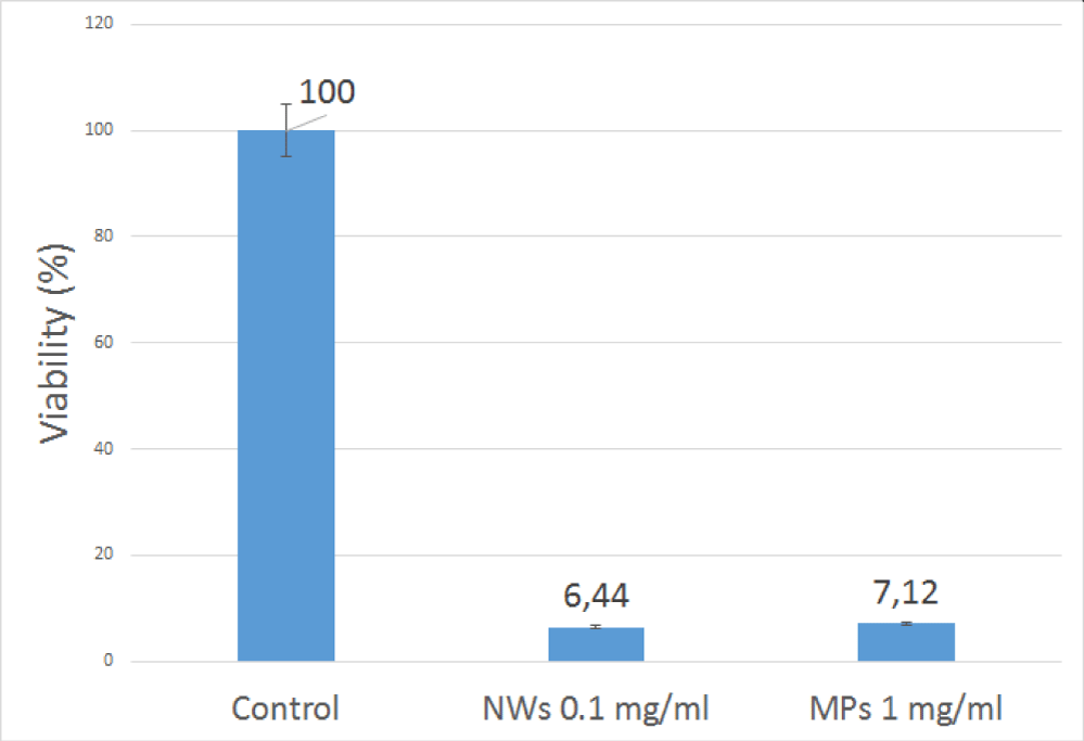
The Fe-Cr-Nb-B MPs and Fe-Co NWs were tested on Human Osteosarcoma Cells (HOS) using various concentrations of particles and found that both are biocompatible in vitro. The MPs and NWs induced no negative effects on the cells tested, as the viability after 24h of co-incubation was over 95% for both types of nanomaterials. Subsequently, we studied the effect of MMA in a rotating 80 Oe magnetic field and frequency of 2 Hz on the viability of HOS. We used MPs and NW with different concentrations, and exposure time of 30 minutes, applied 24 hours after the introduction of the MPs and NWs into cell culture media. The results indicated that with a concentration of 1 mg/ml of MPs and 0.1 mg/ml of NWs, respectively, the cell viability decreased to about 6-7%. The concentration of NWs needed to induce cancer cell death is 10 times lower than the one needed for MPs, for the obtained results to be similar (Figure 1).
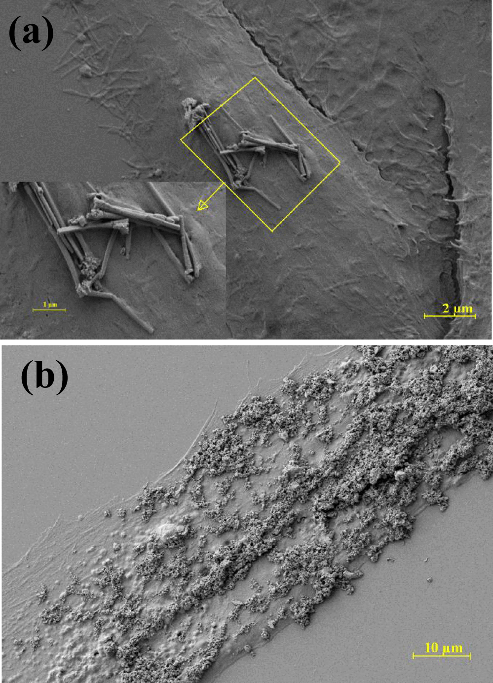
As it can be seen in figure 2a, nanowires adhered in low quantities on the cell membrane, mainly in one agglomeration as opposed with magnetic nanoparticles that tend to not only be more adherent, having a higher load on the cell surface as the figure 2b illustrates, but they were also well distributed on the cell membrane. It is worth mentioning that although NWs lead to the same results using when using lower quantities, due to their higher magnetization and anisotropy, the magnetic nanoparticles were better internalized and metabolized by cells. Therefore, MPs can be better targeted in tissues harder to reach than NWs. The latter are also harder to control, being more suitable for superficially-developed cancer tissues.
Conclusion
It can be summarized that the destruction of cancer cells using Fe-Co NWs is similar with the one caused by Fe-Cr-Nb-B MPs, due to their high magnetization and shape magnetic anisotropy. Although their effects are similar, both types of nanomaterials can be used differently, as the quantity of NWs needed is lower than the one needed for similar results when using MPs, but they are not as adherent as the MPs. Furthermore, MPs have other advantages such as better cell internalization and ease of transportation to the required area, thus being more suitable for applications such as drug targeting, magnetic hyperthermia, contrast agents or cell separation.
Acknowledgment
Work supported by (UEFISCDI) Contract no. PCE20/2021 (PN-III-P4-ID-PCE-2020-2381).
References
- Kim DH, Rozhkova EA, Ulasov IV, Bader SD, Rajh T, Lesniak MS, Novosad V. Biofunctionalized magnetic-vortex microdiscs for targeted cancer-cell destruction. Nat Mater. 2010 Feb;9(2):165-71. doi: 10.1038/nmat2591. Epub 2009 Nov 29. Erratum in: Nat Mater. doi:10.1038/nmat2631. PMID: 19946279; PMCID: PMC2810356.
- Leulmi S, Chauchet X, Morcrette M, Ortiz G, Joisten H, Sabon P, Livache T, Hou Y, Carrière M, Lequien S, Dieny B. Triggering the apoptosis of targeted human renal cancer cells by the vibration of anisotropic magnetic particles attached to the cell membrane. Nanoscale. 2015 Oct 14;7(38):15904-14. doi: 10.1039/c5nr03518j. Epub 2015 Sep 14. PMID: 26364870.
- Goiriena-Goikoetxea M, García-Arribas A, Rouco M, Svalov AV, Barandiaran JM. High-yield fabrication of 60 nm Permalloy nanodiscs in well-defined magnetic vortex state for biomedical applications. Nanotechnology. 2016 Apr 29;27(17):175302. doi: 10.1088/0957-4484/27/17/175302. Epub 2016 Mar 17. PMID: 26984933.
- Contreras MF, Sougrat R, Zaher A, Ravasi T, Kosel J. Non-chemotoxic induction of cancer cell death using magnetic nanowires. Int J Nanomedicine. 2015 Mar 17;10:2141-53. doi: 10.2147/IJN.S77081. PMID: 25834430; PMCID: PMC4370947.
- Chiriac H, Radu E, Țibu M, Stoian G, Ababei G, Lăbușcă L, Herea DD, Lupu N. Fe-Cr-Nb-B ferromagnetic particles with shape anisotropy for cancer cell destruction by magneto-mechanical actuation. Sci Rep. 2018 Aug 1;8(1):11538. doi: 10.1038/s41598-018-30034-3. PMID: 30069055; PMCID: PMC6070495.
- Minuti AE, Stoian G, Herea D-D, Radu E, Lupu N, Chiriac H. Fe-Cr-Nb-B Ferrofluid for Biomedical Applications. Nanomaterials. 2022;12(9):1488. doi: 10.3390/nano12091488.
Content Alerts
SignUp to our
Content alerts.
 This work is licensed under a Creative Commons Attribution 4.0 International License.


