Medicine Group . 2022 August 10;3(8):880-882. doi: 10.37871/jbres1526.
Cystic Posterior Neck Mass in a Premature Newborn
Estephanie Rivero1*, Adel Zauk2 and Zarah J Pua2
2Department of Neonatology, St. Joseph’s University Medical Center, Paterson, NJ, USA
Summary
Case
A female infant born via vaginal delivery at 36 weeks and 5 days gestation to a 22-year-old, Gravida 4, Para 1 mother with a complicated pregnancy. She is a product of a spontaneous monochorionic-diamniotic twin gestation. Maternal serologies including, Hepatitis B, Rubella, HIV and Syphilis were unremarkable, except positive GBS colonization. At 22 weeks and 4 days, the patient’s fetal MRI showed a hypodense focus extending exophytically from the midline scalp while the underlying skin and calvarium appeared intact. Mass appeared to be more like a skin tag with no intracranial extension; unlikely atretic encephalocele.
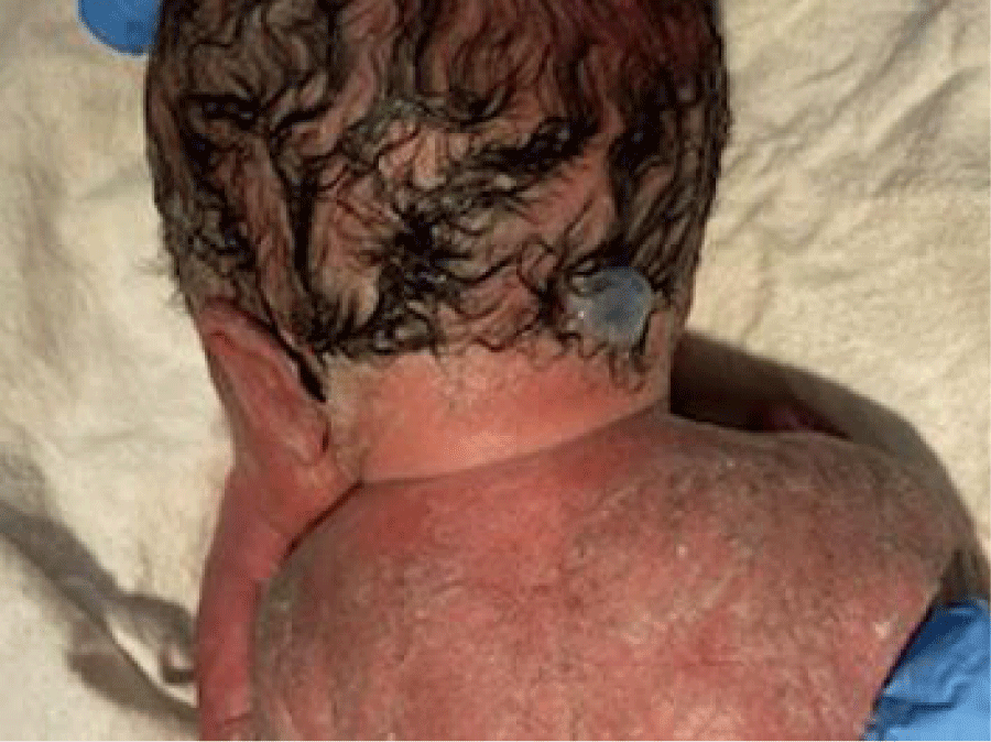
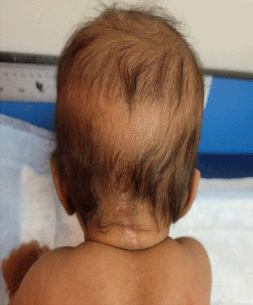
At the delivery, baby had a loose cord wrapped once around the neck and on examination there was a small cystic mass, measuring roughly 2cm, on the back of the head, as shown in figure 1. Mass was wrapped loosely with sterile gauze. Apgar scores of 8 and 9 at 1 and 5 minutes, respectively. Her identical twin sister had an unremarkable physical examination.
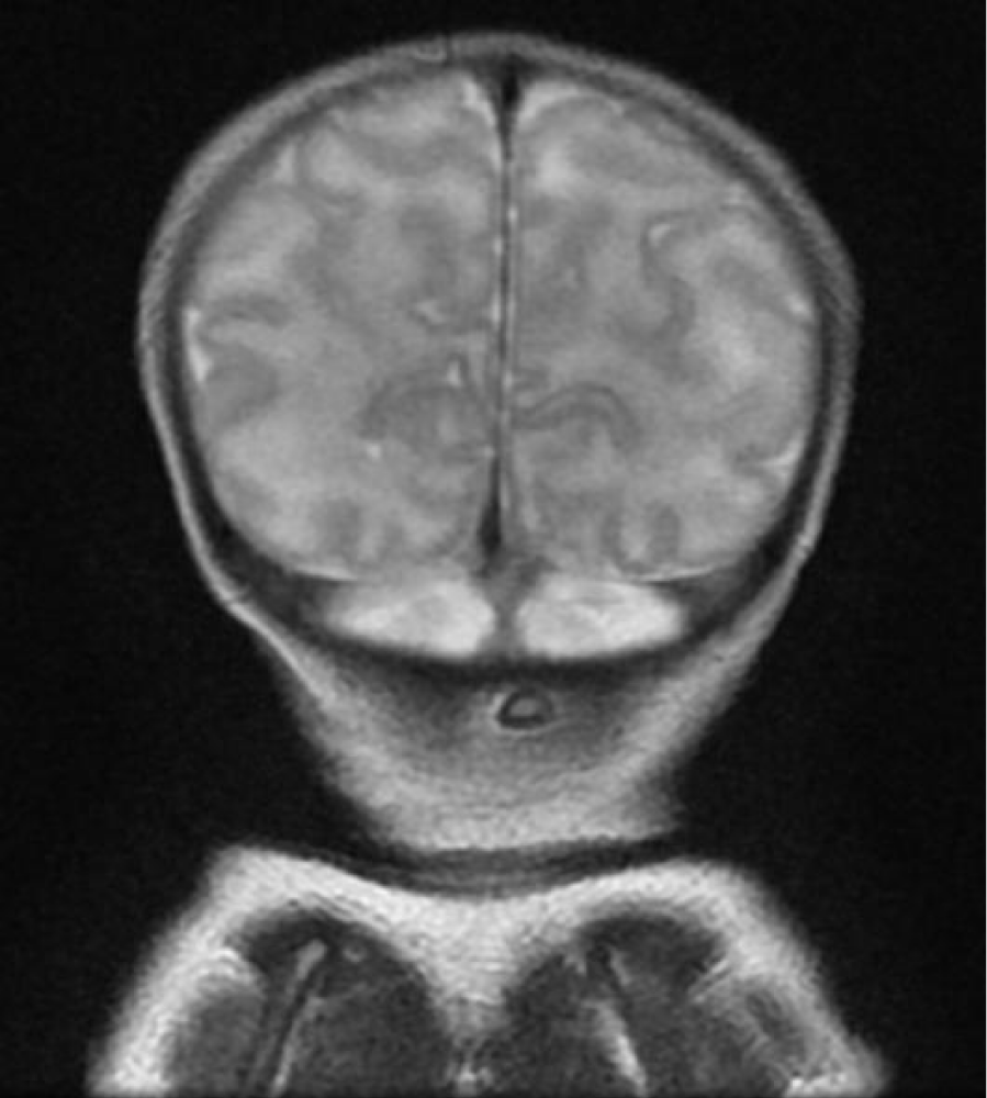
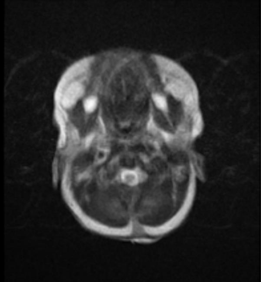
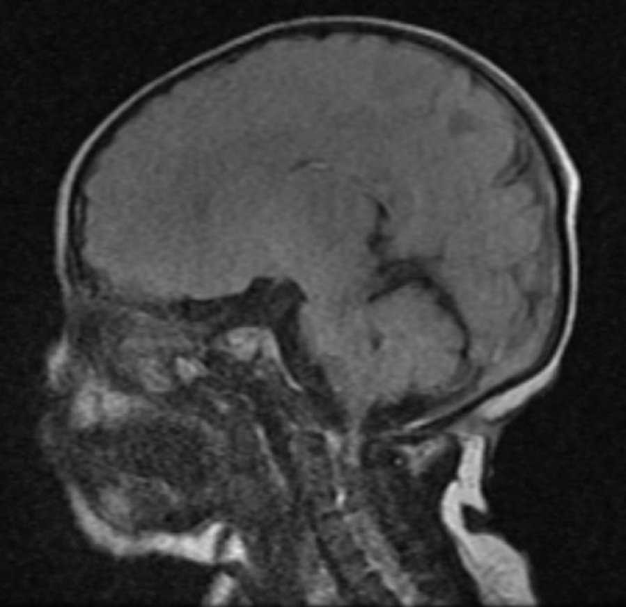
Radiologic imaging was obtained on day one of life to determine the underlying pathology. Some of the MRI images delineate the dermal sinus tract penetrating through the subcutaneous fat and bone, as seen in figures 2-4. The infant was started on ampicillin and gentamicin for prophylaxis due to the possibility of underlying tract into the central nervous system. Neurosurgery was consulted and the infant was taken to surgery the following day. Neurosurgery’s operative findings were indicative of a sinus tract that extended into the dura. The dura was dissected rostrally and caudally with removal that was extending from the occipital dura.
The specimen was sent out for a pathology report, with results of meningeal fibroconnective tissue, covered by a thin layer of epidermis with focal surface erosions. No definitive glial tissue was identified. Patient was discharged on post-operative day two with post-surgical skin care education and follow-up within one month. Follow up at the two months well visit showed a well-healed surgical scar as seen in figure 5.
Discussion
The infant presented with an occult spinal dysraphism, known as dermal sinus, which occurs in 1–3/1000 live births [1]. There has been a significant decrease in the prevalence of spinal dysraphism, due to supplementary folic acid, increased access to antenatal care that includes prenatal ultrasonography and genetic screening [1]. Nevertheless, these anomalies remain prevalent and can present with varying degrees of severity.
A dermal sinus is the result of an abnormal communication between the dermis and intracranial structures because of the dysjunction of cutaneous ectoderm from the neural ectoderm during the 4th week of fetal development [2]. The tract is lined by epithelium and may terminate within the thecal sac [3]. These sinuses can occur at any level of the spine and is rarely associated with complicated vertebral abnormalities [4]. Documented cases have reported tracts from the nasion and occipital area to the lumbar and sacral regions [2]. Most common location is in the lumbar and lumbosacral region (75%) [2]. While most cases have been isolated findings, there is one report of a patient with double dermal sinuses, located over the head and neck. Presentation can range from hyperpigmented patch, hairy nevus, or capillary angioma on the overlying skin. Dorsal spinal tracts can be associated with other anomalies of the central nervous system such as tethered cord, inclusion tumors, and split cord malformations [3]. Neurological examination is typically normal except when there is cord compression from inclusion tumors [5]. Meningitis or abscess may occur as a result of the connection from the skin to the intradural components [6]. Few patients go into adulthood without any preceding illness.
MRI has been used to evaluate the extent of the sinus tract and any associated lesions. However, it has a false-negative rate for the detection of inclusion cysts and the intradural component of the tract [6]. Therefore, surgical intervention is always recommended to prevent any impending infection.
Author Disclosure
Drs. Rivero, Pua, and Zauk, have disclosed no financial relationships relevant to this article. This commentary does not contain a discussion of unapproved or investigative use of a commercial product or device.
References
- Hussein NA, Ahmed KA, Osman NM, Elkess Yacoub GE. Role of ultrasonography in screening of spinal dysraphism in infants at risk. Egypt J Radiol Nucl Med. 2022 Feb 15;53(46). doi: 10.1186/s43055-022-00722-2.
- El Khashab M, Nejat F, Ertiaei A. Double dermal sinuses: A case study. Journal of Medical Case Reports. 2008 Aug 26;2(281). doi: 10.1186/1752-1947-2-281.
- Singh I, Rohilla S, Kumar P, Sharma S. Spinal dorsal dermal sinus tract: An experience of 21 cases. Surg Neurol Int. 2015 Oct 7;(6):S429-34. doi:10.4103/2152-7806.166752.
- French BN. The embryology of spinal dysraphism. Clin Neurosurg. 1983;30:295-340. doi: 10.1093/neurosurgery/30.cn_suppl_1.295. PMID: 6365396.
- Venkataramana NK. Spinal dysraphism. Journal of Pediatric Neurosciences. 2011 Oct 10;6(3):31-40. doi: 10.4103/1817-1745.85707.
- Tisdall MM, Hayward RD, Thompson DN. Congenital spinal dermal tract: How accurate is clinical and radiological evaluation?. Journal of Neurosurgery: Pediatrics. 2015 Jun;15(6):651-6. doi: 10.3171/2014.11.peds14341.
Content Alerts
SignUp to our
Content alerts.
 This work is licensed under a Creative Commons Attribution 4.0 International License.


