Medicine Group . 2022 April 13;3(4):332-334. doi: 10.37871/jbres1444.
An Unexpected Case of Metal Biliary Stent Fracture Detected by Contrast Enhanced 18F-FDG PET/C
Ferdinando Calabria* and Mario Leporace
- 18F-FDG
- PET/CT
- Biliary stent fracture
- Contrast enhanced CT
- Pancreatitis
- Lung cancer
Abstract
18F-FDG PET/CT is an useful diagnostic tool for cancer patients. In our routine practice, we perform a significant number of PET/CT scans in association with contrast enhanced CT, to assess resectability of abdominal tumors or to evaluate distant brain and liver metastases. We documented an unexpected emergency disease during a contrast enhanced 18F-FDG PET/CT. In a 63-year-old male patient, examined during follow-up of lung cancer, no sites of disease relapse were detected at PET/CT; nevertheless, diffuse tracer uptake was detected along the outline of a biliary stent. Correlative contrast enhanced CT diagnosed fracture of a metallic biliary stent without any evidence of cancer involvement. Patient was referred to the local department of surgery for substitution of the biliary stent. Although rare, unexpected emergency diseases requiring urgent management are detected using 18FFDG PET/CT. Correlative CT should be carefully evaluated in order to avoid delays in diagnosis, potentially leading to poorer prognosis.
Introduction
Case report
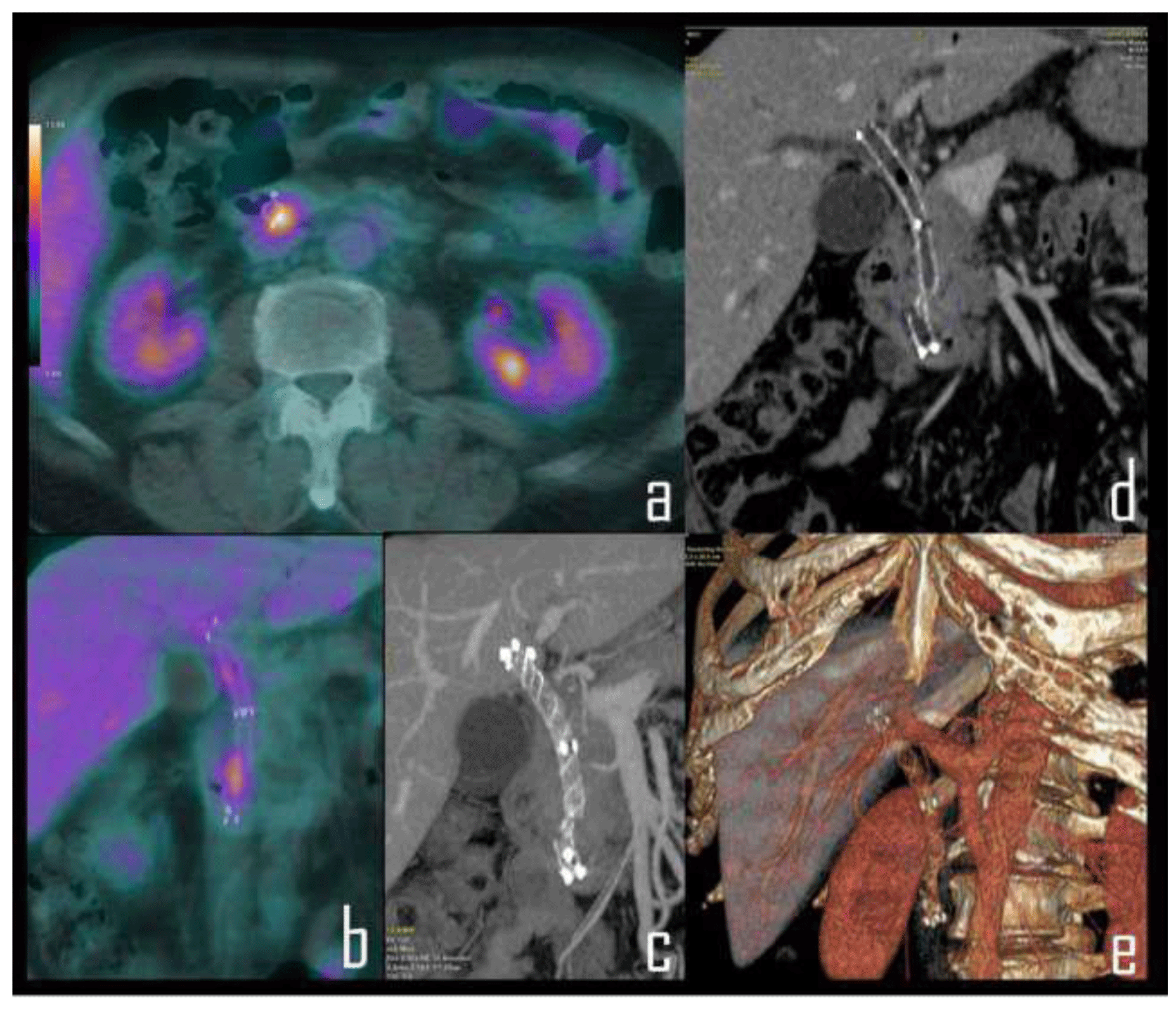
A 63-year-old male patient underwent whole body contrast enhanced 18F-FDG PET/CT in our Department, during follow-up of lung cancer. Patient was previusly submitted (two years before) to upper right lobectomy for lung squamous carcinoma. Patient anamnesis was also positive for chronic pancreatitis. For this reason, he was submitted, 11 months before, to implantation of biliary stent due to the development of fibrosis of the bile duct. No sites of lung cancer relapse were detected at PET/CT. Nevertheless, PET/CT showed focal tracer uptake along the outline of the biliary stent (Figure 1) as evident in axial (a) and coronal (b) PET/CT details. Coronal Maximum Intensity Projection CT (c) and CT detail (d) after contrast administration underlined the fracture of the distal tract of the biliary stent, also evident as a covered self-expandable biliary metal stent in correlative Volume Rendering CT of the upper abdomen (e). Patient was referred to our local department of surgery, where he underwent biliary stent removal and reimplantation. After surgery, histological specimen diagnosed chronic inflammation; confirming the absence of neoplastic involvement. Biliary stents are used to manage benign and malignant obstruction by enabling biliary drainage and decompression, alleviate patient symptoms and improve quality of life [4]. Stent occlusion and stent migration are the most common complications of biliary stent insertion, whereas cholecystitis, cholangitis, pancreatitis, perforation and and bleeding are less common [5]. On the other hand, stent fracture is an extremely rare complication which has only been described in a few case reports [6,7]. PET/CT with 18F-FDG is widely used for staging or restaging cancer patients; it is known that a significant minority of oncology patients may present some emergency conditions which may mimick a malignant neoplasm at PET/CT or can be misdiagnosed to the relative specificity of 18F-FDG. In fact, being an analogue of glucose, 18F-FDG uptake may occur in several conditions other than malignant lesions, as phlogosis and infection. Correlative imaging with contrast enhanced CT can play a role of the utmost importance. Several studies have reviewed unexpected CT findings obtained in the emergency department [8,9]. For the best of our knowledge, no data are reported about the detection of the fracture of a biliary stent during a contrast enhanced 18F-FDG PET/CT performed in a cancer patient. The accurate knowledge of this emergency disease which can be observed in clinical practice of nuclear physicians and radiologists by 18F-FDG PET/CT can favour rapid diagnoses and could enable the need for urgent intervention for some patients. The CT component of the exam, with or without contrast agent, should be carefully evaluated immediately after the images acquisition, in order to avoid delays in diagnosis, potentially leading to poorer prognoses.
Conflict of Interest
The authors do not have conflicts of interest relevant to this article to disclose.
Contributors Statement Page
Dr. Ferdinando Calabria examined PET/CT scan, conceptualized and designed, drafted the initial manuscript and reviewed and revised the manuscript. Dr. Mario Leporace examined contrast enhanced CT, collected data, carried out initial analyses and critically revised the manuscript. All authors approved the final manuscript as submitted and agree to be accountable for all aspects of the work.
References
- Nakagawa K, Aoyagi M, Inaji M, Maehara T, Toriyama H, Kawano Y, Tamaki M, Nariai T, Ohno K. [The usefulness of whole body FDG-PET/CT in patients with brain metastasis]. No Shinkei Geka. 2009 Feb;37(2):159-66. Japanese. PMID: 19227157.
- Hundt W, la Fougère C, Vogtmann J, Steinbach S, Burbelko M, Tiling R. Evaluation of contrast medium enhancement and [(18)F]-FDG uptake of liver metastasis in PET/CT prior to therapy. Eur J Radiol. 2012 Apr;81(4):652-7. doi: 10.1016/j.ejrad.2011.01.026. Epub 2011 Feb 5. PMID: 21300504.
- Toriihara A, Yamaga E, Nakadate M, Oyama J, Tateishi U. Detection of unexpected emergency diseases using FDG-PET/CT in oncology patients. Jpn J Radiol. 2017 Sep;35(9):539-545. doi: 10.1007/s11604-017-0664-5. Epub 2017 Jul 3. PMID: 28674772.
- Alkhiari R, Patel V, Cohen L. Spontaneous fracture of a covered self-expandable biliary metal stent and endoscopic technique for removal. Can J Gastroenterol Hepatol. 2014;28:411-412.
- Catalano O, De Bellis M, Sandomenico F, de Lutio di Castelguidone E, Delrio P, Petrillo A. Complications of biliary and gastrointestinal stents: MDCT of the cancer patient. AJR Am J Roentgenol. 2012 Aug;199(2):W187-96. doi: 10.2214/AJR.11.7145. PMID: 22826420.
- Sriram PV, Ramakrishnan A, Rao GV, Nageshwar Reddy D. Spontaneous fracture of a biliary self-expanding metal stent. Endoscopy. 2004;36:1035-1036.
- Yoshida H, Mamada Y, Taniai N, Mizuguchi Y, Shimizu T, Aimoto T, Nakamura Y, Nomura T, Yokomuro S, Arima Y, Uchida E, Misawa H, Uchida E, Tajiri T. Fracture of an expandable metallic stent placed for biliary obstruction due to common bile duct carcinoma. J Nippon Med Sch. 2006 Jun;73(3):164-8. doi: 10.1272/jnms.73.164. PMID: 16790985.
- Bruzzi JF, Truong MT, Marom EM, Mawlawi O, Podoloff DA, Macapinlac HA, Munden RF. Incidental findings on integrated PET/CT that do not accumulate 18F-FDG. AJR Am J Roentgenol. 2006 Oct;187(4):1116-23. doi: 10.2214/AJR.05.0712. Erratum in: AJR Am J Roentgenol. 2007 Feb;188(2):300. PMID: 16985164.
- Osman MM, Cohade C, Fishman EK, Wahl RL. Clinically significant incidental findings on the unenhanced CT portion of PET/CT studies: frequency in 250 patients. J Nucl Med. 2005 Aug;46(8):1352-5. PMID: 16085594.
Content Alerts
SignUp to our
Content alerts.
 This work is licensed under a Creative Commons Attribution 4.0 International License.


