> Medicine Group. 2021 September 29;2(9):839-845. doi: 10.37871/jbres1319.
Intentional Replantation and 3d Obturation with Biodentine of a Resorbed Root in Young People: A Case Report
Giovanni Messina*, Luca Boschini, Luigi Stagno d’Alcontres, Stefano Milani, Maria Elena Cipollina, Gaia Bonandi and Calogero Bugea
- Biodentine
- 3d obturation
- Replantation
- Root resorption
Abstract
Referred patient 14 years old (in orthodontic treatment) for a suspected resorption on a 2.1 that was exposed to a trauma [1]. After an apical x ray, a cone beam was performed to have a complete diagnosis [2,3]. Almost the 80% of the root is resorbed, after achieving the parents’ consent to the treatment, was planned an intentional replantation [4] and a retrograde approach. In case like this the treatment’s goal is an intentional replantation to stop the root resorption, removing all the resorbing tissue and rebuild the root by biodentine’s use [5]. The tooth was replanted and splinted to the ortho appliances to allow a precise position of replantation. The final X ray control confirmed the correct rebuilding of the root anatomy. After two weeks the endodontic treatment was performed at all.
After 6 months the patient has completed the ortho treatment and the X ray control revealed a good response, the biodentine‘s stability and no sign of tissue inflammation.
Since the good preliminary results at six months of the intentional replantation with Biodentine root rebuilding, this treatment, in young patient, can be considered as a good option to the maintainability of the tooth till the adult age and to long lasting prosthetic treatments.
Introduction
The pediatric patient is particularly at high risk of traumatic events [6-8]. The early loss of a tooth due to traumatic causes in the growing patient is difficult to manage, since prosthetic or implant rehabilitative solutions cannot be performed [9]. In particular implantology in a growing patient is absolutely not indicated for the ankylosis process that causes a misplacement of the tooth at the end of bone developing. For this reason it is essential to try to keep traumatized teeth, even if they are strongly compromised, in young patient as temporary work waiting the complete growth.
The inflammatory-related root resorption is a physiological phenomenon that involve primary dentition‘s roots, granting the permanent teeth eruption [10-12].
In case this process involves the permanent teeth it is considered a pathological one that causes the slow loss of roots substance leading to the tooth loss.
In literature are described different classifications of the resorption process that are based on etiological factors or morological one’s [13-15].
The resorption process are divided in external and internal based on the beginning point of resorption, and are also divided in apical and cervical ones by the localization on the root surface.
Another classification is based on the causes that leaded to it.
Internal resorption can be caused by the chronical presence of inflammatory factor as necrotic pulp tissues or iatrogenic causes (internal bleaching) [15,16]; the external resorption are caused by chronical periodontal inflammation, whitening and orthodontic movements; in some cases there are idiopathic resorptions [15].
There is no standard protocol for the treatment of external resorption. It is suggested a treatment based on the pattern and location of resorption: internal access, external access, and intentional replantation [12].
The external access is performed by a surgical access that allows to repair the defect by using biocompatible materials such the one based on calcium silicate. These material are a “dentinal substitute” and are characterized by the easy manipulation, the resistance to compression, the radiopacity, the excellent biocompatibilty and the color stability [16,18,19].
The success of this treatment is the complete removal of the inflammatory tissue and the total replacement and filling of the defect [20]. Often the area of the resorption does not allow the surgery access and in this case the intentional replantation becomes the first treatment choice [21,22] especially in the growing patient as temporary solution.
The following article describe the treatment of a upper central incisor affected by an external inflammatory sever root resorption that has been totally treated with a 3d root reconstruction of the root by the use of Biodentine and intentionally replanted on a 14 years old patient.
Case Report
A 14 years old young male was referred to the author’s attention for an upper left central incisor root resorption.
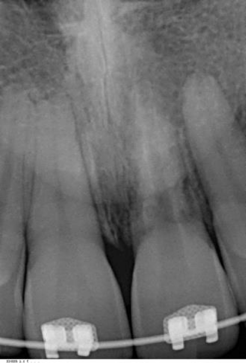
The anamnesis reveals that he lost the incisor traumatically one year before. The tooth was taken by the ground and brought to a dentist for the replantation after a long time from the trauma but the root treatment wasn’t performed after two weeks leading to necrosis. Root resorption appeared one year later during the last check before the brackets were removed (Figure 1).
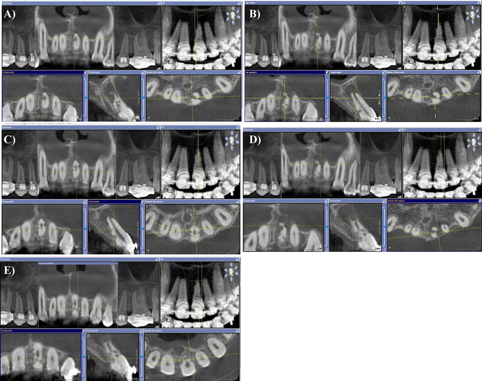
The CBCT showed multiple resorptions of the incisor root and a 7 mm wide periradicular lesion. The tomography showed also a partial loss of the vestibular cortical wall (Figures 2A-E).
The localization of the resorption areas did not allow a conventional surgical approach meanwhile it was not possible to perform an extraction and an implant considering the age and the incomplete skeletal development.
Therefore, with the consent of the patient’s parents, an intentional replantation was performed in order to manage extra orally the multiple lesions.
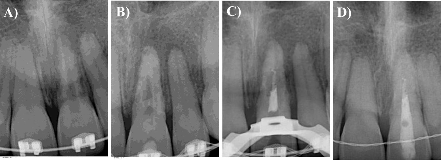
A 3-dimensional reconstruction of the root was performed using a bioceramic material. The surgery was preceded by an orthograde root canal dressing with calcium hydroxide (Figures 3A-D) to reduce the exudate.
The cavity access was performed by a small drill, the irrigation was performed with hypochlorite (Niclor 5%, Ogna) and the instrumentation by Ni-Ti Mtwo (Sweden & Martina, Italy).
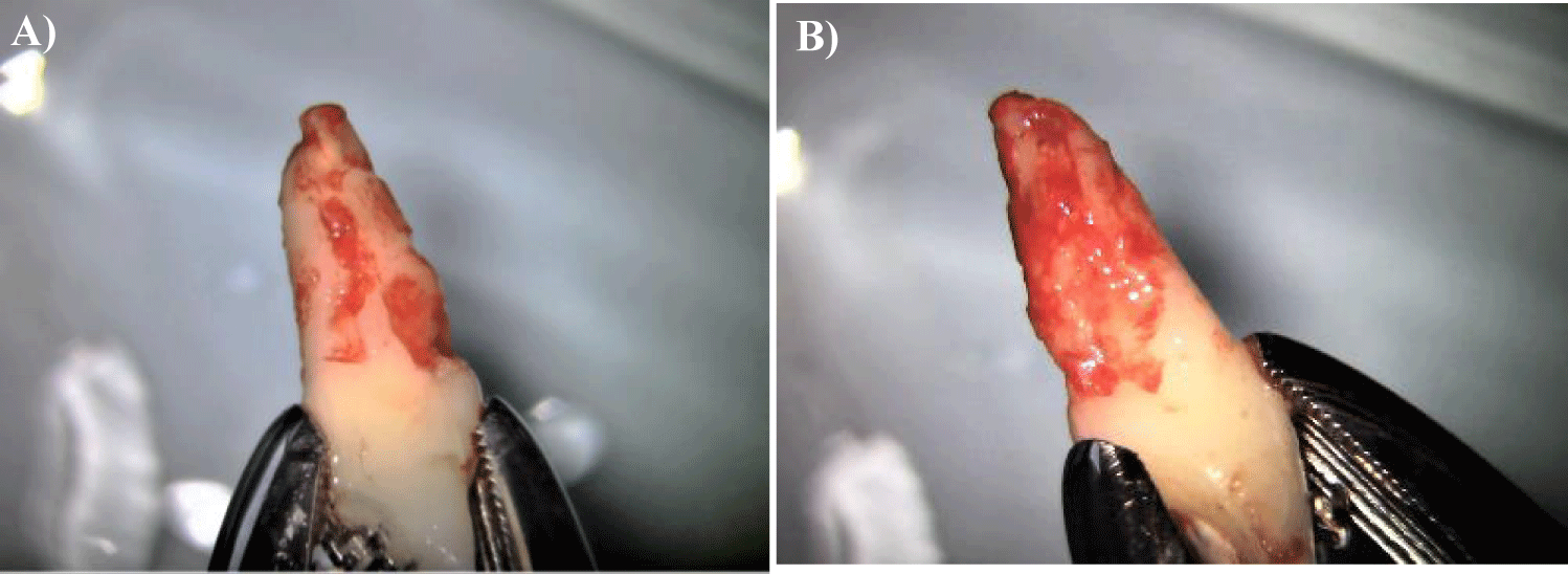
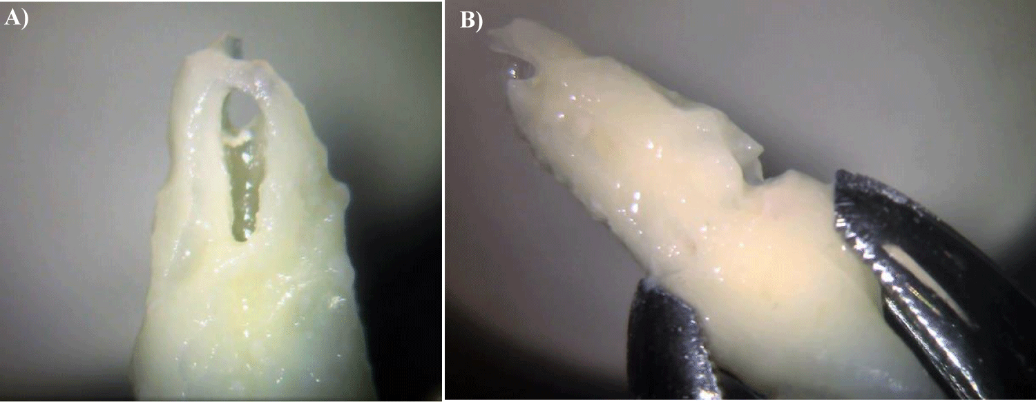
The week after was scheduled the surgery. The skin was disinfected by iodine dye (iodio 7%/5%, Nova Argentia) and the gums by rinsing with a 0.2% clorexidine based mouth wash, using a dental forceps (0100-2, Asa-dental) was performed the atraumatic tooth extraction. During the extraoral procedures the tooth was held by the forceps on the cervical area because for the success of the replantation it is fundamental to safeguard the vitality of the periodontal ligament. For this reason, the ligament must remain hydrated and extra oral time must be as short as possible [23-25]. In this case the root surface with periodontal ligament is very limited due to multiple resorption (Figures 4A,B). In order to avoid the dehydration of this vital ligament areas the root was irrigated with physiological solution at short intervals. To reduce extraoral time during surgery two operators were employed, one endodontist managed the tooth, cleaning and reconstructing the root, while a surgeon managed the socket, removing inflammatory tissues and periapical granuloma.
The cleansing of the resorpted areas and the tridimensional root’s reconstruction was performed by microscope under 0.8-1.2 magnification gradient, and extraoral and intraoral video recordings were performed too.
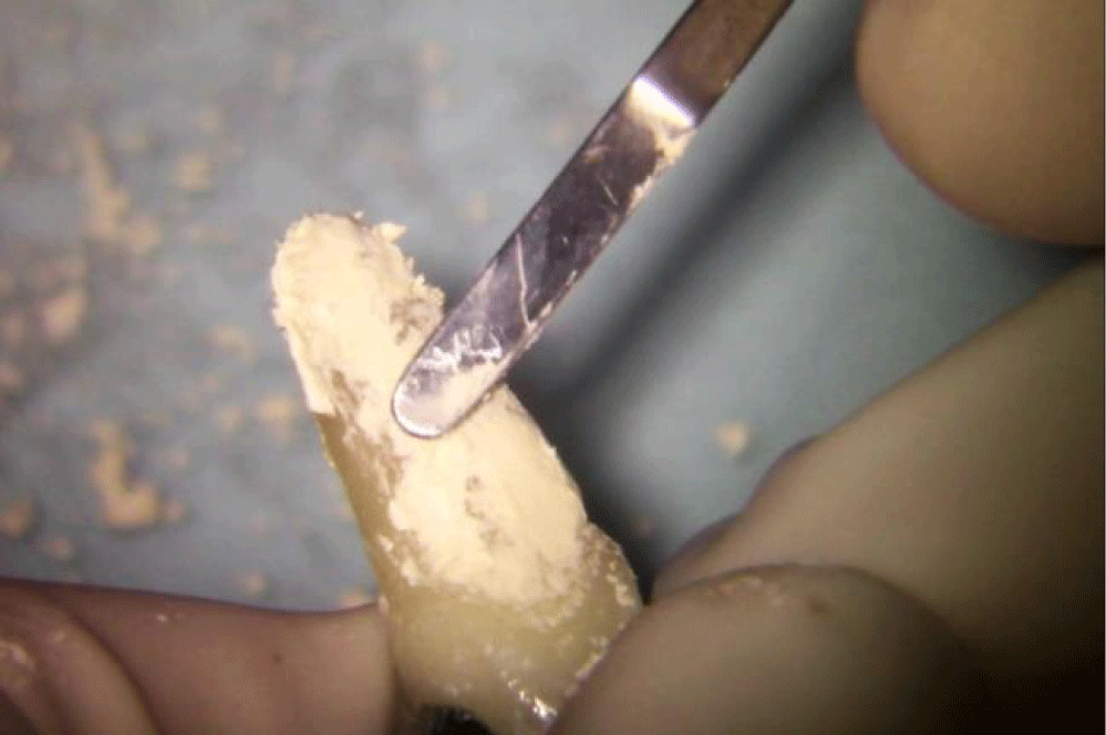
The inflammatory tissues and the infected dentine were removed by the use of ultrasonic tips (Sonic tips SF 56, Komet) mounted on sonic hand piece (monosurgery air power TKD). Once completed the root cleansing the communication between endodontic space and the external root’s part were evident (Figures 5A,B). Afterwards the root was reconstructed by using a bio ceramic cement (Biodentine, Septodont) considering the easy handling.
During the reconstruction procedure we achieved a complete 3D morphology reconstruction of the root from the apical third to the CEJ (Figure 6). The apical third of the root canal was open due to the resorption, so it was filled with Biodentine during 3D reconstruction. Completed the removal of the apical inflammation tissue and after the 3D root reconstruction the tooth 2.1 was replanted and fixed by the use of orthodontic appliance and a passive metallic knot on the ortho wire and completed the therapy the X-ray was taken (Figures 3A-D). No orthodontic forces were applied.
After 15 days the endodontic treatment was performed using 5% hypochlorite for the canal disinfection and distilled water for the final rinse, the canal was sealed with gutta-percha (Meta-biomed) and resin cement (AH-Plus, Dentsply). The cavity access was filled by composite (Tetric Evoceram, Ivoclar), a control X-ray was taken for the final check (Figures 3A-D).
After 4 months the orthodontic appliance on the upper and lower arch was dismantled. The tooth presented physiological mobility and there were no sign of inflammation and pain, so a fixed retainer, by the use of composite and metallic wire from 1.3 and 2.3, was applied and a removable retainer for the lower arch was given to the patient. The tooth was completely removed by protrusive contacts.
The 6 and 12 month follow up shows a tooth in good healing, without clinical or radiographic signs of inflammation. The tooth maintains a physiological mobility; this demonstrates the presence of a functional support tissue without the appearance of ankyloses (Figure 7).
Discussion
The dental traumas are most likely frequent in pediatric age, more often in the male subjects and especially in the front teeth [26].
The more frequent and severe traumas are considered the dislocation (lateral, extrusive and intrusive) and the dental loss, these traumas will get worse like pulp necrosis, ankyloses and external root resorption [27-29].
The root resorption starts with the activation of clastic cells trough an inflammatory process. It develops because the toxin and bacteria passage through dentinal tubules from the pulp to the external root surface or from damage to the periodontal attachment. The inflammatory root resorption results to be more frequent in young patient and more frequent in dental traumas cases with long term delayed treatment [30,31]. The treatment timing and the kind of treatment are fundamental in case of dental loss caused by trauma [32]. The prognosis is better if the time for the replantation of the loss tooth is quick [33]. The way the loss tooth is preserved before replantation is also really important.
The endodontic guidelines are clear in these cases: the root canal should be performed after 15 days in case of preservation of periodontal attachment, and extraorally and immediately in case of complete loss of it and complete root formation [32].
In the presented case the initial management of the dental trauma wasn’t correct. The young patient’s parents kept the tooth in a dry handkerchief compromising the periodontal attachment (clearly not aware of the mistake). The replantation has been performed, but the tooth was not endodontically treated even if the root was already matured. The consequent pulp necrosis caused a periodontal inflammation let starting a massive root resorption process that has been discovered only 1 year after the trauma.
The treatment proposed in these cases were based on the age of the patient and the lack of complete skeletal development. The extraction and the rehabilitation with a dental implant was not suitable in this particular case because of the young age of the patient.
There are not clear guidelines in case of root resorptions, so the treatment is based on the clinical condition of the case and the patient, and the single operator experience [29]. Even if has been proposed a new classification of external root resorption [15], there are not still operative guidelines for the different kind of resorption.
The clinical treatment options are: the orthograde root canal, endo-surgery and intentional replantation based on the single clinical case.
When the root resorption compromised areas not reachable with the conventional endodontic surgery, the intentional root replantation is the only and more extreme clinical treatment option [17].
In the referred case the perioradicular inflammation and the presence of fistula, requested an immediate treatment that aimed to maxime the subsistence of the tooth as main goal, to get to the minimal age for the conventional prosthetic implant treatment. Since the conventional surgery approach would be too demolitive and difficult because of the palatal access required by the lesion the intentional replantation treatment was the first choice.
The golden standard for this kind of clinical choice are MTA and Biodentine because they are bioactive and promote the growth of periodontal attachment over the material itself [34,35].
The Mta was not chosen for this case because of the grey color, and the more difficult manipulation and usage and the missing possibility of layering composite over it. The Biodentine was chosen for the white color, the high antibacterial action (basic pH) and the possibility to layer composite over it in case of gum recession.
To reduce the pH and dued to the active periapical lesion a calcium hydroxide dressing was applied before the replantation in the root canal. The root canal treatment was finished after 2 weeks from the intentional replantation. The tooth extraction was performed atraumatically by the use of pliers and syndesmotomy. The root resulted highly damaged under the inflammatory tissues and the root canal was also exposed especially in the apical third so it was decided to fill completely it by the use of Biodentine and to recreate a 3D reconstruction of the whole root anatomy to improve root adaption to the socket and to avoid the direct contact of the bone on the resorpted areas.
Biodentine was not applied on the healthy part of the root trying to avoid of compromising the last areas of periodontal attachment left.
The root canal of the coronal third of the canal not sealed on the surgical session was performed by the use of hypochlorite at 5% not heated or activated by US to avoid modification of the Biodentine previously applied and sealed by warm gutta-percha.
Even if in this case the follow up is only 12 months the post-treatment x-rays shows up healing of the periradicular lesion and bone formation.
The 3-dimensional root treatment by the use of Biodentine performed extraorally could be indicated in case of high resorpted roots when the orthograde treatment and the endodontic surgery are not a suitable option. Even if the prognosis of this treatment is not certain in a long term, in young patients it’s a valuable option of treatment to space maintaining and periodontal tissues trophism.
Results
Following a previous trauma, the traumatized tooth of the referred case developed multiple external root resorption that hindered a conventional endodontic surgery [36]. It was decided to approach with an intentional replantation in order to extend the permanence of the tooth while awaiting the complete skeletal development of the patient. A 3-dimensional reconstruction of the root was performed with Biodentine to stop the periradicular inflammatory processes. After one year the tooth is in a state of health and, although this is a short-term evaluation, this result bodes well in the possibility of reaching adult age to get to conventional prosthetic implant rehab.
In the pediatric patient the intentional replantation associated with a 3-dimensional root reconstruction can also be suggested as extreme solution to extend the permanence of resorbed teeth.
References
- Santos BO, Mendoca DS, Sousa DL, Neto JJ, Araujo RB. Root resorption after dental traumas: Classification and clinical, radiographic and histologic aspects. Rev Bras Odontol 2011;8:439-45.
- Deliga Schröder ÂG, Westphalen FH, Schröder JC, Fernandes Â, Westphalen VPD. Accuracy of Digital Periapical Radiography and Cone-beam Computed Tomography for Diagnosis of Natural and Simulated External Root Resorption. J Endod. 2018 Jul;44(7):1151-1158. doi: 10.1016/j.joen.2018.03.011. Epub 2018 Jun 5. PMID: 29884337.
- Bhuva B, Barnes JJ, Patel S. The use of limited cone beam computed tomography in the diagnosis and management of a case of perforating internal root resorption. Int Endod J. 2011 Aug;44(8):777-86. doi: 10.1111/j.1365-2591.2011.01870.x. Epub 2011 Mar 4. PMID: 21371054.
- Tsukiboshi M. Autotransplantation of teeth: requirements for predictable success. Dent Traumatol. 2002 Aug;18(4):157-80. doi: 10.1034/j.1600-9657.2002.00118.x. PMID: 12442825.
- Malkondu Ö, Karapinar Kazandağ M, Kazazoğlu E. A review on biodentine, a contemporary dentine replacement and repair material. Biomed Res Int. 2014;2014:160951. doi: 10.1155/2014/160951. Epub 2014 Jun 16. PMID: 25025034; PMCID: PMC4082844.
- Wallace A, Rogers HJ, Zaitoun H, Rodd HD, Gilchrist F, Marshman Z. Traumatic dental injury research: on children or with children? Dent Traumatol. 2017 Jun;33(3):153-159. doi: 10.1111/edt.12299. Epub 2016 Aug 9. PMID: 27385489.
- Ritwik P, Massey C, Hagan J. Epidemiology and outcomes of dental trauma cases from an urban pediatric emergency department. Dent Traumatol. 2015 Apr;31(2):97-102. doi: 10.1111/edt.12148. Epub 2014 Nov 25. PMID: 25425231.
- Rodrigues Campos Soares T, de Andrade Risso P, Cople Maia L. Traumatic dental injury in permanent teeth of young patients attended at the federal University of Rio de Janeiro, Brazil. Dent Traumatol. 2014 Aug;30(4):312-6. doi: 10.1111/edt.12087. Epub 2013 Dec 2. PMID: 24289752.
- Nagori SA, Bhutia O, Roychoudhury A, Pandey RM. Immediate autotransplantation of third molars: an experience of 57 cases. Oral Surg Oral Med Oral Pathol Oral Radiol. 2014 Oct;118(4):400-7. doi: 10.1016/j.oooo.2014.05.011. Epub 2014 Jun 2. PMID: 25183229.
- N Nasseh H, El Noueiri B, Pilipili C, Ayoub F. Evaluation of Biodentine Pulpotomies in Deciduous Molars with Physiological Root Resorption (Stage 3). Int J Clin Pediatr Dent. 2018 Sep-Oct;11(5):393-394. doi: 10.5005/jp-journals-10005-1546. Epub 2018 Oct 1. PMID: 30787552; PMCID: PMC6379528.
- Demars C, Fortier JP. Propos sur l’endodontie des dents tem-poraires. Actual Odontostomatol (Paris). 1981;35(134):213-224.
- Fortier JP, Demars-Fremault C. Abrégé de pédodontie. Masson, Paris: 1987.
- Trope M. Root resorption of dental and traumatic origin: classification based on etiology. Pract Periodontics Aesthet Dent. 1998 May;10(4):515-22. PMID: 9655059.
- Tronstad L. Root resorption--etiology, terminology and clinical manifestations. Endod Dent Traumatol. 1988 Dec;4(6):241-52. doi: 10.1111/j.1600-9657.1988.tb00642.x. PMID: 3078294.
- Fuss Z, Tsesis I, Lin S. Root resorption--diagnosis, classification and treatment choices based on stimulation factors. Dent Traumatol. 2003 Aug;19(4):175-82. doi: 10.1034/j.1600-9657.2003.00192.x. PMID: 12848710.
- Pruthi PJ, Dharmani U, Roongta R, Talwar S. Management of external perforating root resorption by intentional replantation followed by Biodentine restoration. Dent Res J (Isfahan). 2015 Sep-Oct;12(5):488-93. doi: 10.4103/1735-3327.166235. PMID: 26604965; PMCID: PMC4630715.
- Espona J, Roig E, Durán-Sindreu F, Abella F, Machado M, Roig M. Invasive Cervical Resorption: Clinical Management in the Anterior Zone. J Endod. 2018 Nov;44(11):1749-1754. doi: 10.1016/j.joen.2018.07.020. Epub 2018 Sep 19. PMID: 30243659.
- Neelakantan P, Ahmed HMA, Wong MCM, Matinlinna JP, Cheung GSP. Effect of root canal irrigation protocols on the dislocation resistance of mineral trioxide aggregate-based materials: A systematic review of laboratory studies. Int Endod J. 2018 Aug;51(8):847-861. doi: 10.1111/iej.12898. Epub 2018 Feb 23. PMID: 29377170.
- Patel S, Foschi F, Condon R, Pimentel T, Bhuva B. External cervical resorption: part 2 - management. Int Endod J. 2018 Nov;51(11):1224-1238. doi: 10.1111/iej.12946. Epub 2018 Jun 9. PMID: 29737544.
- Heithersay GS. Treatment of invasive cervical resorption: an analysis of results using topical application of trichloracetic acid, curettage, and restoration. Quintessence Int. 1999 Feb;30(2):96-110. PMID: 10356561.
- Patel S, Mavridou AM, Lambrechts P, Saberi N. External cervical resorption-part 1: histopathology, distribution and presentation. Int Endod J. 2018 Nov;51(11):1205-1223. doi: 10.1111/iej.12942. Epub 2018 Jun 1. PMID: 29704466.
- Eftekhar L, Ashraf H, Jabbari S. Management of Invasive Cervical Root Resorption in a Mandibular Canine Using Biodentine as a Restorative Material: A Case Report. Iran Endod J. 2017 Summer;12(3):386-389. doi: 10.22037/iej.v12i3.16668. PMID: 28808471; PMCID: PMC5527220.
- Kim E, Jung JY, Cha IH, Kum KY, Lee SJ. Evaluation of the prognosis and causes of failure in 182 cases of autogenous tooth transplantation. Oral Surg Oral Med Oral Pathol Oral Radiol Endod. 2005 Jul;100(1):112-9. doi: 10.1016/j.tripleo.2004.09.007. PMID: 15953925.
- Andreasen JO. Interrelation between alveolar bone and periodontal ligament repair after replantation of mature permanent incisors in monkeys. J Periodontal Res. 1981 Mar;16(2):228-35. doi: 10.1111/j.1600-0765.1981.tb00970.x. PMID: 6453985.
- Hammarström L, Blomlöf L, Lindskog S. Dynamics of dentoalveolar ankylosis and associated root resorption. Endod Dent Traumatol. 1989 Aug;5(4):163-75. doi: 10.1111/j.1600-9657.1989.tb00354.x. PMID: 2517781.
- Lauridsen E, Hermann NV, Gerds TA, Kreiborg S, Andreasen JO. Pattern of traumatic dental injuries in the permanent dentition among children, adolescents, and adults. Dent Traumatol. 2012 Oct;28(5):358-63. doi: 10.1111/j.1600-9657.2012.01133.x. Epub 2012 Jul 17. PMID: 22805514.
- Andreasen FM, Pedersen BV. Prognosis of luxated permanent teeth--the development of pulp necrosis. Endod Dent Traumatol. 1985 Dec;1(6):207-20. doi: 10.1111/j.1600-9657.1985.tb00583.x. PMID: 3867505.
- Andreasen FM, Andreasen JO. Luxation injuries of permanent teeth: general findings. In: Andreasen, JO; Andreasen, FM; Andersson, L; editors. Textbook and color atlas of traumatic injuries to the teeth. 4th ed. Odder: Blackwell Munksgaard; 2007. p. 372-403.
- Soares Ade J, Gomes BP, Zaia AA, Ferraz CC, de Souza-Filho FJ. Relationship between clinical-radiographic evaluation and outcome of teeth replantation. Dent Traumatol. 2008 Apr;24(2):183-8. doi: 10.1111/j.1600-9657.2007.00528.x. PMID: 18352921.
- Lima TFR, Silva EJNLD, Gomes BPFA, Almeida JFA, Zaia AA, Soares AJ. Relationship between Initial Attendance after Dental Trauma and Development of External Inflammatory Root Resorption. Braz Dent J. 2017 Jan-Apr;28(2):201-205. doi: 10.1590/0103-6440201701299. PMID: 28492750.
- de Oliveira NG, de Souza Araújo PR, da Silveira MT, Sobral APV, Carvalho MV. Comparison of the biocompatibility of calcium silicate-based materials to mineral trioxide aggregate: Systematic review. Eur J Dent. 2018 Apr-Jun;12(2):317-326. doi: 10.4103/ejd.ejd_347_17. PMID: 29988207; PMCID: PMC6004803.
- Bastos JV, Ilma de Souza Côrtes M, Andrade Goulart EM, Colosimo EA, Gomez RS, Dutra WO. Age and timing of pulp extirpation as major factors associated with inflammatory root resorption in replanted permanent teeth. J Endod. 2014 Mar;40(3):366-71. doi: 10.1016/j.joen.2013.10.009. Epub 2013 Nov 14. PMID: 24565654.
- Andersson L, Andreasen JO, Day P, Heithersay G, Trope M, DiAngelis AJ, Kenny DJ, Sigurdsson A, Bourguignon C, Flores MT, Hicks ML, Lenzi AR, Malmgren B, Moule AJ, Tsukiboshi M. Guidelines for the Management of Traumatic Dental Injuries: 2. Avulsion of Permanent Teeth. Pediatr Dent. 2017 Sep 15;39(6):412-419. doi: 10.1111/j.1600-9657.2012.01125.x. PMID: 29179383.
- Hupp JG, Mesaros SV, Aukhil I, Trope M. Periodontal ligament vitality and histologic healing of teeth stored for extended periods before transplantation. Endod Dent Traumatol. 1998 Apr;14(2):79-83. doi: 10.1111/j.1600-9657.1998.tb00815.x. PMID: 9558520.
- Ahangari Z, Nasser M, Mahdian M, Fedorowicz Z, Marchesan MA. Interventions for the management of external root resorption. Cochrane Database Syst Rev. 2015 Nov 24;2015(11):CD008003. doi: 10.1002/14651858.CD008003.pub3. PMID: 26599212; PMCID: PMC7185846.
- Heithersay GS. Management of tooth resorption. Aust Dent J. 2007 Mar;52(1 Suppl):S105-21. doi: 10.1111/j.1834-7819.2007.tb00519.x. PMID: 17546866.
Content Alerts
SignUp to our
Content alerts.
 This work is licensed under a Creative Commons Attribution 4.0 International License.


