> Medicine Group. 2021 September 09;2(9):777-778. doi: 10.37871/jbres1309.
Reflection on the benignity or pathology of the presence of “U waves” in the electrocardiogram
Iríbar Diéguez IK1*, Lucas Cabornero J2 and Lopetegui Caño M3
2Cardiology Service of the Integrated Health Organization - Regional Hospital Bidasoa of Hondarribia (Guipúzcoa), Spain
3Resident Physician of Family and Community Medicine, Integrated Health Organization Bidasoa, Hondarribia Health Center (Guipúzcoa), Spain
Discussion
We know from the U wave of the electrocardiogram (ECG) the positive wave in precordial leads (negative in aVR), of low amplitude and low frequency, located in the TP segment just after the T wave [1]. It is not always noticeable, and it is easier to see in patients with a heart rate prone to bradycardia (it is rarely perceived with heart rates above 95 beats per minute) [2,3].
It can be a physiological finding (secondary to a slow repolarization of the Purkinje fibers, a repolarization of the papillary muscles, or postpotentials caused by stretching of the left ventricular wall during diastole), or appear in pathological contexts [1-6].
Positive in cases of: hypercalcemia, hypokalaemia (visualized even in mild hypokalaemia: K + around 3.5 milliequivalents / liter), thyrotoxicosis, or in patients treated with digitalis, quinidine, amiodarone or phenothiazines.
Negative in cases of: arterial hypertension, mitral or aortic valve disease, right ventricular hypertrophy, or ischemic heart disease (in a patient with acute chest pain, a negative U wave in the precordial leads or inferior leads represents an ischemic injury until prove otherwise [5]; and exercise-induced inversion of the U wave is also highly predictive of significant coronary artery disease) [6].
The only way to suspect if a U wave is pathological will be through the symptoms and history of the patient, and the chronic treatment of him.
Therefore, we must rule out with complementary studies that a U wave represents a pathological situation when:
- Its width is > 2 millimeters (more visible in V2-4).
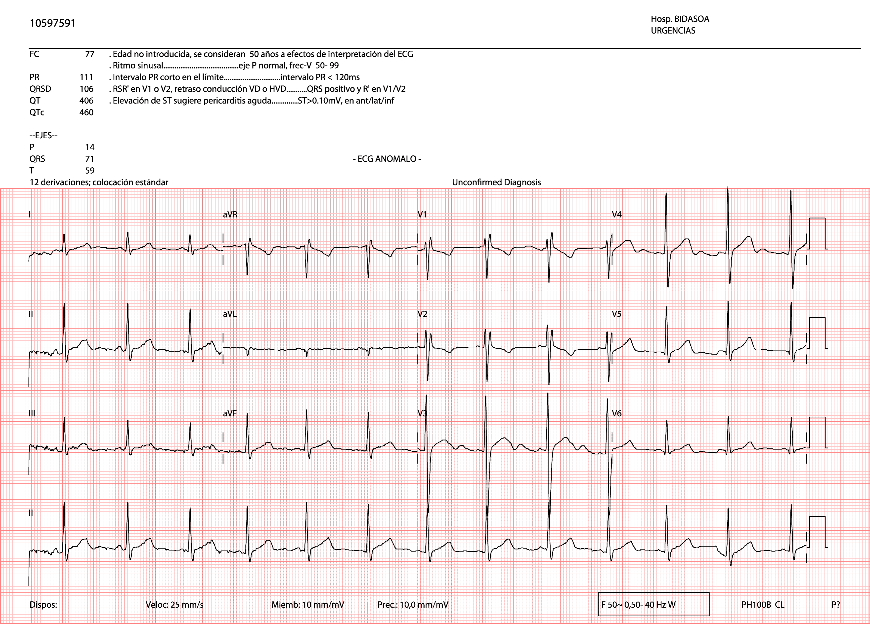
- It is inverted in precordial leads (Figure 2) [4].
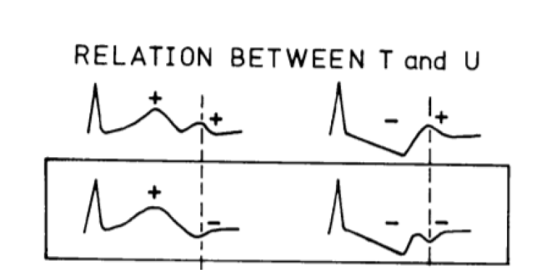
- It merges with the T wave (more common if it coexists with a markedly long QT. Not to be confused with cases in which it appears together with tachycardia: in these, the U wave appears before the end of the T wave and gives an image of T “nicked” with false prolongation of the QT space).
- Its amplitude is greater than that of the previous T wave.
- Happens in elderly patients.
Although the U wave remains a mystery as to its genesis, the clinical observation of this small “neglected” wave can have great clinical significance in very different pathologies.
Taking this information into account can be especially useful in care settings where other complementary tests are not available, such as Primary Care consultations or Continuous Care Points.
References
- Mas Casals A, Lobos Bejarano JM. ¿A qué puede corresponder cada alteración de una onda, complejo o intervalo? AMF 2012;8(10):597-600.
- Pérez-Lescure Picarzo J. Guía rápida para la lectura sistemática del ECG pediátrico. Rev Pediatr Aten Primaria 2006;8:319-326. https://bit.ly/3jY3za0
- Millán JJ. Ondas “U” en electrocardiogramas. 2016. https://bit.ly/3hj0BLQ.
- Kishida H, Cole JS, Surawicz B. Negative U wave: the clinical significance and the consideration of pathogenesis. In: Rosenbaum MB, Elizari MV (eds). Frontiers of Cardiac Electrophysiology. Desarrollos en Medicina Cardiovascular, Springer. Dordrecht. 1983;19:100-119. Doi: 10.1007/978-94-009-6781-65
- Dr Smith, in Dr Smith ECG blog. 2018. https://bit.ly/3yW835A.
- Pérez Riera AR, Ferreira C, Filho CF, Ferreira M, Meneghini A, Uchida AH, Schapachnik E, Dubner S, Zhang L. The enigmatic sixth wave of the electrocardiogram: the U wave. Cardiol J. 2008;15(5):408-21. PMID: 18810715.
Content Alerts
SignUp to our
Content alerts.
 This work is licensed under a Creative Commons Attribution 4.0 International License.


