> Medicine Group. 2021 September 06;2(9):768-770. doi: 10.37871/jbres1307.
A Young Female with Mass Originating from Middle Lobe of Lung-Choriocarcinoma: Extragonadal Germ Cell Tumor
Muhammad Umer Salim*, Mohammad Rabie Alrohaibani, Mohammed Hassan Shabrawishi, Sobhy Atalla Ibrahim, Abed Sabry Elfar and Asmaa Ezzat Mousa
- Choriocarcinoma
- Middle lobe of lung
- Extragonadal germ cell tumor
- Bleomycin
- Etoposide
- Cisplatin
Abstract
Extragonadal germ cell tumors are the rare tumors originate commonly from the mediastinum and the reason is still unknown however; there are theories, which suggest that the gonadal cells in the mediastinum represent reverse migration from the gonads.
A young female presented with mass originating from the middle lobe of lung and right sided pleural effusion that was drained, and biopsy was taken from the mass; diagnosed as choriocarcinoma. She received (BEP) Bleomycin, Etoposide and Cisplatin chemotherapy; responded well and her symptoms improved.
Introduction
Extra Gonadal Germinal Cell Tumors (EGGCTs) are rare tumors that predominantly affect young males. Seminomas account for 30-40% of these tumors, and Nonseminomatous Germ Cell Tumors (NS-GCTs) account for 60-70% [1]. Nonseminomatous germ cell tumors include the following:
• Yolk-sac tumors
• Embryonal carcinomas
• Choriocarcinomas
• Teratomas
• Nonteratomatous combined germ cell tumors
The most common site of extra gonadal germ cell tumors is the mediastinum (50- 70%) followed by the retro peritoneum (30-40%), the pineal gland (5%), and the sacrococcygeal area (less than 5%) [1].
The reason to report this case was the tumor was not arising from the mediastinum but from the right middle lobe, which is unusual location of this tumor.
Case Report
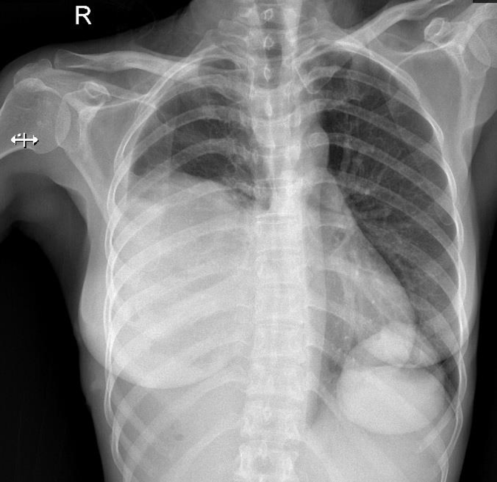
A 39-years-old married woman presented with low-grade fever, non-productive cough and difficulty in breathing for 6 months. There was no previous history of Tuberculosis (TB) or TB contact, weight loss or hemoptysis. She informed that she had not contacted her husband for 6 years as he was out of the country. On examination, she had neither clubbing of the fingers nor palpable lymph nodes. Chest examination showed decreased movements on the right side of chest with reduced expansion; there was dullness at right inframammary area anteriorly and interscapular and infrascapular region posteriorly with absent breath sounds and decreased vocal resonance. Abdominal, muscular, neurological, breast and pelvic examination was unremarkable.
Her β-HCG 17723 U/L (normal range: 0-1 U/L), alpha-fetoprotein 3.81 µg/L (normal range 0-8.5 µg/L).
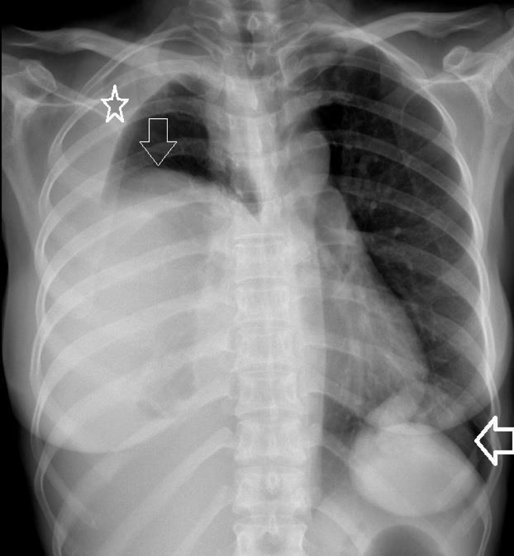
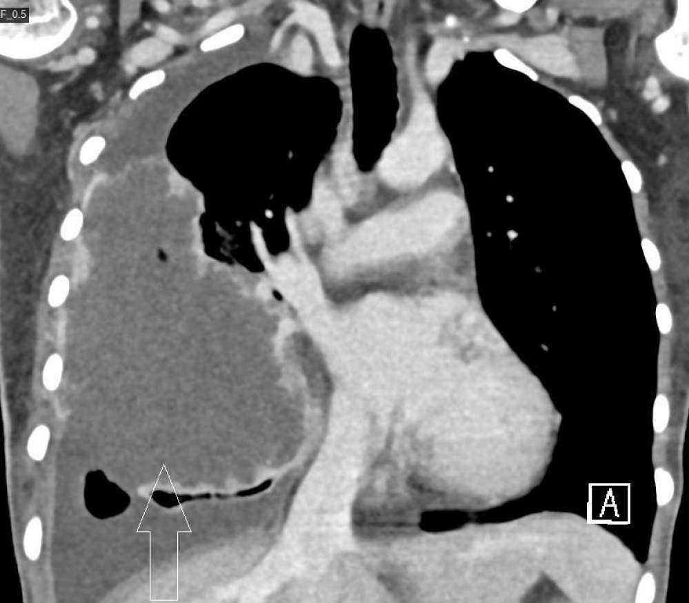
Chest x-ray is given in figure 1. CT scan chest with contrast shows giant lobulated, peripherally enhancing cystic lesion occupying most of the right lung with epicenter of the lesion noticed in the right middle lobe with fluid content. No calcification, fat or enhancing soft tissue component is appreciated within the lesion. No adjacent bone erosion or chest wall invasion. It measures about 9.7 × 6.4 × 10.8 cm in AP, TS, CC dimensions respectively, causing mild leftward mediastinal shift and collapsed remnant part of the left lung.
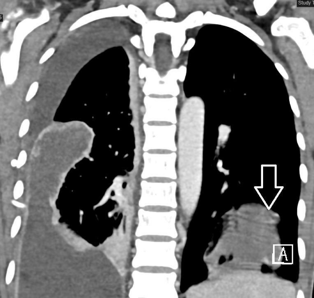
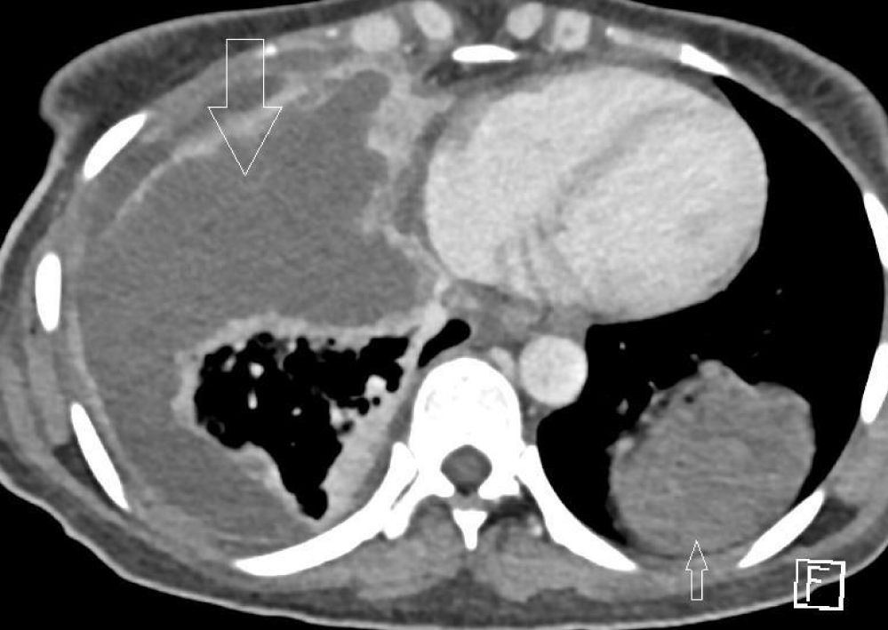
The tracheobronchial tree is patent. No significant enlarged mediastinal, hilar or axillary lymph nodes. Mediastinal vascular structures appear unremarkable. No pericardial effusion. Osseous structures demonstrate no suspicious focal lesion (Figure 2). Another similar cystic lesion noticed in the lower lobe of the left lung measuring about 4.8 × 5.2 × 5.7 cm (Figure 3 & 4).
Her pleural effusion was drained and thoracic surgery performed open lung biopsy from a big mass originating from the middle lobe was found. The histopathology report came out to be choriocarcinoma with immunohistochemistry positive for PanCK, CK-7 and βhCG and negative for TTF-1, CK20, p63, CK 5/6, inhibin, synaptophysin, S100 and SMA.
Then she was referred to oncology for chemotherapy (BEP) Bleomycin, Etoposide and Cisplatin chemotherapy. She has been following up in the oncology clinic and showing good response (Figure 5).
Discussion
It is rare for the choriocarcinoma to be originating from mediastinum without involvement of the gonads (e.g., ovaries) but in our patient, choriocarcinoma originated from the right middle lobe without involvement of gonads.
These tumors can be found anywhere on the midline, particularly the retroperitoneum, the anterior mediastinum, the sacrococcyx, and the pineal gland. Other less common sites include the orbit, suprasellar area, palate, thyroid, submandibular region, anterior abdominal wall, stomach, liver, vagina, and prostate. The classic theory suggests that Germ Cell Tumors (GCTs) in these areas are derived from local transformation of primordial germ cells misplaced during embryogenesis.
For patients receiving intensive chemotherapy, 5-year survival rates of 40-65% have been reported. Extragonadal seminomas carry the best survival rates [2]. Overall survival of patients with seminomatous extragonadal GCTs has been reported to range from 88%-100%, whereas suvival of patients with mediastinal non-seminomatous extragonadal GCTs is 40-45% [3,4].
All of the following indicate good prognosis for non-seminomatous germ cell tumor (choriocarcinoma):
• Testis/retroperitoneal primary
• No non-pulmonary visceral metastases
• Good tumor marker levels - AFP < 1000 ng/mL, β-hCG < 1000 IU/L, and LDH < 1.5 times the upper limit of normal.
So, it was concluded that this rare tumor of mediastinum or gonadal origin can also have middle lobe lung origin as mentioned in our case report.
References
- Ronchi A, Cozzolino I, Montella M, Panarese I, Zito Marino F, Rossetti S, Chieffi P, Accardo M, Facchini G, Franco R. Extragonadal germ cell tumors: Not just a matter of location. A review about clinical, molecular and pathological features. Cancer Med. 2019 Nov;8(16):6832-6840. doi: 10.1002/cam4.2195. Epub 2019 Sep 30. PMID: 31568647; PMCID: PMC6853824.
- Busch J, Seidel C, Zengerling F. Male Extragonadal Germ Cell Tumors of the Adult. Oncol Res Treat. 2016;39(3):140-4. doi: 10.1159/000444271. Epub 2016 Feb 23. PMID: 27032104.
- Bokemeyer C, Nichols CR, Droz JP, Schmoll HJ, Horwich A, Gerl A, Fossa SD, Beyer J, Pont J, Kanz L, Einhorn L, Hartmann JT. Extragonadal germ cell tumors of the mediastinum and retroperitoneum: results from an international analysis. J Clin Oncol. 2002 Apr 1;20(7):1864-73. doi: 10.1200/JCO.2002.07.062. PMID: 11919246.
- Makino T, Konaka H, Namiki M. Clinical Features and Treatment Outcomes in Patients with Extragonadal Germ Cell Tumors: A Single-center Experience. Anticancer Res. 2016 Jan;36(1):313-7. PMID: 26722059.
Content Alerts
SignUp to our
Content alerts.
 This work is licensed under a Creative Commons Attribution 4.0 International License.


