> General Science. 2021 July 05;2(7):538-542. doi: 10.37871/jbres1273.
Juvenile Nasopharyngeal Angiofibroma: From Diagnosis to Surgical Approach
Joana Borges da Costa1*, André Carção1, Delfim Duarte2 and Miguel Viana1
2Director of Otolaryngology Department of Hospital Pedro Hispano, Portugal
- Juvenile nasopharyngeal angiofibroma
- Benign tumor
- Embolization
Abstract
Juvenile Nasopharyngeal Angiofibroma (JNA) is a rare benign tumor with primary involvement of the nasopharynx in 98% of cases. It is responsible for 0.5% of tumors of the head and neck, occurring in 1/150,000 individuals. Patients between 14 and 25 years old are particularly affected, with a predominance almost exclusively of males. Despite having a benign nature, AFJ has the potential to grow and involve neighboring structures, which highlights the importance of an attempted diagnosis and therapeutic intervention. This work serves to make a JNA theoretical review and present an endoscopic resection video of a JNA in a young male after a pre-op embolization of the tumour, which was essential for the success of the surgical intervention. JNA is an aggressive and locally invasive tumor that can recur after surgery, so an early diagnosis, adequate staging and the appropriate therapeutic plan are essential for the resolution of the clinical situation.
Introduction
Juvenile Nasopharyngeal Angiofibroma (JNA) is a rare benign tumor responsible for 0.5% of tumors of the head and neck, occurring in 1/150.000 individuals [1,2]. Upward of 98% of tumors primarily involve the nasopharynx which justifies the term of nasopharyngeal angiofibroma, though extra-nasopharyngeal presentations have also been reported [3,4]. Patients between 14 and 25 years old are particularly affected, with a distinct predominance almost exclusively of males [5].
The tumor was first described by Hippocrates in the 5th century BC and in 1940 it was designated as juvenile angiofibroma by Friedberg [6]. The etiopathogenesis of the tumor is not completely understood but, since a vast majority of the cases do occur in males, it may be explained by high Androgen Receptor (AR) expression suggesting that JNA is androgen dependent [6]. There are also a hamartoma and vascular malformation theories, once the JNA histologic origin involves endothelial cells or fibroblasts [7].
JNA typically appears in the pterygopalatine canal [8] and, in the early stages of growth, it extends to the nasopharynx and nasal cavity through the sphenopalatine hole, so the first clinical manifestations of the tumor are usually complaints of unilateral nasal obstruction and recurrent epistaxis [9]. Despite having a benign nature, AFJ has the potential to grow and involve neighbouring structures, so based on clinical and radiological characteristics, it can be classified as type I when the lesions are fundamentally located in the nasal cavity, paranasal sinus, nasopharynx or pterygopalatine fossa; type II is a tumor that extends into the infratemporal fossa, buccopharyngeal space or orbital cavity; and type III is when there is an intracranial involvement of the middle fossa [10].
JNA is, therefore, a clinical entity that must be diagnosed and intervened as early as possible, so this work serves to make a brief theoretical review about the pathology and to present the case of a JNA and its endoscopic surgical resection video.
Case Report
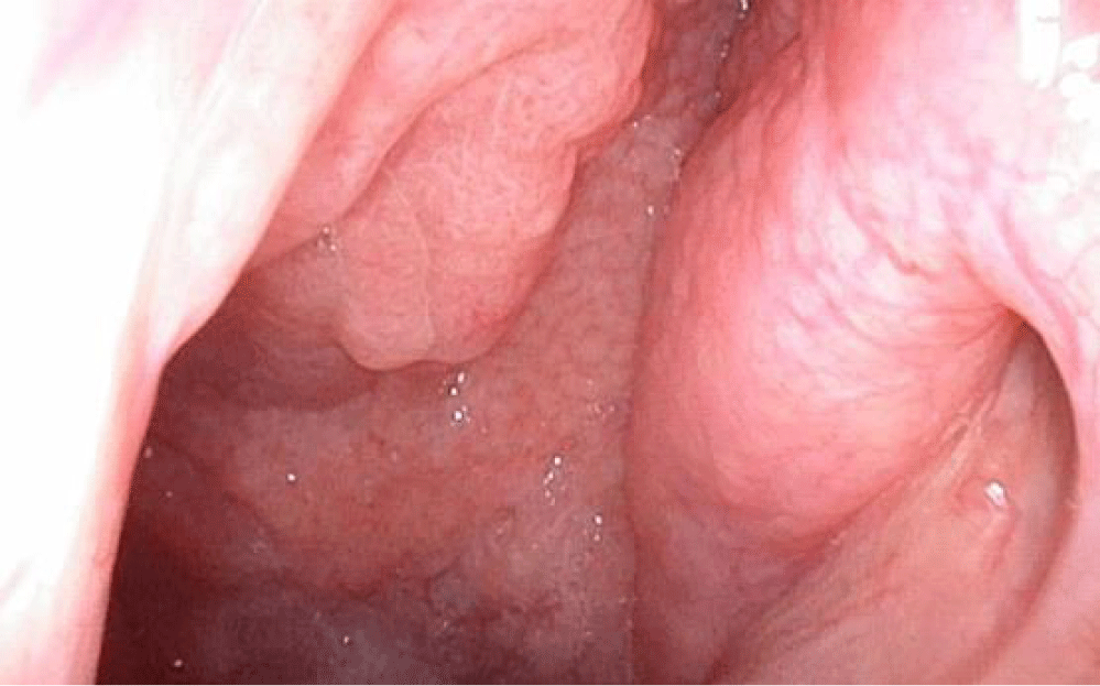
A 14-year-old male patient, with no relevant medical personal or familiar antecedents, was referred to an otolaryngology consultation with progressive complaints of right nasal obstruction over 1 year and several episodes of right-sided epistaxis that required electrocautery in the emergency department and anterior nasal packing with Merocel®. The anterior rhinoscopy have not shown any lesion but a flexible nasal endoscopic exam subsequently demonstrated a large, vascular and angiomatous mass filling the right posterior nasal passage and nasopharynx, occupying the superomedial portion of right choana and extending to the left side (Figure 1). The remainder of the head and neck exam and laboratory workup was within normal limits and the patient denied other symptoms.
After observing the lesion described, a cranial Computed Tomography (CT) and a Magnetic Resonance Imaging (MRI) were performed that showed a massive soft tissue formation that occupied the sphenoid sinus, median-paramedian to the left, surrounding the medial wall of the right chamber of the sphenoid sinus, insinuating itself in the ethmoidal complex, in the nasal cavity and pharyngeal cavum, causing airway obliteration (Video 1). This lesion presented an erosive nature, globose configuration, rounded contours and defined limits, showing a tomodensitometric pattern overlapping the muscular planes. These nonspecific characteristics raised the hypothesis of corresponding to chronic inflammatory disease or nasopharyngeal polyposis, not excluding, however, other diagnosis as a soft tissue or vascular tumor.
Video 1: Coronal view of cranial computed tomography showing the nasossinusal lesion.
Given the clinical presentation, the image aspect and the fact that it was a young male patient, an AFJ was the most likely diagnostic hypothesis and the patient was proposed to surgical resection. An analytical examination including coagulation was performed and went back normal.
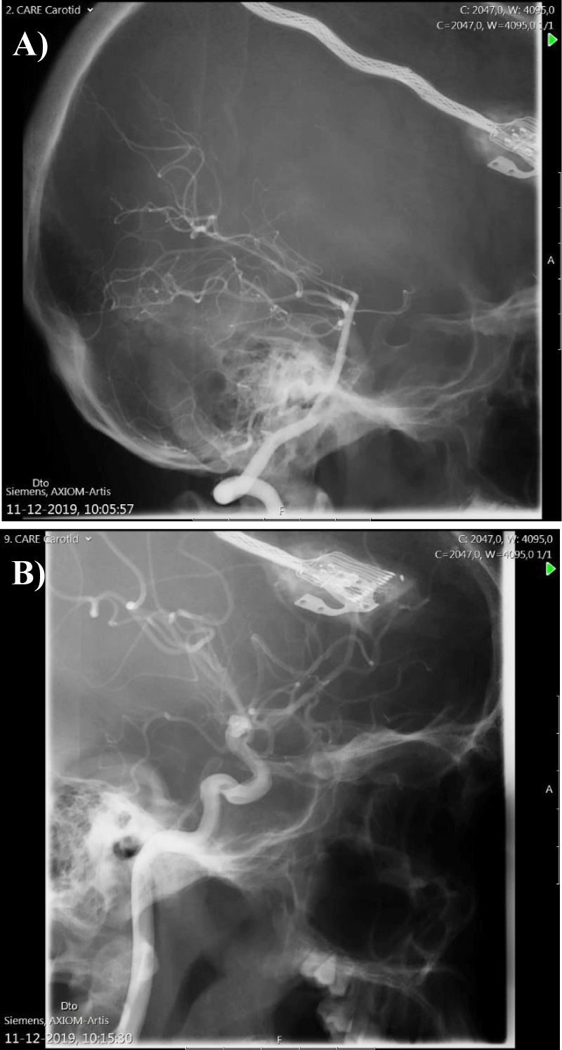
Two weeks after the diagnosis, the patient was submitted to a bilateral angiography. In the arteriography of the right maxillary artery, an extensive nasopharyngeal tumor blush was identified, mainly fed by branches of the sphenopalatine artery (Figure 2a,b). No nutritional afferences were identified in the right ascending pharyngeal artery or in the left external carotid artery. The angio of the internal carotid arteries excluded anatomical variants and confirmed the normal filling of the choroidal crescent, bilaterally. There was a slight filling of the tumor bed via the right vidian artery.
Given the high risk of blood loss from vascular tumors surgical resection, it was decided to proceed with vascular embolization through the right external carotid artery, selectively embolizing the maxillary artery termination, with significant tumor devascularization.
The patient was then submitted to an endoscopic resection of the angiofibroma through a right medial maxillectomy type 2, anterior and posterior ethmoidectomy and a sphenoidotomy. It was possible to observe the lesion occupying the right nasal fossa, with origin in pterygopalatine fossa. The tumor was dissected and haemostatic clips were placed on the vascular pedicle of the mass, which allowed the whole removal of the tumor, without a catastrophic bleeding (Video 2). The surgical procedure and the immediate post-operatory period went without any complication.
Video 2: Video of the surgery (cirurgia).
Avail: https://www.youtube.com/watch?v=SNYmtdMUCNw
The patient was medicated with prophylactic antibiotic over the first week of post-op and with oseltamivir for the influenza virus flu that the patient developed during hospitalization.
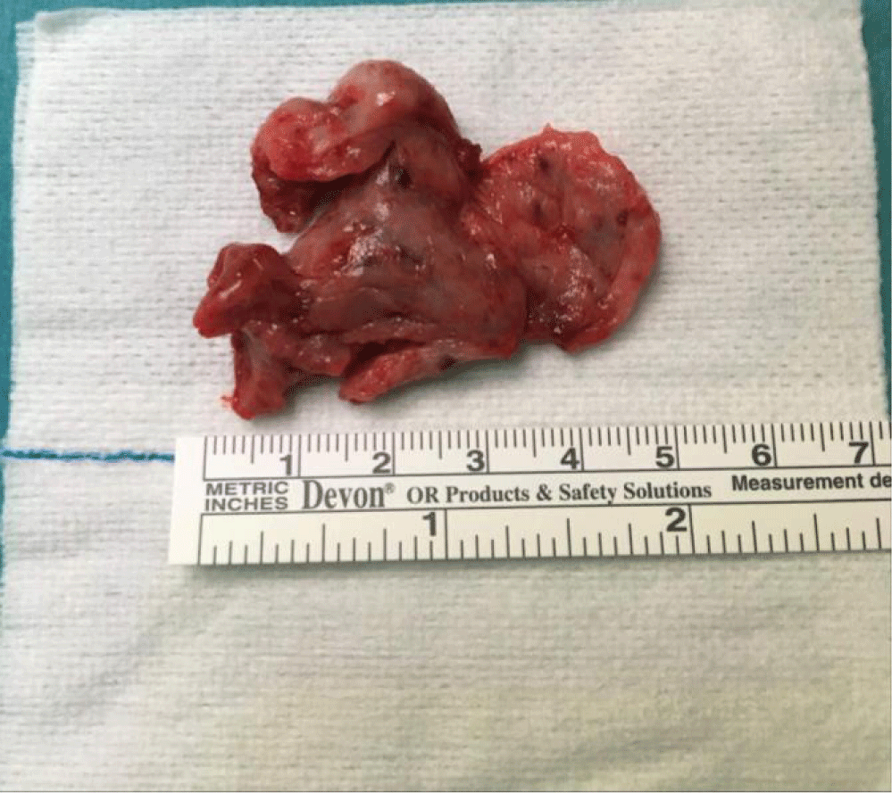
The histopathological examination revealed a lesion with 18 g and 36 mm of larger diameter (Figure 3), with characteristics of angiofibroma and the presence of mucosal flaps covered by ciliated columnar epithelium with foci of mononucleated inflammatory infiltrate. There were no signs of malignancy.
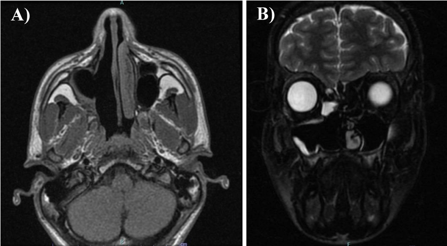
The patient has been followed up monthly in ENT consultation since surgery, to remove nasal crusts and to search for recurrence signs that are absent to date. Six months after surgical intervention, the patient is totally asymptomatic, with complete resolution of nasal obstruction and occasional epistaxis complaints and a flexible nasal endoscopy showed a significant patency of the right maxillary sinus, ethmoidal cells and sphenoid sinus, absence of crusts, purulent secretions, blood loss or signs of tumor recurrence (Video 3). It was also performed an MRI that showed signs of previous surgical intervention on the right nasal cavity, without apparent signs of locoregional recurrence of the disease (Figure 4a,b).
Discussion
Juvenile Nasopharyngeal Angiofibroma (JNA) is a rare tumor first described by Hippocrates in the fifth century that in most cases occurs in a characteristic manner: unilaterally in adolescent males [11]. The tumor usually originates in the lateral wall of the nasal cavity, close to the superior border of the sphenopalatine foramen [12] and the growth initiates in the submucosa of the nasopharynx floor, further spreading into the septum and posterior half of the nasal fossa and causing a typical triad of airway obstruction, recurrent epistaxis and mass in the nasal cavity [11]. Although benign, it is a locally aggressive tumor and may invade the surrounding tissues and bone through pressure resorption. Therefore, continuous grow may cause the involvement of sphenoidal sinus, nasal fossa, middle turbinate, pterygopalatine fossa and the posterior wall of the maxillary sinus, as seen in the presented case, and in the worst-case scenario, it may invade the infratemporal and middle cranial fossa [11]. In the cases of orbital and intracranial extension, signs of proptosis, cranial nerve palsies and loss of vision may occur and advanced stages can present with facial dysmorphism, eustachian tube obstruction and conductive hearing loss, recurrent otitis media, anosmia and eye pain [13]. There are also some reports of angiofibromas arising outside the nasopharynx, most commonly in the maxillary and ethmoid sinus, that usually develop at older ages and occur more frequently in women that typical JNA [14].
Several staging systems for JNA have been proposed by Chandler, et al. [15] which helped to determine the tumor site and extent but Fisch classification is currently accepted [16].
Aside from direct visualization of complete history, clinical examination and the presence of a nasopharyngeal mass on nasal endoscopy, the diagnosis and staging of JNA can usually be made through CT and MRI of the paranasal sinuses [17]. These techniques help to stablish the exact site, extension and relation of the tumor to the adjacent structures, allow to precisely stage JNA and to determine the treatment options, operative approach and prognosis [18,19]. Sometimes, imaging reveals widening of the pterygomaxillary fissure with anterior bowing of the posterior maxillary sinus wall (Holman Miller sign) that, though classically associated with JNA, is an unspecific radiological finding [20] and was not present in the reported case. MRI is superior to CT for detecting the intracranial tumor [17]. These features along with the specific age and sex predilection help to differentiate JNA from other differential diagnosis as antrochoanal, inflammatory or angiomatous polyps, soft tissue neoplasms as papilloma, lymphoma, neurofibroma, maxillary malignancies, nasopharyngeal cyst and carcinomas, pyogenic granuloma or adenoid hypertrophy [21,22].
Angiography is a useful complementary tool in the diagnosis of vascular tumors, once that the tumor location, size and feeding vessels are clearly demonstrated by this technique. Traditionally, JNA originate from one or more of several ipsilateral vascular networks, usually branches of internal maxillary artery, as sphenopalatine, descending palatine and posterior superior alveolar branches. JNA may also be supplied by the ascending pharyngeal artery [23]. In the reported case, angiography was used not only for identification of vascular supply but also for embolization of the tumor, facilitating the surgical removal of the mass and minimizing the risk of haemorrhage.
Early diagnosis and treatment are fundamental for a good prognosis in JNA. However, this tumor is relatively innocuous and typically presents with unspecific and long-term symptoms, which often delays the diagnosis and increases the likelihood of an advanced stage tumor at diagnosis. Advanced lesions with orbital and intracranial extension are difficult to treat and usually present higher recurrence rates. When diagnosed early, the patients can be treated with a combination of preoperative embolization of the tumor and surgical resection, providing a much better prognosis, as in the described case report. Recurrent tumor must be managed individually, considering several factors as tumor location and patient age. Recurrence rates depend on the clinical stage of the tumor but are generally higher in patients with anterior and/or posterior lateral tumor extension [24]. As most tumor recurrences tend to occur within 3 to 4 months postoperatively, it has been recommended that patients undergo scheduled surveillance visits monthly for the first 6 months with an initial MRI at 6 months after surgery [25].
References
- Patterson CN. Juvenile nasopharyngeal angiofibroma. Otolaryngol Clin North Am. 1973 Oct;6(3):839-61. PMID: 4378176.
- Gullane PJ, Davidson J, O’Dwyer T, Forte V. Juvenile angiofibroma: a review of the literature and a case series report. Laryngoscope. 1992 Aug;102(8):928-33. doi: 10.1288/00005537-199208000-00014. PMID: 1323003.
- De Vincentiis G, Pinelli V. Rhinopharyngeal angiofibroma in the pediatric age group. Clinical-statistical contribution. Int J Pediatr Otorhinolaryngol. 1980 Jun;2(2):99-122. doi: 10.1016/0165-5876(80)90012-9. PMID: 6323331.
- Windfuhr JP, Remmert S. Extranasopharyngeal angiofibroma: etiology, incidence and management. Acta Otolaryngol. 2004 Oct;124(8):880-9. doi: 10.1080/00016480310015948. PMID: 15513521.
- Coutinho-Camillo CM, Brentani MM, Nagai MA. Genetic alterations in juvenile nasopharyngeal angiofibromas. Head Neck. 2008 Mar;30(3):390-400. doi: 10.1002/hed.20775. PMID: 18228521.
- Schick B, Rippel C, Brunner C, Jung V, Plinkert PK, Urbschat S. Numerical sex chromosome aberrations in juvenile angiofibromas: genetic evidence for an androgen-dependent tumor? Oncol Rep. 2003 Sep-Oct;10(5):1251-5. PMID: 12883689.
- Nonogaki S, Campos HG, Butugan O, Soares FA, Mangone FR, Torloni H, Brentani MM. Markers of vascular differentiation, proliferation and tissue remodeling in juvenile nasopharyngeal angiofibromas. Exp Ther Med. 2010 Nov;1(6):921-926. doi: 10.3892/etm.2010.141. Epub 2010 Aug 26. PMID: 22993619; PMCID: PMC3446741.
- Liu ZF, Wang DH, Sun XC, Wang JJ, Hu L, Li H, Dai PD. The site of origin and expansive routes of juvenile nasopharyngeal angiofibroma (JNA). Int J Pediatr Otorhinolaryngol. 2011 Sep;75(9):1088-92. doi: 10.1016/j.ijporl.2011.05.020. Epub 2011 Jun 29. PMID: 21719122.
- Martins MB, de Lima FV, Mendonça CA, de Jesus EP, Santos AC, Barreto VM, Santos RC Júnior. Nasopharyngeal angiofibroma: Our experience and literature review. Int Arch Otorhinolaryngol. 2013 Jan;17(1):14-9. doi: 10.7162/S1809-97772013000100003. PMID: 25991988; PMCID: PMC4423317.
- Yi Z, Fang Z, Lin G, Lin C, Xiao W, Li Z, Cheng J, Zhou A. Nasopharyngeal angiofibroma: a concise classification system and appropriate treatment options. Am J Otolaryngol. 2013 Mar-Apr;34(2):133-41. doi: 10.1016/j.amjoto.2012.10.004. Epub 2013 Jan 16. PMID: 23332298.
- Makhasana JA, Kulkarni MA, Vaze S, Shroff AS. Juvenile nasopharyngeal angiofibroma. J Oral Maxillofac Pathol. 2016 May-Aug;20(2):330. doi: 10.4103/0973-029X.185908. PMID: 27601836; PMCID: PMC4989574.
- Bremer JW, Neel HB 3rd, DeSanto LW, Jones GC. Angiofibroma: treatment trends in 150 patients during 40 years. Laryngoscope. 1986 Dec;96(12):1321-9. doi: 10.1288/00005537-198612000-00001. PMID: 3023771.
- Alves FR, Granato L, Soares M, Lambert E. Surgical approaches to juvenile nasopharyngeal angiofibroma - case report and literature review. Intl ArchOtorhinolaryngol. 2006;2(10):162-166.
- Huang RY, Damrose EJ, Blackwell KE, Cohen AN, Calcaterra TC. Extranasopharyngeal angiofibroma. Int J Pediatr Otorhinolaryngol. 2000 Nov 30;56(1):59-64. doi: 10.1016/s0165-5876(00)00404-3. PMID: 11074117.
- Koshy S, George M, Gupta A, Daniel RT. Extended osteoplastic maxillotomy for total excision of giant multicompartmental juvenile nasopharyngeal angiofibroma. Indian J Dent Res. 2008 Oct-Dec;19(4):366-9. doi: 10.4103/0970-9290.44526. PMID: 19075445.
- Fisch U. The infratemporal fossa approach for nasopharyngeal tumors. Laryngoscope. 1983 Jan;93(1):36-44. doi: 10.1288/00005537-198301000-00007. PMID: 6185810.
- Lloyd GA, Phelps PD. Juvenile angiofibroma: imaging by magnetic resonance, CT and conventional techniques. Clin Otolaryngol Allied Sci. 1986 Aug;11(4):247-59. doi: 10.1111/j.1365-2273.1986.tb01926.x. PMID: 3028678.
- Mishra S, Praveena NM, Panigrahi RG, Gupta YM. Imaging in the diagnosis of juvenile nasopharyngeal angiofibroma. J Clin Imaging Sci. 2013 Mar 22;3(Suppl 1):1. doi: 10.4103/2156-7514.109469. PMID: 23878770; PMCID: PMC3716018.
- Weinstein MA, Levine H, Duchesneau PM, Tucker HM. Diagnosis of juvenile angiofibroma by computed tomography. Radiology. 1978 Mar;126(3):703-5. doi: 10.1148/126.3.703. PMID: 203978.
- Som PM, Shugar JM, Cohen BA, Biller HF. The nonspecificity of the antral bowing sign in maxillary sinus pathology. J Comput Assist Tomogr. 1981 Jun;5(3):350-2. doi: 10.1097/00004728-198106000-00007. PMID: 6263958.
- Acharya S, Naik C, Panditray S, Dany SS. Juvenile Nasopharyngeal Angiofibroma: A Case Report. J Clin Diagn Res. 2017 Apr;11(4):MD03-MD04. doi: 10.7860/JCDR/2017/23729.9630. Epub 2017 Apr 1. PMID: 28571176; PMCID: PMC5449822.
- Beham A, Kainz J, Stammberger H, Auböck L, Beham-Schmid C. Immunohistochemical and electron microscopical characterization of stromal cells in nasopharyngeal angiofibromas. Eur Arch Otorhinolaryngol. 1997;254(4):196-9. doi: 10.1007/BF00879273. PMID: 9151019.
- Wu AW, Mowry SE, Vinuela F, Abemayor E, Wang MB. Bilateral vascular supply in juvenile nasopharyngeal angiofibromas. Laryngoscope. 2011 Mar;121(3):639-43. doi: 10.1002/lary.21337. Epub 2010 Oct 26. PMID: 21344446.
- Szymańska A, Szymański M, Czekajska-Chehab E, Szczerbo-Trojanowska M. Two types of lateral extension in juvenile nasopharyngeal angiofibroma: diagnostic and therapeutic management. Eur Arch Otorhinolaryngol. 2015 Jan;272(1):159-66. doi: 10.1007/s00405-014-2965-y. Epub 2014 Mar 6. PMID: 24599598; PMCID: PMC4282713.
- Douglas R, Wormald PJ. Endoscopic surgery for juvenile nasopharyngeal angiofibroma: where are the limits? Curr Opin Otolaryngol Head Neck Surg. 2006 Feb;14(1):1-5. doi: 10.1097/01.moo.0000188859.91607.65. PMID: 16467630.
Content Alerts
SignUp to our
Content alerts.
 This work is licensed under a Creative Commons Attribution 4.0 International License.


