> Medicine Group. 2021 May 11;2(5):315-319. doi: 10.37871/jbres1233.
Erroneous Recording of Maternal Heart Rate as Fetal Heart Rate During Second Stage of Labour: Isn’t it Time to Stop this?
Ms. Ferha Saeed1, Dr. Sanduni Abeysuriya2* and Mr. Edwin Chandraharan3
2MBChB, Newham University Hospital, Barts Health NHS Trust London, UNITED KINGDOM
3MBBS, MS (Obs & Gyn), DFFP, DCRM, FSLCOG, FRCOG
Introduction
Electronic Fetal Heart Rate (FHR) monitoring is recommended to assess fetal well-being during labour in high risk pregnancies. This Cardiotocograph (CTG) monitoring relies on the ultrasound technology with the limitation of signal loss in 15% to 40% of the cases [1]. In the earlier versions of these CTG monitors, fetal heart tracings were generally of reasonable quality with many artefacts and some degree of occasional large signal noise. Subsequent models were improved by signal modulation and autocorrelation. Although, these new methodologies of signal processing have reduced the signal loss, the issues of inadvertent monitoring of the maternal heart rate as fetal heart rate and inaccurate evaluations of baseline fetal heart rate (i.e. doubling or halving) continue to pose difficulties during intrapartum fetal heart rate monitoring.
Fetal signal ambiguity occurs when the electronic fetal heart rate monitoring equipment erroneously picks up and displays the maternal heart rate as the fetal heart. In some situations, heart rate of an unintended fetus (i.e. of the same twin) may be recorded twice, in error, on the monitor. Misinterpretation of CTG tracings due to maternal heart rate accelerations has been reported in the scientific literature [13], and Nageotte describes five of the most common FHR monitoring errors due to Maternal Heart Rate Accelerations (MHRA) [2].
Erroneous monitoring of the maternal heart rate occurs more frequently during the second stage of labour which is affected by higher fetal signal loss due to maternal movements, more frequent MHRA and more fetal heart decelerations [11]. The descent of fetal head in the pelvis during advanced labour, especially in the second stage, with the close proximity of maternal iliac vessels to the abdominal transducer provide the best canvas for this seamless transition from monitoring the fetal to the maternal heart rate. This is because the transducer is able to pick stronger pulsatile signals emanating from the maternal iliac blood vessels as opposed to the fetal heart chambers, which are situated deeper within the maternal birth passage. The likelihood of erroneously picking up the maternal heart rate is even more likely in cases where the fetal heart rate is beyond the ‘range’ of the abdominal transducer as in women with high Body Mass Index (BMI) and the second twin, prior to the birth of the first twin.
The ambiguity and misinterpretation of the fetal heart rate can lead to unnecessary interventions such as fetal blood sampling, operative deliveries (i.e. perceived bradycardia) or conversely, failure to recognise ongoing fetal compromise due to false reassurance by the repetitive accelerations of the maternal heart rate. Therefore, clinicians should be familiar with fetal as well as maternal physiology during in the second stage of labour so as to recognize the CTG features associated with the erroneous recording of the maternal heart during the second stage of labour. Studies have shown that the use of Fetal Scalp Electrode (FSE) and concomitant use of the fetal ECG (ST-Analyser or STAN) monitoring help to decrease the likelihood of erroneous monitoring of MHR as FHR [3-6]. In addition, maternal monitoring with ECG or Pulse oximetry can allow a continuous comparison throughout labour [7,8]. In cases of suspicion of erroneous monitoring, an abdominal ultrasound may be used to detect fetal heart movement and rate. This article discusses the pathophysiological mechanisms behind the features of the fetal and maternal heart rate recordings on the CTG trace, their significance during the second stage of labour and practical tips to avoid erroneous monitoring so as to optimise the outcomes.
CTG recording during the second stage of labour: what is the problem?
Second stage of labour is not only stressful for the mother and fetus but also poses difficulties for the clinicians as they attempt to fulfil the wishes of a woman to assume different birth positions whilst trying to record the FHR at the same time. Clinicians are trained to assess the CTG traces based on “pattern recognition” as a snap shot and may fail to appreciate maternal and fetal physiological dynamics during active pushing and the resultant changes on their respective heart rates [9]. Poor quality tracings are commonly associated with maternal postures and movements and as these are common during second stage of labour, obtaining a continuous recording of the FHR may be fraught with difficulties.
The erroneous recording of the maternal heart rate on the CTG trace can often be recognized due to a significant difference between the two baseline heart rates. However, this ambiguity becomes more evident when either the fetal heart rate is at the lower limits of the normal range i.e. 100-110 beats per minute (post term fetus) or this can occur when there is a relative maternal tachycardia and during active pushing with MHRA. This is because active pushing involves contraction of the uterine and voluntary muscles leading to an increase in maternal efforts and resultant maternal tachycardia. This is similar to undertaking an anaerobic exercise. CTG machines are designed to pick up the closest possible heart rate pattern resembling a fetal heart rate and therefore, can pick up the any available and detectable options including a high maternal heart rate. This can occur especially when fetal heart rate is at or below the lower limit of the normal range. If such erroneous monitoring of MHR leads to an incorrect diagnosis of ‘fetal bradycardia’, then, due to a suspicion of fetal compromise can lead to an unnecessary operative intervention with potential complications to the woman.
Conversely, false reassurance due to features of the MHR, when there is an ongoing fetal bradycardia during the second stage and continued maternal pushing may lead to fetal acidaemia and brain damage or even an intrapartum stillbirth or neonatal death. Medicine and Healthcare Products Regulatory Authority (MHRA) in the UK released a Medical Device Alert (MDA) on the erroneous monitoring of the maternal heart rate as fetal heart rate for the clinical users of CTG monitors after the adverse outcomes in the presence of normal CTG trace [10].
Understanding the pathophysiological changes during the second stage of labour
Mother: During the active second stage of labour, the maternal heart rate increases up to the levels comparable to that seen during moderate to heavy physical exercises. During pushing, the valsalva manoeuvre increases intra-thoracic pressure and hence, decreases the venous return and hence, the cardiac output. This initiates maternal tachycardia to ensure haemodynamic stability. Second stage of labour is associated with 50% increase in the maternal cardiac output because each uterine contraction causes an auto-transfusion of approximately 500 ml of blood back into the maternal systemic circulation. This causes an increase venous return to the heart and short term increase in maternal heart rate (maternal heart rate accelerations) and increased blood pressure. This physiological increase in MHR can be easily misinterpreted as fetal accelerations on the CTG Trace.
Other contributory factors responsible for maternal tachycardia are pain of uterine contractions as well as maternal anxiety leading to an increase in the circulating catecholamines and positional changes (supine versus left lateral recumbent position). Epidural anaesthesia can also lead to a decrease in systemic vascular resistance and a compensatory increase in the maternal heart rate. Women receiving intravenous oxytocin may also have a higher heart rate due to a decrease in the peripheral arterial resistance.
Fetus: Fetal heart rate, in contrast, is more likely to drop rather than accelerate during the second stage of labour. Firstly, during the active phase of second stage; maternal tachycardia, shorter diastole and inadequate filling of maternal heart chambers can lead to a reduced oxygenation of the placental bed. This in turn can result in reduced fetal oxygenation and hence, increased risk of fetal heart rate decelerations. Secondly, the compression of the fetal head during the active phase of second stage of labour causes an increase in intracranial pressure and resultant parasympathetic stimulation and a drop in the fetal heart rate. Thirdly, umbilical cord compression can lead to fetal systemic hypertension resulting in a baroreceptor-mediated drop in fetal heart rate causing decelerations [11]. Hence, a fetus is very unlikely to exhibit accelerations in active second stage of labour, when the hypoxic stress is maximal.
Characteristics of maternal and fetal heart rates: How to differentiate?
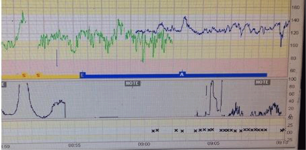
Baseline HR: The normal range of the resting maternal heart rate baseline is 60 to 100 beats per minute [12]. However with pushing efforts it is estimated to increase up to maximal heart rate of 180 beats per minute [13]. This can potentially mimic the full range of fetal heart rate (110-160 bpm) for different gestational ages which can be the same rate as the upper normal range for maternal heart rate in labour. The fetal sympathetic system matures before the parasympathetic systems resulting in a higher heart rate for preterm fetuses. The normal range for a preterm FHR baseline is between 120 and 160 bpm depending upon the gestational age, higher the rate for more preterm fetuses [14,15]. Conversely, the baseline for a term fetus should be between 100 and 140 bpm. Therefore, the baseline heart rate cannot be used to distinguish between the maternal and fetal heart rates due to this degree of overlap between them (Figure 1).
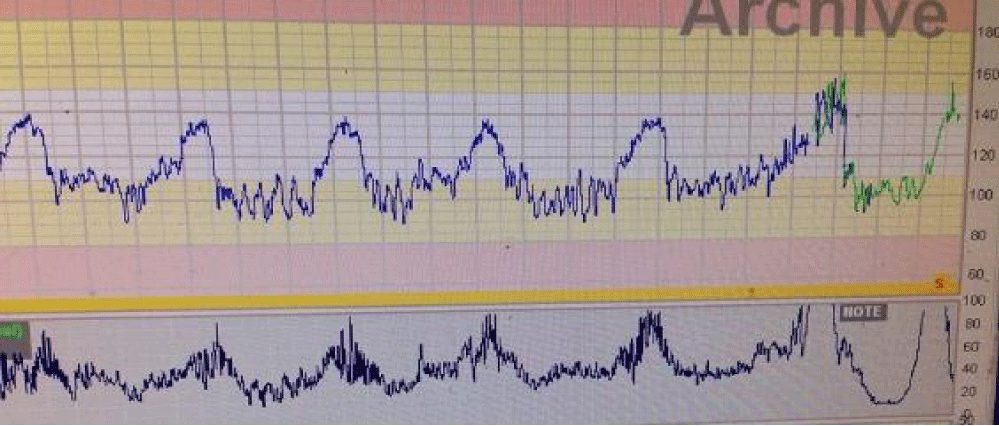
Maternal HR accelerations: Maternal Heart Rate Accelerations (MHRA) mainly occur late during the second stage of labour, especially during active maternal pushing. This is due to the increase in maternal venous return during a uterine contraction, the increase in exertion during active pushing, and maternal anxiety and pain. These MHRA coincide and peak with the contraction producing a single hump and remain present throughout the contractions (Figure 2). MHRA can be differentiated from fetal accelerations by their higher amplitude (i.e. > 15 bpm) and longer duration (i.e. > 15 seconds) as the labour progresses [16]. Cases have been reported where these were misinterpreted as fetal accelerations [17,18]. Therefore, if heart rate accelerates during pushing, other modalities should be used to confirm the source of the trace.
Fetal HR accelerations: Spontaneous FHR accelerations are associated with a transient increase in fetal heart rate with in-utero fetal movements and represent fetal wellbeing [19]. Accelerations are mediated through the fetal somatic nervous system and recorded as a more than 15 bpm increase from the baseline lasting for more than 15 seconds in term fetuses. In pregnancies less than 32 weeks, the minimum amplitude required for the characterization of the accelerations is 10 bpm [20]. The absence of accelerations is not an abnormal feature in intrapartum FHR traces [21]. Fetal accelerations rarely occur during a uterine contraction and their presence should be interpreted with caution especially if they are present after a segment of trace with late or chemoreceptor mediated decelerations. There has been some debate about the value of a “provoked accelerations” (i.e. following fetal scalp stimulation) being able to predict a non-acidaemic fetus. It has been reported that the absence of a provoked acceleration does not predict an acidaemic fetus however presence of provoked acceleration can predict non-acidaemic fetuses [22].
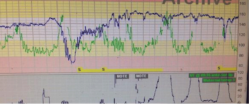
A high index of suspicion should be raised when the fetal heart pattern is showing accelerations coinciding with uterine contractions especially with active pushing in second stage of labour, a sudden change of baseline or an improved CTG pattern should always be investigated because the fetus often shows decelerations during this stage (Figure 3). During active pushing loss of contact or poor tracing is a very common scenario. During this phase maternal accelerations may overlap fetal heart signals and may appear either as a baseline or as fetal decelerations.
Good practice points to avoid erroneous monitoring of the MHR during labour
It is a good practice to check the presence of a fetal heart beat by independent means (e,g. Pinard stethoscope, hand-held Doppler) before starting a continuous fetal electronic monitoring and use maternal pulse oximetry throughout labour, especially during the second stage of labour. An alert may not be generated if the FHR trace is constantly showing MHR and therefore, the recording of maternal pulse makes it easier to detect whether MHR is erroneously recorded as FHR. Simultaneous maternal heart rate traces using ECG monitoring throughout labour especially in the second stage can provide a more precise and continuous maternal heart rate and will also avoid fetal or maternal movement artefacts [23].
In the case of suspicions in second stage of labour, clinicians should confirm fetal heart rate using independent means e.g. Pinard or hand held Doppler. Application of Fetal Scalp Electrode (FSE) will most probably resolve this uncertainty. The possibilities of this switch happening with fetal scalp electrodes are less frequent as it acquires the fetal electrocardiogram using R-R intervals and ventricular depolarization. This, however, does not exclude maternal signals fully which still can happen in the event of fetal death by accidental cervical placement of the electrode. A bedside ultrasound to locate the fetal heart and confirm the baseline rate should be used more liberally in second stage of labour. Table 1 illustrates the features that should prompt an immediate action to exclude erroneous recording of the maternal heart rate as fetal heart rate.
| Table 1: Features suggestive of erroneous recording of the maternal heart rate as FHR. | |
| Feature | Explanation |
| A sudden shift in the baseline FHR | Transducer has picked signals from maternal iliac vessels and this has resulted in the ‘sudden drop’ in the baseline HR. Maternal pulse should be immediately checked to ensure that it is not identical to the recorded baseline HR |
| Sudden improvement of the CTG Trace | FHR shows repetitive decelerations during second stage of labour due to repetitive head or cord compression. If the transducer starts picking up maternal heart signals, these decelerations will suddenly disappear |
| Presence of ‘high amplitude’ accelerations | FHR accelerations with amplitudes > 30 bpm that last more than 15 seconds are not usually seen during active second stage of labour |
| Accelerations coinciding with uterine contractions | Fetal oxygen saturation is lowest immediately after a contraction and therefore, a fetus is very unlikely to accelerate during a contraction when the oxygenation is reduced. In contrast, mother performs ‘work’ during active pushing and therefore, is likely to show accelerations during contractions. |
| ‘Double Drum’ Pattern showing multiple recordings on the CTG Trace | Similar to listening intermittently from 2 drums beating at the same time with different intensities, the CTG Trace shows intermittent recording of maternal and fetal heart rates depending on which signal is stronger, leading to confusion. |
Conclusion
Erroneous monitoring of the MHR as fetal during second stage of labour may lead to unnecessary operative interventions or poor perinatal outcomes due to the failure to recognise ongoing fetal compromise. Fetal monitoring equipment should be improved with probes obtaining maternal ECG within the display along with alarm systems to warn when maternal and fetal heart rate baselines are very close to each other. However, clinicians should have a high index of suspicion in second stage of labour. The use of fetal scalp electrode, maternal pulse oximetry, maternal ECG, fetal ECG (STAN) and the use of a bedside ultrasound machine, when appropriate, to exclude erroneous monitoring of MHR as FHR should be employed. In the era of modern technological advances, it is no longer acceptable to monitor the wrong person (i.e. the mother) during second stage of labour, when the hypoxic stress to a fetus is at its maximum.
References
- Spencer JA, Belcher R, Dawes GS. The influence of signal loss on the comparison between computer analyses of the fetal heart rate in labour using pulsed Doppler ultrasound (with autocorrelation) and simultaneous scalp electrocardiogram. Eur J Obstet Gynecol Reprod Biol. 1987 May;25(1):29-34. doi: 10.1016/0028-2243(87)90089-x. PMID: 3297840.
- Nageotte MP. Avoiding 5 common mistakes in FHR monitoring. Contemp Ob Gyn. 2007;52(5):50-55.
- Gonçalves H, Rocha AP, Ayres-de-Campos D, Bernardes J. Internal versus external intrapartum foetal heart rate monitoring: the effect on linear and nonlinear parameters. Physiol Meas. 2006 Mar;27(3):307-19. doi: 10.1088/0967-3334/27/3/008. Epub 2006 Feb 6. PMID: 16462016.
- Nunes I, Ayres-de-Campos D, Costa-Santos C, Bernardes J. Differences between external and internal fetal heart rate monitoring during the second stage of labor: a prospective observational study. J Perinat Med. 2014 Jul;42(4):493-8. doi: 10.1515/jpm-2013-0281. PMID: 24445232.
- Nurani R, Chandraharan E, Lowe V, Ugwumadu A, Arulkumaran S. Misidentification of maternal heart rate as fetal on cardiotocography during the second stage of labor: the role of the fetal electrocardiograph. Acta Obstet Gynecol Scand. 2012 Dec;91(12):1428-32. doi: 10.1111/j.1600-0412.2012.01511.x. Epub 2012 Sep 18. PMID: 22881463.
- Reinhard J, Hayes-Gill BR, Schiermeier S, Hatzmann H, Heinrich TM, Louwen F. Intrapartum heart rate ambiguity: a comparison of cardiotocogram and abdominal fetal electrocardiogram with maternal electrocardiogram. Gynecol Obstet Invest. 2013;75(2):101-8. doi: 10.1159/000345059. Epub 2013 Jan 17. PMID: 23328351.
- Neilson DR Jr, Freeman RK, Mangan S. Signal ambiguity resulting in unexpected outcome with external fetal heart rate monitoring. Am J Obstet Gynecol. 2008 Jun;198(6):717-24. doi: 10.1016/j.ajog.2008.02.030. Epub 2008 Apr 2. PMID: 18377859.
- RCOG. The use of electronic fetal monitoring. The use of cardiotocography in intrapartum fetal surveillance. Evidence-based clinical guideline number 8. Clinical Effectiveness Support Unit. London: RCOG Press; 2001.
- Chauhan SP, Klauser CK, Woodring TC, Sanderson M, Magann EF, Morrison JC. Intrapartum nonreassuring fetal heart rate tracing and prediction of adverse outcomes: interobserver variability. Am J Obstet Gynecol. 2008 Dec;199(6):623.e1-5. doi: 10.1016/j.ajog.2008.06.027. Epub 2008 Jul 30. PMID: 18667185.
- Medical Device Alert Ref MDA/2010/054. Device-Fetal monitor/cardiotocograph. Medicines and Healthcare Products Regulatory Agency; 2010.
- McDonnell S, Chandraharan E. Fetal Heart Rate Interpretation in the Second Stage of Labour: Pearls and Pitfalls. British Journal of Medicine & Medical Research. 2015;7(12): 957-970.
- Bhuinneain MN, McKenna P, O’Herlihy C, Sugrue D. The domain analysis of maternal heart rate variability in normal pregnancy-A longitudinal study [Abstract No. 667]. American Journal of Obstetrics and Gynecology. 2000;182:200.
- Söhnchen N, Melzer K, Tejada BM, Jastrow-Meyer N, Othenin-Girard V, Irion O, Boulvain M, Kayser B. Maternal heart rate changes during labour. Eur J Obstet Gynecol Reprod Biol. 2011 Oct;158(2):173-8. doi: 10.1016/j.ejogrb.2011.04.038. Epub 2011 Jun 8. PMID: 21641105.
- Murray M. Antepartal and intrapartal fetal monitoring (2nd Ed.). Albuquerque, NM: Learning Resources International. 1997.
- Afors K, Chandraharan E. Use of continuous electronic fetal monitoring in a preterm fetus: clinical dilemmas and recommendations for practice. J Pregnancy. 2011;2011:848794. doi: 10.1155/2011/848794. Epub 2011 Sep 13. PMID: 21922045; PMCID: PMC3172974.
- Sherman DJ, Frenkel E, Kurzweil Y, Padua A, Arieli S, Bahar M. Characteristics of maternal heart rate patterns during labor and delivery. Obstet Gynecol. 2002 Apr;99(4):542-7. doi: 10.1016/s0029-7844(01)01785-9. PMID: 12039107.
- Yamashiro V, Scales P, Ng H. Fetal heart rate monitoring casebook: heart rate monitoring in a case of antepartum stillbirth. J Perinatol. 1988 Summer;8(3):276-81. PMID: 3225671.
- Herbert WN, Stuart NN, Butler LS. Electronic fetal heart rate monitoring with intrauterine fetal demise. J Obstet Gynecol Neonatal Nurs. 1987 Jul-Aug;16(4):249-52. doi: 10.1111/j.1552-6909.1987.tb01581.x. PMID: 3650324.
- Hoh JK, Park MI, Park YS, Koh SK. The significance of amplitude and duration of fetal heart rate acceleration in non-stress test analysis. Taiwan J Obstet Gynecol. 2012 Sep;51(3):397-401. doi: 10.1016/j.tjog.2012.07.014. PMID: 23040924.
- Lauletta AL, Nomura RM, Miyadahira S, Francisco RP, Zugaib M. Transient accelerations of fetal heart rate analyzed by computerized cardiotocography in the third trimester of pregnancy. Rev Assoc Med Bras (1992). 2014 May-Jun;60(3):270-5. doi: 10.1590/1806-9282.60.03.017. PMID: 25004274.
- Cahill Alison G, Odibo Anthony, Roehl Kimberly, Macones George. Electronic fetal heart rate patterns in the second stage of labor: Utility of the NICHD nomenclature. American Journal of Obstetrics and Gynecology. 2012;274-S275. https://bit.ly/3bhlAvn
- Holzmann M, Wretler S, Nordström L. Absence of accelerations during labor is of little value in interpreting fetal heart rate patterns. Acta Obstet Gynecol Scand. 2016 Oct;95(10):1097-103. doi: 10.1111/aogs.12939. Epub 2016 Jul 26. PMID: 27301645.
- Gonçalves H, Pinto P, Silva M, Ayres-de-Campos D, Bernardes J. Electrocardiography versus photoplethysmography in assessment of maternal heart rate variability during labor. Springerplus. 2016 Jul 15;5(1):1079. doi: 10.1186/s40064-016-2787-z. PMID: 27462527; PMCID: PMC4945517.
Content Alerts
SignUp to our
Content alerts.
 This work is licensed under a Creative Commons Attribution 4.0 International License.


