> Medicine Group. 2021 Mar 12;2(3):136-138. doi: 10.37871/jbres1203.
Cranioplasty Flap Lifting Caused by Intracranial Hypertension Literature Review
Yakhya CISSE1*, Jean Michel NZISABIRA1, Abdoulaye DIOP2, Ansoumane DONZO1, Louncény Fatoumata BARRY1, Rokhaya DIAJHETE3, Nantenin DOUMBIA1, Papa Ndiouga LO1, Aissatou KEBE1, Fatou SENE1, Alioune Badara THIAM1, Momar Codé BA1 and Seydou Boubakar BADIANE1
2Neurosurgery Unit, Ziguinchor Regional Hospital - Ziguinchor, Sénégal
3Geriatric Department - Fann University Hospital Center - Dakar, Senegal
- Cranioplasty flap
- Hydrocephalus
- Intracranial hypertension
Abstract
Cranioplasty is a neurosurgical technique that replaces a bone defect in the skull with hard replacement tissue. It is indicated in particular after a decompressive craniectomy performed in severe head trauma in order to control intracranial hypertension refractory to medical treatment. Cranioplasty is sometimes associated with a significant number of complications, including hydrocephalus. In this article, we report the case of a cranioplasty flap lifting on intracranial hypertension following postoperative hydrocephalus and discuss the clinical relevance with a review of the literature.
Introduction
Cranioplasty is a neurosurgical procedure that restores the shape and function of the skull with replacement hard tissue. This technique, performed after decompressive craniectomy in severe head trauma, often improves the clinical condition of patients [1]. However, it is sometimes associated with a large number of complications including hydrocephalus [2-4]. In this article, we report the case of cranioplasty flap lifting caused by intracranial hypertension in an adult having undergone a decompressive craniectomy for an acute post-traumatic subdural hematoma.
Case Report
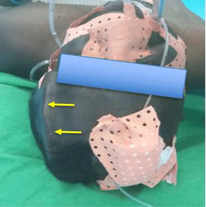
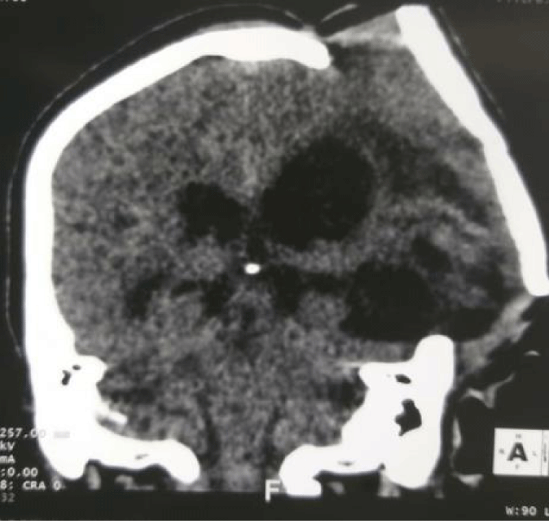
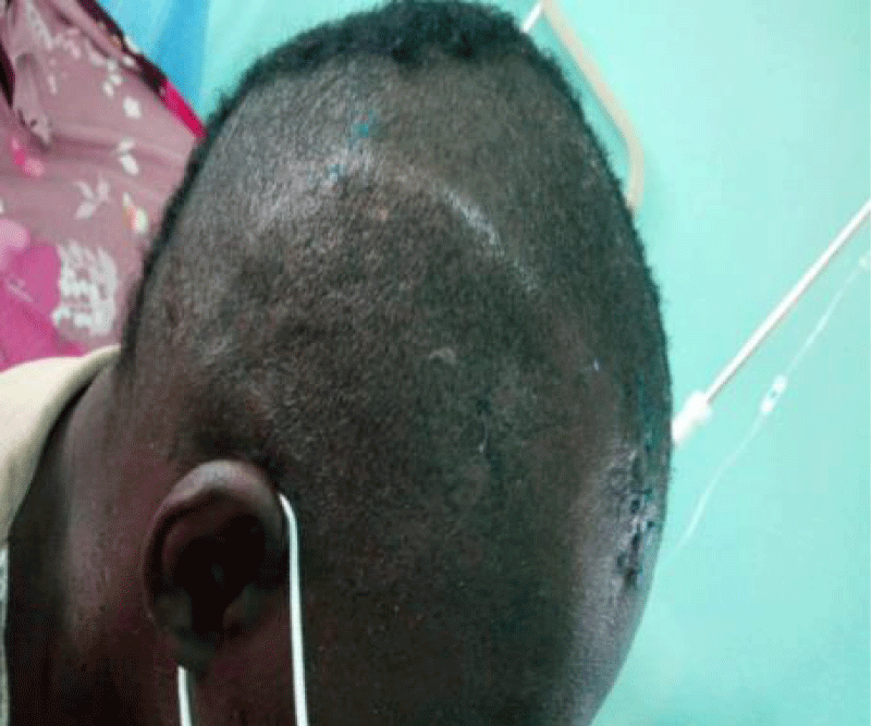
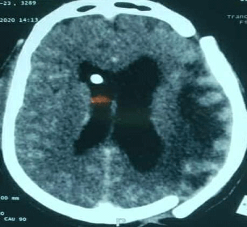
The patient is 57 years old, male, with no specific history admitted to the emergency room for severe head trauma by road accident. At the clinical examination he presented altered consciousness, Glasgow Coma Scale 7 (E1 V1 M5) with right hemibody deficit. Brain CT revealed an acute left hemispherical subdural hematoma with a significant shift. A left hemispherical decompressive craniectomy was performed urgently to control intracranial hypertension. There was significant clinical improvement and the patient discharged from hospitalization after a few days. A left hemispherical cranioplasty was performed after 3 months using surgical cement. Two months after cranioplasty, the patient is readmitted to hospital for convulsive seizures of the right half of the body. Glasgow at 8 (E4V1M3), anisocoria with photo reactive left mydriasis and cranioplasty uplift (Figure 1). Brain CT showed hydrocephalus, loss of fixation of the cranioplasty flap with expansion of the brain parenchyma out of the cranial box (Figure 2). An External Ventricular Drainage (EVD) was placed urgently with clinical improvement with regaining of consciousness and arrest of seizures. A Ventriculoperitoneal Shunt (VPS) was placed afterwards (Figure 3) and the patient discharged from hospitalization after a few days (Figure 4). We didn’t fix the cranioplasty after the ventriculoperitoneal shunt. Nevertheless we plan to replace the cranioplasty flap by an other and fix it.
Discussion
Hydrocephalus is defined as an active dilatation of the ventricles due to a disorder of the hemodynamics of CSF [5]. Overproduction or defective absorption may be the cause. In our case the patient developed a postoperative hydrocephalus (Following a cranioplasty after decompressive craniectomy). In fact, the resistance to the flow of the CSF is reduced and the cerebral compliance increased after a decompressive craniectomy. When in addition there is hydrocephalus preoperatively and the craniectomy is important, it can facilitate the irreversibility of the ventricular dilation over weeks or months [6]. The decompressive craniectomy, in our patient, was performed urgently. It made it possible to control intracranial hypertension with clinical improvement. Indeed, decompressive craniectomy reduces cerebral blood flow and improves oxygenation at the level of the decompressed hemisphere and at the contralateral level, while the metabolism is little modified. This throughput reduction may participate in the reduction of the ICP [7].
Cranioplasty may be a factor promoting hydrocephalus by restoring resistance to CSF resorption [7,8]. Several authors support this unconfirmed hypothesis. In a retrospective and non-randomized study by Bonis, et al. [9] 9 patients developed hydrocephalus after decompressive craniectomy crossing the midline out of a total of 26 patients. This study does not confirm with certainty that the hydrocephalus was related to decompressive craniectomy. Waziri, et al. [10] found 15 postoperative communicating hydrocephalus in a total of 17 patients and five of these patients received ventriculoperitoneal CSF diversion. He concluded that the diagnosis of hydrocephalus in the hemicraniectomy population is necessarily subjective. Nasi, et al. [11] found 37 postoperative hydrocephalus in 130 patients and 34 of these patients received ventriculoperitoneal CSF diversion.
In fact, after cranioplasty, the dilated ventricles can, via the cortical mantle, obstruct the subarachnoid space and thus constitute a resistance to the resorption of the CSF. However, injuries caused by head trauma can also lead to hydrocephalus.
The cranioplasty component was raised by intracranial hypertension caused by the increase in the intracranial fluid compartment [6,12]. Waziri and colleagues have recently suggested that decompressive craniectomy may play a role in the ‘‘flattening’’ of the normally dicrotic CSF pulse waveform seen in patients who undergo DC, due to the transmission of the pressure pulse out through the open cranium [10].
Conclusion
Cranioplasty may be a contributing factor to hydrocephalus in that it increases resistance to CSF resorption. It is important to take this into account when performing any cranioplasty.
Authors’ Contributions
All the authors contributed to this work.
References
- Honeybul S, Ho KM. The current role of decompressive craniectomy in the management of neurological emergencies. Brain Inj. 2013;27(9):979-91. doi: 10.3109/02699052.2013.794974. Epub 2013 May 10. PMID: 23662706.
- Chang V, Hartzfeld P, Langlois M, Mahmood A, Seyfried D. Outcomes of cranial repair after craniectomy. J Neurosurg. 2010 May;112(5):1120-4. doi: 10.3171/2009.6.JNS09133. PMID: 19612971.
- Coulter IC, Pesic-Smith JD, Cato-Addison WB, Khan SA, Thompson D, Jenkins AJ, Strachan RD, Mukerji N. Routine but risky: a multi-centre analysis of the outcomes of cranioplasty in the Northeast of England. Acta Neurochir (Wien). 2014 Jul;156(7):1361-8. doi: 10.1007/s00701-014-2081-1. Epub 2014 Apr 22. PMID: 24752723.
- Kan P, Amini A, Hansen K, White GL Jr, Brockmeyer DL, Walker ML, Kestle JR. Outcomes after decompressive craniectomy for severe traumatic brain injury in children. J Neurosurg. 2006 Nov;105(5 Suppl):337-42. doi: 10.3171/ped.2006.105.5.337. PMID: 17328254.
- Davson H. Formation and drainage of the cerebrospinal fluid. Sci Basis Med Annu Rev. 1966:238-59. PMID: 5327139.
- Kim SW, Lee SM, Shin H. Hydrocephalus Developed after Cranioplasty: Influence of Cranioplasty on the CSF Circulation. Journal of Korean Neurosurgical Society. 2006:193-5.
- Hochwald GM, Epstein F, Malhan C, Ransohoff J. The rôle of the skull and dura in experimental feline hydrocephalus. Dev Med Child Neurol Suppl. 1972;27:65-9. doi: 10.1111/j.1469-8749.1972.tb09776.x. PMID: 4509415.
- Schaller B, Graf R, Sanada Y, Rosner G, Wienhard K, Heiss WD. Hemodynamic and metabolic effects of decompressive hemicraniectomy in normal brain. An experimental PET-study in cats. Brain Res. 2003 Aug 22;982(1):31-7. doi: 10.1016/s0006-8993(03)02900-7. PMID: 12915237.
- De Bonis P, Pompucci A, Mangiola A, Rigante L, Anile C. Post-traumatic hydrocephalus after decompressive craniectomy: an underestimated risk factor. J Neurotrauma. 2010 Nov;27(11):1965-70. doi: 10.1089/neu.2010.1425. PMID: 20812777.
- Waziri A, Fusco D, Mayer SA, McKhann GM 2nd, Connolly ES Jr. Postoperative hydrocephalus in patients undergoing decompressive hemicraniectomy for ischemic or hemorrhagic stroke. Neurosurgery. 2007 Sep;61(3):489-93; discussion 493-4. doi: 10.1227/01.NEU.0000290894.85072.37. PMID: 17881960.
- Nasi D, Dobran M, Di Rienzo A, di Somma L, Gladi M, Moriconi E, Scerrati M, Iacoangeli M. Decompressive Craniectomy for Traumatic Brain Injury: The Role of Cranioplasty and Hydrocephalus on Outcome. World Neurosurg. 2018 Aug;116:e543-e549. doi: 10.1016/j.wneu.2018.05.028. Epub 2018 May 14. PMID: 29772371.
- Shapiro K, Fried A, Takei F, Kohn I. Effect of the skull and dura on neural axis pressure-volume relationships and CSF hydrodynamics. J Neurosurg. 1985 Jul;63(1):76-81. doi: 10.3171/jns.1985.63.1.0076. PMID: 4009278.
Content Alerts
SignUp to our
Content alerts.
 This work is licensed under a Creative Commons Attribution 4.0 International License.


