Medicine Group . 2023 July 26;4(6):1143-1152. doi: 10.37871/jbres1779.
Arthroscopic Treatment for Chronic Ankle Instability
Antonio Zanini, Manuel Bondi* and Andrea Pizzoli
- Ankle instability
- Ankle sprain
- Ankle arthroscopy
- Ankle distraction
- Ligament repair
- Level IV: Case series.
Abstract
Background: Ankle sprain is the most frequent sports trauma. After repeated sprains, Chronic Instability (CAI) occurs and usually requires a surgical procedure. New surgical strategies to treat symptomatic CAI are evolving. In this article we describe a fully arthroscopic technique for CAI treatment.
Methods: January 2017 to January 2020, we arthroscopically treated 26 patients for CAI, 21 of these were included in the study. The patient’s average age was 33, 43 years.
Median Follow-Up (FU) was 22, 38 months. Using a suture passer or a grasper and knotless anchors, the ligaments were repaired with an arthroscopic technique and reinforced with arthroscopic plication technique.
Results: At FU the AOFAS mean was 96, 29; FADI mean was 131, 38; VAS mean was 1, 00.
All patients reported subjective improvement in their ankle instability after the arthroscopic repair.
Conclusion: Chronic ankle instability can be successfully treated by an arthroscopic repair reinforced with arthroscopic plication technique. It is possible obtain significant functional outcomes with high patient satisfaction.
Introduction
Ankle sprains are one of the most common injuries among the general population and is a frequent disabling condition in athletes with significant limitations in performance and training programs. It represents 45% of all trauma in basketball and 31% of all trauma in soccer [1]. Conservative treatment is mainly chosen [2,3], but chronic ankle instability develops in 20% to 30% of patients and surgical treatment is necessary [4,5]. Chronic Ankle Instability (CAI) is generally caused by an Anterior Talofibular Ligament (ATFL) tear that is an intra-articular structure [6].
For several years the gold standard technique consists in repairing the ligament (Broström technique [7]) and strengthening the repair by adding extensor retinaculum (Gould technique [4,8]).
The Broström repair was first described in 1966. It concerned direct repair of the torn ends of the ATFL and CFL by midsubstance suturing [9].
To maintain stability and reduce complications, there is an increasing interest in ligament augmentation using suture tape to treat this condition [10]. Cho BK, et al. [11] reported that, in 34 young female patients, ligament repair using suture tape yielded satisfactory functional results.
Thermal shrinkage, ligament repair, and techniques of ligament reconstruction have been described [12-16].
In 2013, Vega J, et al. [17] proposed an anatomic and arthroscopic all-inside ATFL repair with a knotless anchor (2.9 mm × 15 mm), without mini tape. Satisfactory clinical data were observed in the 16 patients of the study, and no major or neurological complications were reported. The absence of nerve-related complications may be explained by the fully intra-articular technique [9].
In another recent study Vega J, et al. [18], described the arthroscopic all-inside repair of the ATFL and CFL; it will be important to wait for an adequate follow up to determine the results.
As we say, when ankle sprains, especially inversion injuries, are not treated well, 10–30% of them can progress to CAI [19]. Consequently, if lateral ankle instability is not treated adequately and is neglected for a long period with continued unbalanced loading in the medial ankle, medial compartment Ankle Osteoarthritis (OA) may develop [20]. Patients that have degenerative cartilage deterioration may experience recurrent medial ankle pain and swelling, with persistent pain even during normal walking.
CAI combined with early medial ankle OA has been known to be associated with a poor functional status compared with CAI with intact ankle cartilage. This highlights the importance of treating ankle sprain, even in acute cases.
The aim of this study is to describe an arthroscopic repair of the ATFL and capsular-ligament augmentation by plications, and to report the early results of the technique in patients treated for chronic ankle instability. It was hypothesized that this arthroscopic technique can effectively repair the ATFL through a reproducible technique, even in the hands of a less experienced surgeon, and that the proposed procedure would yield excellent results in patients with chronic ankle instability.
Material and Methods
In our Sports and Traumatology Hospital structure, from January 2017 to January 2020, 157 patients were arthroscopically treated for ankle problems by the same surgeon. From this group 26 patients underwent ankle arthroscopy for CAI. Five patients were excluded from the study for insufficient follow-up time.
In the 21 patients included in the study, the average age was 33, 43 years (range, 20-52) at the time of surgery. Of these 14 patients were male and 7 female; 7 right ankle, 14 left (Table 1).
| Table 1: Data patients. | |
| Data | |
| Age (years) | 33.43 ± 8.10 |
| Weight (kg) | 72.90 ± 10.80 |
| Gender (M/F) | 14/7 |
| Side (D/S) | 7/14 |
| Traumatic event | 16 sport |
| 5 accident | |
According to the traumatic events, 16 patients were injured during sports, 5 patients had an accidental traumatic event. All the athletes were agonists and practiced respectively 3 volleyball, 4 rugby, 4 basket and 5 football (Table 1).
Criterion for inclusion was an ankle impingement syndrome due to ankle sprain graded I-III, the presence of unilateral, ongoing pain when bearing weight, palpatoric pain of the anterior talocrural joint space and pain provoked by maximum dorsiflexion of the ankle, recurrent swelling in sports and hard works, a feeling of the ankle giving way, and weakness. Before surgery all patients were subject to conservative treatment with non-steroid drugs (only if VAS > 4), heel lifting and physiotherapy without success for at least 3 months, but in many cases till 24 months or more.
The exclusion criteria concerned the presence of rheumatic and diabetic pathologies, previous surgery on the same ankle, dementia, neuropathic arthropathies; patients with concomitant deltoid ligament injury or chronic ankle instability and poor quality of the remnant ligament-tissue that made not possible grasping the ligament with suture passer, or when ligament-tissue remnant was absent; follow up period less than 12 months.
Physical examination demonstrated positive anterior drawer test, and talar tilt in all patients, concomitant anterolateral pain for soft tissue impingement.
In postoperative period, a rigid bandage is maintained for a 2 weeks, then replaced by a bivalve brace for others 4 weeks, to avoid foot inversion and eversion. The gradual mobility in flexion and extension are started after 2 weeks and weight bearing is allowed after 3 weeks.
Subsequently, the program provides for the recovery of complete range of motion by protected weight-bearing, better in water, proprioception and muscle, with particular attention to flexion-extension recovery.
Clinical data were collected at the time of follow-up with the American Orthopaedic Foot and Ankle Society (AOFAS) scale, with the Foot and Ankle Disability Index (FADI) scale and VAS. Patients were also assessed with use of the Coughlin Score [21], which classifies the results, in relation to the patient’s subjective satisfaction, as excellent, good, fair, or poor. The clinical evaluation of all patients was performed by the same surgeon (M.B.), who did not perform any of the surgical procedures.
One of the most widely used PRO measures for foot and ankle conditions is the American Orthopedic Foot and Ankle Society Score (AOFAS), developed in 1994, reviewed over the years by national and international scientific societies [22]. The surveys include a mixture of questions that are both subjective and objective in nature. The pain category which asks patients a single question about their level of pain is subjective, while the alignment category (to be answered by the physician) is objective. The function category, however, consists of 5-7 questions and requires completion by both the patient and the physician.
The Foot and Ankle Disability Index (FADI) is a region-specific self-report of function, firstly described in 1999, reviewed over the years by national and international scientific societies [23]. The FADI contains 4 pain related items and 22 activity related items.
The VAS scale [24] owes its origin to some visual-analogue scales developed in the field of psychology to measure well-being.
To date, it is one of the best-known one-dimensional outcome measures for measuring pain intensity. It corresponds to the visual representation of the amplitude of pain felt by the patient and consists of a predetermined line 10 cm long, where the left end corresponds to "no pain", while the right end to "worst possible pain". The patient is asked to draw a mark on the line representing the level of pain experienced.
According to Coughlin score, at patients were asked to express their degree of satisfaction to the post-operative on a 5-point scale (0 = unsatisfied, 1 = barely satisfied, 2 = satisfied, 3 = very satisfied, 4 = excellent).
Preoperative Magnetic Resonance Imaging (MRI) of the ankle was obtained and showed ATFL and CFL lesions in all patients but in five patients the MRI was reported as showing chronic injury with no ligament disruption. Osteochondral involvement at the medial talar dome was observed in 8 patients. This shows the importance of arthroscopic approach compared to open surgery.
Anterior soft tissue impingement was always present.
Descriptive results were presented as median and range. Statistical analysis was performed using Microsoft Office Excel. Clinical examination-related data analysis was performed to evaluate the increase in scores at follow-up. Comparison of results using AOFAS, FADI, VAS, and Coughlin scores was done using t-tests. The differences were considered significant when the p-value was < 0.05.
Surgical Technique
The surgery is performed under loco-regional anesthesia, using a pneumatic tourniquet at the limb root (we do not consider its decisive use). The patients are lying on their back on the operating table. We use an infusion pump, but it is also possible without it, and a 4.5 mm arthroscope with 30° optics.
Cutaneous landmarks were highlighted, and the superficial peroneal nerve was identified when possible with forced inversion of the ankle and 4th toe plantarflexion [25]. Sometimes, when the nerve was not visible with this method, we used arthroscopic trans illumination through the medial portal as effective test in localizing the superficial peroneal nerve. The standard anterolateral and anteromedial portals are used, without the use of an ankle distractor in order to perform inversion and subversion movements, as well as flexion-extension. In the initial diagnostic phase, in order to obtain an adequate visualization of the astragalo-tibial articular surface and of the lateral and medial recesses, joint debridement and accurate synovectomy are practiced, often the cause of anterior impingement and therefore of pain, also by means of a motorized instrument. This turns out to be a surgical time that lengthens the surgery, but which we consider very important [5].
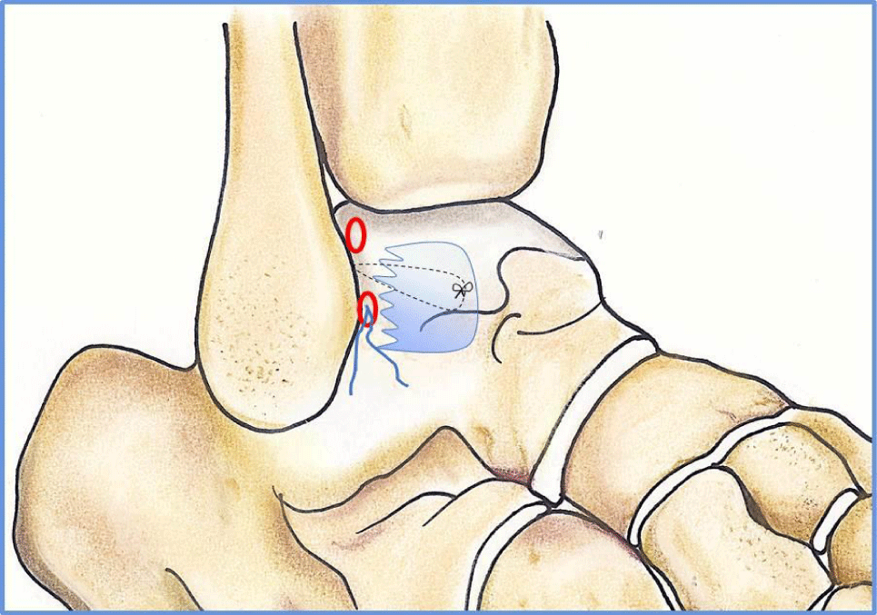
An accessory anterolateral portal was placed 1-1.5 cm proximal and anterior to the fibular tip. The ATFL was identified and completely detached off the fibular origin with the shaver help introduced through the anterolateral portal.
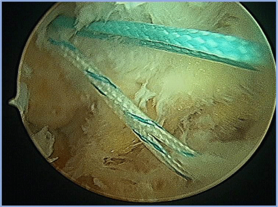
A capsular and periosteum thermal shrinkage by radiofrequency, in the fibular tip was done allow anchor fixation stability. The radiofrequency terminal was introduced through the anterolateral portal, and under direct arthroscopic visualization. Important is not remove the cortical bone, to allow greater anchor hold (Figure 1).
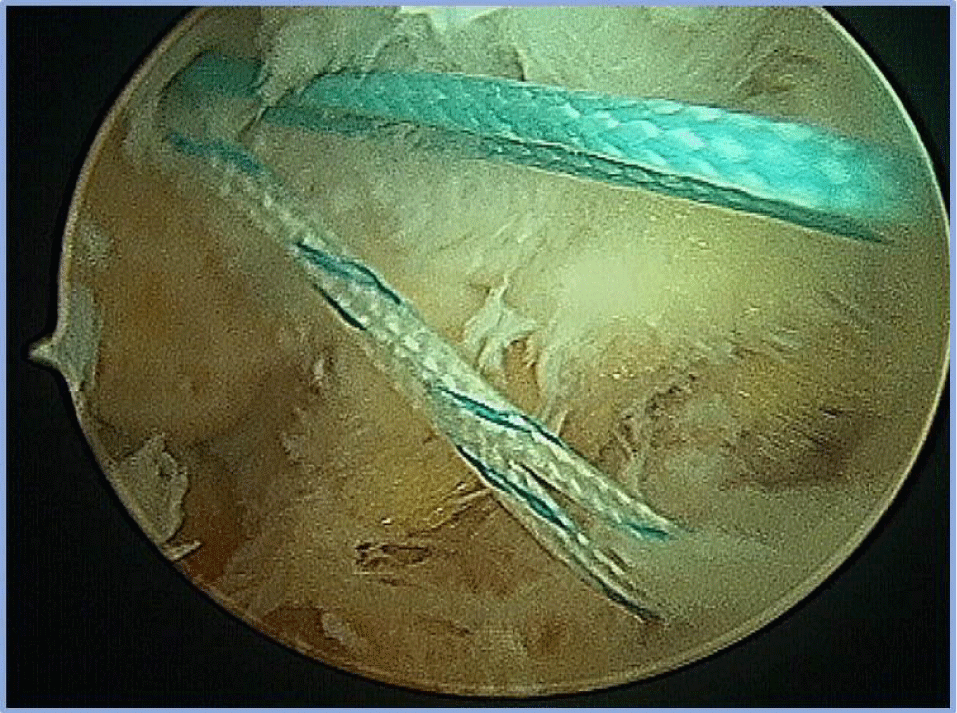
The Iconix 2.3 double wire soft anchor (Stryker®) was placed into the fibula through a previously drilled bone tunnel. The placement of the anchor in the fibula was determined by the ATFL footprint, which is located just distal to the fibular insertion of the anterior tibio-fibular ligament.
It must be checked that the implant is correctly positioned, the anchor must disappear under the cortical bone (Figure 2). The 4 wires can remain in the accessory anterolateral portal.
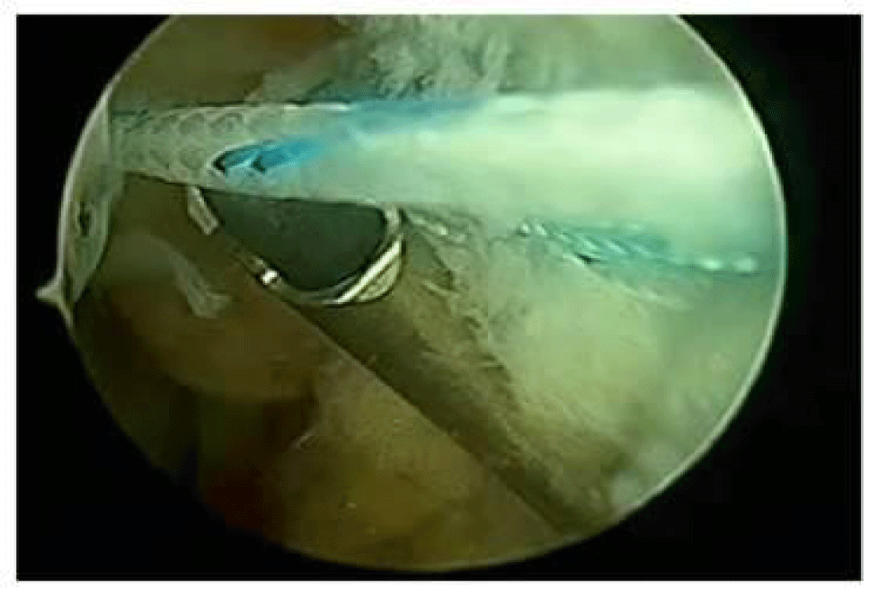
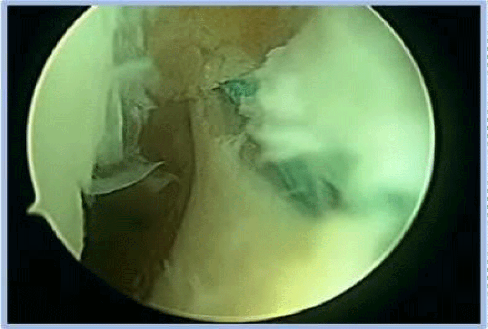
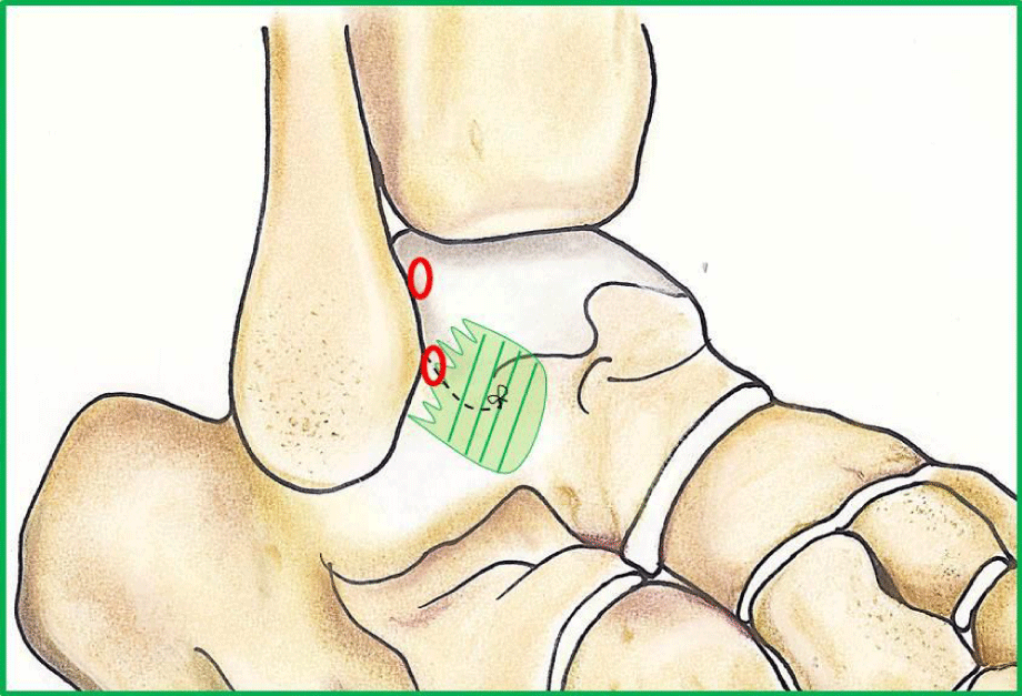
With the ATFL detached from him insertion, the ligamentous tissue was grasped with the reusable suture passer or with a suture grasper, according to surgeon’s experience (Figure 3). Sometimes we use also a shuttle release in PDS wire by needle introducing. It was introduced through the anterolateral portal, and under direct arthroscopic visualization, the maximum possible amount of ligamentous tissue was penetrated from lateral to medial. The wires of the same color are recovered and a sliding knot is made to shift and bring the ligament close to the fibula. This suture must have a horizontal direction (approximately parallel to the ground); ligamentous residue is necked to deperiosteum area (Figure 4). Then, the sutures were cut with arthroscopic scissors. This procedure is performed intracapsularly (Figure 5).
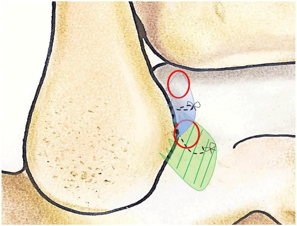
With the suture passer introduced through the anterolateral portal, the upper lateral capsule near fibula was penetrated one of the wire left is recovered and extracted from the portal.
The same procedure was done through the accessory anterolateral portal with the inferior lateral capsule penetration, adding it to the residual ligamentous previously sutured, and the recovery of the last wire left (Figure 6).
A subcutaneous mini grasper for small joints was introduced through the accessory anterolateral portal and the wire, previously positioned in the anterolateral portal, is recovered and a sliding knot is made to obtain a capsular plication [15,16] (Figure 7). This suture must have an oblique direction; the nerve is not involved because we are above it.
Then, the sutures were cut with arthroscopic scissors.
Results
Patients were clinical re-evaluated between January 2021 and December 2022, according to the health restrictions of the COVID period.
The average follow-up for the 21 patients consisted in 34, 57 months (ranged from 18 to 48) after arthroscopic surgery.
Additional ankle arthroscopic procedures included: anterior ankle soft-tissue impingement (probably scar fibrosis) debridement in all cases (100%), anterior tibial osteophyte resection in 6 (28, 6%), osteochondral debridement and micro fracturing in 8 (38, 1%), loose body removal in 2 (9, 5%) (probable fibula tip fracture passed by unknown).
The AOFAS mean score was 96, 29 (ranged from 90 to 98), the mean value of the FADI score was 131, 38 (ranged from 126 to 134) and the mean value of VAS score was 1.00 (ranged from 0 to 3) (Table 2).
| Table 2: Clinical scores at the follow up. | |
| Data | |
| Mean follow up | 34.57 ± 9.21 |
| AOFAS score | 96.29 ± 1.98 |
| FADI score | 131.38 ± 1.86 |
| VAS score | 1.00 ± 0.89 |
| Unsatisfied | 0 |
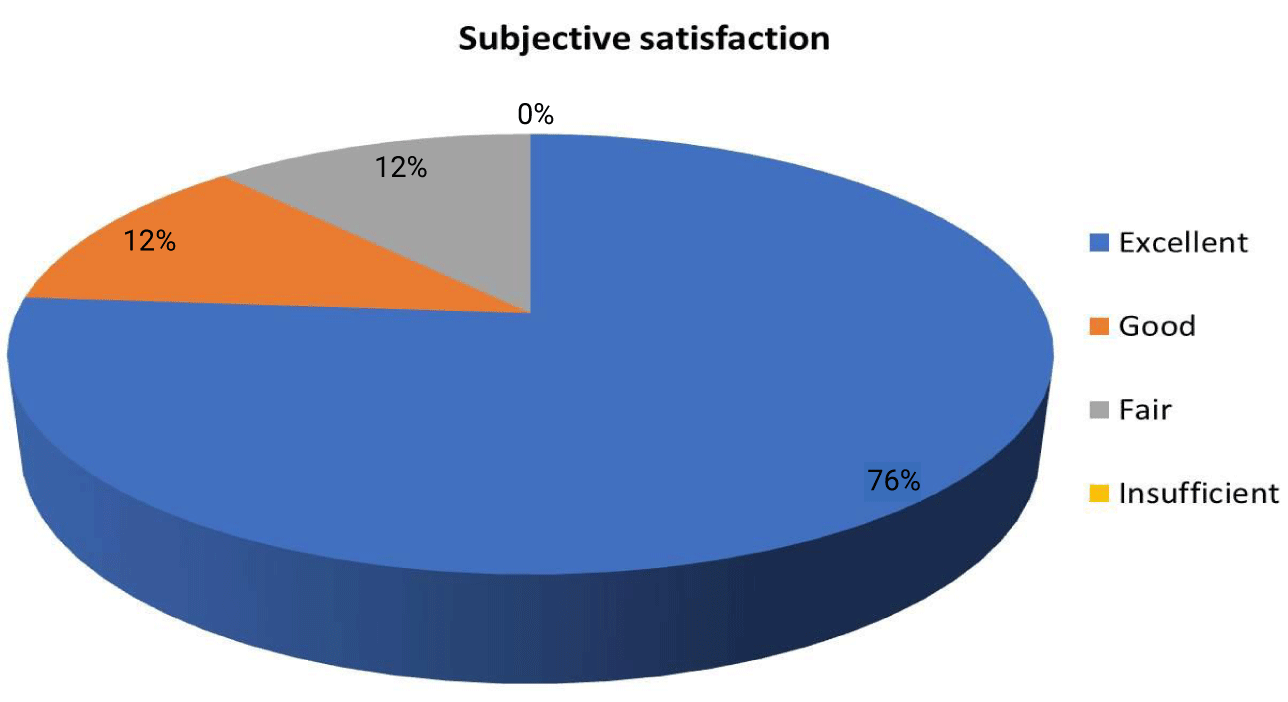
Nineteen patients (88, 1%) provided an excellent or very good opinion on the success of the surgery; only two patients (11, 9%) reported a sufficient opinion (Figure 8).
At final follow-up, all patients reported improvement in their symptoms of ankle instability. On clinical examination, the anterior drawer test was negative and no taller tilt was elicited in any of the patients.
Two (9, 5%) patients had ankle plantar-flexion deficit compared to the contralateral side which ranged from 10° to 20°. As described by Vega J, et al. [18], this mild plantarflexion deficit was not considered a complication but a consequence of the technique itself as it caused no functional limitations and may be index of good results. But we think that is important inform the patient before the surgery.
In the 2 cases of the sufficient opinion (11, 9%) we noticed no return to sport due to post-operative lack physiotherapy; these patients developed a subtalar joint rigidity.
In other 2 cases did we have paresthesia’s resolved on our own within a month.
No patients required an arthroscopic debridement in second look.
Discussion
The 20% of acute lateral ligament injuries result in chronic and require surgical treatment [4].
Correct diagnosis is essential to rule out associated injuries, such as cartilage involvement, osteochondral deficit, fractures or impingements.
It is unlikely that patients come to the orthopedic doctor due to ankle instability, but more easily due to associated injuries, we believe it is incorrect to treat osteochondral lesions without taking care of capsule-ligament instability which could be the only real cause of joint symptomatology.
Many techniques have been described and generally classified into anatomical or nonanatomical repairs. Non-anatomical repairs provide poor long-term results and are not recommended as the first choice for treatment [7,10] in sports but keep for hard works.
Arthroscopic techniques of repairing the ATFL have been described in which some surgeons perform only Broström and others add Gould augmentation percutaneously or by a mini open technique [26,27]. Guillo S, et al. [28] described Broström with Gould augmentation technique performed with direct control and visualization by arthroscopy and endoscopy.
To date, the Broström technique is an anatomic repair technique and has been a standard procedure for chronic lateral ankle instability [4,29].
The modified Broström repair, which is an anatomic reconstruction, has shown satisfactory outcomes, but there are still some complications, including ankle instability and ligament tear [29]. Porter M, et al. [30] reported that, of 25 patients who underwent the modified Broström repair, 3 patients experienced recurrent ankle injuries.
Kirk KL, et al. [31] reported that Broström repairs can only provide approximately 50% of the strength of the native, uninjured ligament. As a result, the modified Broström repair with augmentation using suture tape was proposed.
Xu DL, et al. [32] compared Broström with augmentation using suture tape group and modified Broström Repair group. In his study both methods have satisfactory clinical outcomes for chronic lateral ankle instability, and the suture tape may provide better strength in the repaired ligament. Some studies have reported similar outcomes of ankle function recovery during follow-up [33].
Intra-articular pathology that can contribute to pain and dysfunction has been observed in 66-93% of unstable ankles [34-36]. Arthroscopic treatment of associated intra-articular pathology before an open Broström procedure has been described in literature [35,37], but is more difficult recognize the residual ATFL for edema tissue.
Recently, arthroscopic techniques to treat ankle instability have been proposed instead of a combined procedure [17,38].
The clinical results of the arthroscopic techniques have proven similar or superior to open procedures [39]. The first technique to use arthroscopy for lateral ligament repair had good results, but also some possible complications, due to nerve entrapment by the suture during the percutaneous gesture [40-42].
According to these fact, a safe zone for the percutaneous passage has been described [15,16]. Our technique is fully arthroscopic and avoids percutaneous suture passage, proving safer than percutaneous techniques.
In our case series, we had no neurological complications. This is possible because the portal is placed just lateral to the peroneus tertius tendon, and medial to the superficial peroneal nerve when visible transcutaneously [17,43]. Sometimes the presence of anatomical variations of the nerve [44] demands caution during portal placement [45].
It is estimated that 20% of chronic ankle instability results from an injury to both the ATFL and CFL [9].
The need of repairing the CFL, when both the ATFL and CFL are injured, has been questioned in the literature with clinical as well as biomechanical studies that report excellent results after repairing of only the ATFL when both the ATFL and CFL are injured [46-48]. According to Vega, et al. [18] studies, these good clinical results can be anatomically explained by the presence of arciform fibers connecting the inferior ATFL with the CFL [6].
The anterolateral joint capsule usually appears thickened in patients with chronic ankle instability, suggesting a potential stabilizing role in the unstable ankle.
We believes that it could be potentially used as a biological reinforcement of the ligament repair; according to this we utilized the plication technique [15,16] in augmentation to the ATFL reconstruction.
Our results and others clinical studies suggest that isolated ATFL repair yields excellent results in cases of CAI with injury of both ATFL and CFL [12,46-48]. We prefer to add the capsular plication to ensure greater joint stability by reducing capsular redundancy.
Despite a rather flat learning curve, a thorough command of ankle arthroscopic skills and arthroscopic anatomy is mandatory.
According to the different ankle sizes, the anatomical conformations, the residues tissue that can be found, authors believe it is important that the surgeon knows more techniques about surgical ligament reconstruction, so that he can adapt to the specific case to be addressed. We routinely use the technique described in this article in CAI cases.
Limitations of this study include a small number of patients and the lack of a control group, but to remedy this we utilized those reported in literature. Another limitation is that the AOFAS score used is not a validated outcome scale for the evaluation of ankle instability. A specific clinical score to assess ankle instability would have increased the validity of the study.
Conclusion
The arthroscopic repair of the ATFL and capsular-ligament augmentation by plications is a viable option for surgical treatment of chronic ankle instability in those patients with major ankle instability and injury of the ATFL. With arthroscopic technique there is a minimal risk of damage to surrounding anatomical structures.
Conflicts of Interest
The authors declare that they have no conflict of interest.
Funding
This research did not receive any specific grant from funding agencies in the public, commercial, or not-for-profit sectors.
References
- van den Bekerom MP, Kerkhoffs GM, McCollum GA, Calder JD, van Dijk CN. Management of acute lateral ankle ligament injury in the athlete. Knee Surg Sports Traumatol Arthrosc. 2013 Jun;21(6):1390-5. doi: 10.1007/s00167-012-2252-7. Epub 2012 Oct 30. PMID: 23108678.
- Jotoku T, Kinoshita M, Okuda R, Abe M. Anatomy of ligamentous structures in the tarsal sinus and canal. Foot Ankle Int. 2006 Jul;27(7):533-8. doi: 10.1177/107110070602700709. PMID: 16842721.
- Zanini A, Bondi M, Bettinsoli PF, Pizzoli A. Return to sport after ankle lesions in arthroscopy and sport injuries: Applications in high-level athletes. Springer International Publishing Switzerland; 2015. p.425-431. doi: 10.1007/978-3-319-14815-1_54.
- Guillo S, Bauer T, Lee JW, Takao M, Kong SW, Stone JW, Mangone PG, Molloy A, Perera A, Pearce CJ, Michels F, Tourné Y, Ghorbani A, Calder J. Consensus in chronic ankle instability: aetiology, assessment, surgical indications and place for arthroscopy. Orthop Traumatol Surg Res. 2013 Dec;99(8 Suppl):S411-9. doi: 10.1016/j.otsr.2013.10.009. Epub 2013 Nov 20. PMID: 24268842.
- Bondi M, Zanini A, Pizzoli A. Ankle arthroscopy after ankle sprains. International Journal of Orthopaedics. 2019;6(5):1183-1188.
- Vega J, Malagelada F, Manzanares MC, Dalmau M. The lateral fibulotalocalcaneal ligament complex: An ankle stabilizing isometric structure. Knee Surg Sports Traumatol Arthrosc. 2020;28(1):8-17. doi: 10.1007/s0016 7-018-5188-8.
- Brown AJ, Shimozono Y, Hurley ET, Kennedy JG. Arthroscopic Repair of Lateral Ankle Ligament for Chronic Lateral Ankle Instability: A Systematic Review. Arthroscopy. 2018 Aug;34(8):2497-2503. doi: 10.1016/j.arthro.2018.02.034. Epub 2018 May 2. PMID: 29730218.
- Aydogan U, Glisson RR, Nunley JA. Extensor retinaculum augmentation reinforces anterior talofibular ligament repair. Clin Orthop Relat Res. 2006 Jan;442:210-5. doi: 10.1097/01.blo.0000183737.43245.26. PMID: 16394763.
- Broström L. Sprained ankles. VI. Surgical treatment of "chronic" ligament ruptures. Acta Chir Scand. 1966 Nov;132(5):551-65. PMID: 5339635.
- Matsui K, Burgesson B, Takao M, Stone J, Guillo S, Glazebrook M; ESSKA AFAS Ankle Instability Group. Minimally invasive surgical treatment for chronic ankle instability: a systematic review. Knee Surg Sports Traumatol Arthrosc. 2016 Apr;24(4):1040-8. doi: 10.1007/s00167-016-4041-1. Epub 2016 Feb 11. PMID: 26869032.
- Cho BK, Park KJ, Kim SW, Lee HJ, Choi SM. Minimal Invasive Suture-Tape Augmentation for Chronic Ankle Instability. Foot Ankle Int. 2015 Nov;36(11):1330-8. doi: 10.1177/1071100715592217. Epub 2015 Jun 25. PMID: 26112405.
- Maffulli N, Del Buono A, Maffulli GD, Oliva F, Testa V, Capasso G, Denaro V. Isolated anterior talofibular ligament Broström repair for chronic lateral ankle instability: 9-year follow-up. Am J Sports Med. 2013 Apr;41(4):858-64. doi: 10.1177/0363546512474967. Epub 2013 Feb 6. PMID: 23388673.
- Coughlin MJ, Schenck RC Jr, Grebing BR, Treme G. Comprehensive reconstruction of the lateral ankle for chronic instability using a free gracilis graft. Foot Ankle Int. 2004 Apr;25(4):231-41. doi: 10.1177/107110070402500407. PMID: 15132931.
- Pagenstert G, Valderrabano V, Hintermann B. Lateral ankle ligament reconstruction with free plantaris tendon graft. Tech Foot Ankle Surg. 2005;4:104-112. doi: 10.1097/01.btf.0000152574.09654.80.
- Zanini A, Bondi M, Pizzoli A. Il trattamento dell’instabilità di caviglia: Le plicature. Medicina e Chirurgia della Caviglia e del Piede. 2017;41(3):53-60. doi: 10.23736/S2284-2993.17.01771-X.
- Zanini A, Bondi M, Bettinsoli PF, Pizzoli A. Capsular plications repair. In: Ankle joint arthroscopy. Springer International Publishing Switzerland; 2020. p.135-142. doi: 10.1007/978-3-030-29231-7.
- Vega J, Golanó P, Pellegrino A, Rabat E, Peña F. All-inside arthroscopic lateral collateral ligament repair for ankle instability with a knotless suture anchor technique. Foot Ankle Int. 2013 Dec;34(12):1701-9. doi: 10.1177/1071100713502322. Epub 2013 Aug 26. PMID: 23978706.
- Vega J, Malagelada F, Dalmau-Pastor M. Arthroscopic all-inside ATFL and CFL repair is feasible and provides excellent results in patients with chronic ankle instability. Knee Surg Sports Traumatol Arthrosc. 2020 Jan;28(1):116-123. doi: 10.1007/s00167-019-05676-z. Epub 2019 Aug 20. PMID: 31432243.
- Karlsson J, Lansinger O. Chronic lateral instability of the ankle in athletes. Sports Med. 1993 Nov;16(5):355-65. doi: 10.2165/00007256-199316050-00006. PMID: 8272690.
- Golditz T, Steib S, Pfeifer K, Uder M, Gelse K, Janka R, Hennig FF, Welsch GH. Functional ankle instability as a risk factor for osteoarthritis: using T2-mapping to analyze early cartilage degeneration in the ankle joint of young athletes. Osteoarthritis Cartilage. 2014 Oct;22(10):1377-85. doi: 10.1016/j.joca.2014.04.029. Epub 2014 May 9. PMID: 24814687.
- Coughlin MJ. Treatment of bunionette deformity with longitudinal diaphyseal osteotomy with distal soft tissue repair. Foot Ankle. 1991 Feb;11(4):195-203. doi: 10.1177/107110079101100402. PMID: 1855704.
- Leigheb M, Janicka P, Andorno S, Marcuzzi A, Magnani C, Grassi F. Italian translation, cultural adaptation and validation of the "American Orthopaedic Foot and Ankle Society's (AOFAS) ankle-hindfoot scale". Acta Biomed. 2016 May 6;87(1):38-45. PMID: 27163894.
- Leigheb M, Rava E, Vaiuso D, Samaila EM, Pogliacomi F, Bosetti M, Grassi FA, Sabbatini M. Translation, cross-cultural adaptation, reliability, and validation of the italian version of the Foot and Ankle Disability Index (FADI). Acta Biomed. 2020 May 30;91(4-S):160-166. doi: 10.23750/abm.v91i4-S.9544. PMID: 32555091; PMCID: PMC7944815.
- Dixon JS, Bird HA. Reproducibility along a 10 cm vertical visual analogue scale. Ann Rheum Dis. 1981 Feb;40(1):87-9. doi: 10.1136/ard.40.1.87. PMID: 7469530; PMCID: PMC1000664.
- Stephens MM, Kelly PM. Fourth toe flexion sign: a new clinical sign for identification of the superficial peroneal nerve. Foot Ankle Int. 2000 Oct;21(10):860-3. doi: 10.1177/107110070002101012. Erratum in: Foot Ankle Int 2000 Dec;21(12):995. PMID: 11128019.
- Nery C, Raduan F, Del Buono A, Asaumi ID, Cohen M, Maffulli N. Arthroscopic-assisted Broström-Gould for chronic ankle instability: a long-term follow-up. Am J Sports Med. 2011 Nov;39(11):2381-8. doi: 10.1177/0363546511416069. Epub 2011 Jul 29. PMID: 21803979.
- Guillo S, Odagiri H. All-Inside Endoscopic Broström-Gould Technique. Arthrosc Tech. 2019 Dec 18;9(1):e79-e84. doi: 10.1016/j.eats.2019.09.003. PMID: 32021778; PMCID: PMC6993130.
- Bell SJ, Mologne TS, Sitler DF, Cox JS. Twenty-six-year results after Broström procedure for chronic lateral ankle instability. Am J Sports Med. 2006 Jun;34(6):975-8. doi: 10.1177/0363546505282616. Epub 2006 Jan 6. PMID: 16399935.
- Ellis SJ, Williams BR, Pavlov H, Deland J. Results of anatomic lateral ankle ligament reconstruction with tendon allograft. HSS J. 2011 Jul;7(2):134-40. doi: 10.1007/s11420-011-9199-y. Epub 2011 Mar 25. PMID: 22754413; PMCID: PMC3145865.
- Porter M, Shadbolt B, Ye X, Stuart R. Ankle Lateral Ligament Augmentation Versus the Modified Broström-Gould Procedure: A 5-Year Randomized Controlled Trial. Am J Sports Med. 2019 Mar;47(3):659-666. doi: 10.1177/0363546518820529. Epub 2019 Jan 30. PMID: 30699039.
- Kirk KL, Campbell JT, Guyton GP, Parks BG, Schon LC. ATFL elongation after Brostrom procedure: a biomechanical investigation. Foot Ankle Int. 2008 Nov;29(11):1126-30. doi: 10.3113/FAI.2008.1126. PMID: 19026207.
- Xu DL, Gan KF, Li HJ, Zhou SY, Lou ZQ, Wang Y, Li GQ, Ruan CY, Hu XD, Chen YL, Ma WH. Modified Broström Repair With and Without Augmentation Using Suture Tape for Chronic Lateral Ankle Instability. Orthop Surg. 2019 Aug;11(4):671-678. doi: 10.1111/os.12516. PMID: 31456322; PMCID: PMC6712379.
- Yoo JS, Yang EA. Clinical results of an arthroscopic modified Brostrom operation with and without an internal brace. J Orthop Traumatol. 2016 Dec;17(4):353-360. doi: 10.1007/s10195-016-0406-y. Epub 2016 Apr 23. PMID: 27108426; PMCID: PMC5071235.
- Choi WJ, Lee JW, Han SH, Kim BS, Lee SK. Chronic lateral ankle instability: the effect of intra-articular lesions on clinical outcome. Am J Sports Med. 2008 Nov;36(11):2167-72. doi: 10.1177/0363546508319050. Epub 2008 Jul 31. PMID: 18669983.
- Hintermann B, Boss A, Schäfer D. Arthroscopic findings in patients with chronic ankle instability. Am J Sports Med. 2002 May-Jun;30(3):402-9. doi: 10.1177/03635465020300031601. PMID: 12016082.
- Lee J, Hamilton G, Ford L. Associated intra-articular ankle pathologies in patients with chronic lateral ankle instability: arthroscopic findings at the time of lateral ankle reconstruction. Foot Ankle Spec. 2011 Oct;4(5):284-9. doi: 10.1177/1938640011416355. Epub 2011 Sep 16. PMID: 21926361.
- Hua Y, Chen S, Li Y, Chen J, Li H. Combination of modified Broström procedure with ankle arthroscopy for chronic ankle instability accompanied by intra-articular symptoms. Arthroscopy. 2010 Apr;26(4):524-8. doi: 10.1016/j.arthro.2010.02.002. PMID: 20362833.
- Matsui K, Takao M, Miyamoto W, Innami K, Matsushita T. Arthroscopic Broström repair with Gould augmentation via an accessory anterolateral port for lateral instability of the ankle. Arch Orthop Trauma Surg. 2014 Oct;134(10):1461-7. doi: 10.1007/s00402-014-2049-x. Epub 2014 Jul 12. PMID: 25015792.
- Guelfi M, Zamperetti M, Pantalone A, Usuelli FG, Salini V, Oliva XM. Open and arthroscopic lateral ligament repair for treatment of chronic ankle instability: A systematic review. Foot Ankle Surg. 2018 Feb;24(1):11-18. doi: 10.1016/j.fas.2016.05.315. Epub 2016 May 12. PMID: 29413768.
- Kim ES, Lee KT, Park JS, Lee YK. Arthroscopic anterior talofibular ligament repair for chronic ankle instability with a suture anchor technique. Orthopedics. 2011 Apr 11;34(4). doi: 10.3928/01477447-20110228-03. PMID: 21469637.
- Acevedo JI, Mangone P. Arthroscopic brostrom technique. Foot Ankle Int. 2015 Apr;36(4):465-73. doi: 10.1177/1071100715576107. Epub 2015 Mar 5. PMID: 25743426.
- Corte-Real NM, Moreira RM. Arthroscopic repair of chronic lateral ankle instability. Foot Ankle Int. 2009 Mar;30(3):213-7. doi: 10.3113/FAI.2009.0213. PMID: 19321097.
- Vega J, Dalmau-Pastor M, Malagelada F, Fargues-Polo B, Peña F. Ankle Arthroscopy: An Update. J Bone Joint Surg Am. 2017 Aug 16;99(16):1395-1407. doi: 10.2106/JBJS.16.00046. PMID: 28816902.
- Ucerler H, Ikiz 'A. The variations of the sensory branches of the superficial peroneal nerve course and its clinical importance. Foot Ankle Int. 2005 Nov;26(11):942-6. doi: 10.1177/107110070502601108. PMID: 16309608.
- Guelfi M, Vega J, Malagelada F, Dalmau-Pastor M. The arthroscopic all-inside ankle lateral collateral ligament repair is a safe and reproducible technique. Knee Surg Sports Traumatol Arthrosc. 2020 Jan;28(1):63-69. doi: 10.1007/s00167-019-05427-0. Epub 2019 Mar 4. PMID: 30830298.
- Ko KR, Lee WY, Lee H, Park HS, Sung KS. Repair of only anterior talofibular ligament resulted in similar outcomes to those of repair of both anterior talofibular and calcaneofibular ligaments. Knee Surg Sports Traumatol Arthrosc. 2020 Jan;28(1):155-162. doi: 10.1007/s00167-018-5091-3. Epub 2018 Aug 6. PMID: 30083968.
- Lee KT, Lee JI, Sung KS, Kim JY, Kim ES, Lee SH, Wang JH. Biomechanical evaluation against calcaneofibular ligament repair in the Brostrom procedure: a cadaveric study. Knee Surg Sports Traumatol Arthrosc. 2008 Aug;16(8):781-6. doi: 10.1007/s00167-008-0557-3. Epub 2008 Jun 17. PMID: 18560805.
- Lee KT, Park YU, Kim JS, Kim JB, Kim KC, Kang SK. Long-term results after modified Brostrom procedure without calcaneofibular ligament reconstruction. Foot Ankle Int. 2011 Feb;32(2):153-7. doi: 10.3113/FAI.2011.0153. PMID: 21288414.
Content Alerts
SignUp to our
Content alerts.
 This work is licensed under a Creative Commons Attribution 4.0 International License.


