Medicine Group . 2022 September 15;3(9):1042-1044. doi: 10.37871/jbres1552.
Images of a Highly Metastatic Ependymoma of the Cauda
Arsen Seferi1 and Gentian Vyshka2*
2Biomedical and Experimental Department, Faculty of Medicine, University of Medicine in Tirana, Albania
- Ependymoma
- Extraneural metastases
- Pleural effusion
- Spinal MRI
Abstract
Images from a spinal ependymoma extending from twelfth thoracic to the third lumbar vertebral level will be discussed, together with the history and the final outcome of a highly metastatic neuroepithelial tumor. The young female patient some weeks after laminectomy was diagnosed with multiple metastases, in the form of miliary mottling in both lungs, as well as with left pleural involvement with massive effusion and a solid formation in the right mammary gland. Although ependymomas rarely metastasize, a tight follow-up regimen and thorough screening is advisable, since case reports of multiple metastases via diverse routes (seeding, hematogenous) are becoming frequent and of concern.
Images in Clinical Medicine
Primary tumors of nervous system rarely metastasize, and while doing so generally they spread locally per continuitatem, or via lymphatics. This is very much true for glial tumors, and in fact glioblastomas rarely metastasize, for reasons that are still very much debatable [1]. Metastases generally follow surgical intervention; spontaneous metastases are an extreme rarity [2].
On the other hand, neuroepithelial neoplasms might have a good chance of metastasizing, even remotely [3]. Reports of metastasizing ependymomas in the majority deal with pediatric ages, since there is a clear age profile of this type of tumor, among others [4].
A twenty-one years old female patient appeared for consultancy following gait difficulties, throbbing pain in both lower extremities, and urinary hesitancy. Upon examination she showed no pathological reflexes, with a left foot drop and Achillean bilateral hyporeflexia.
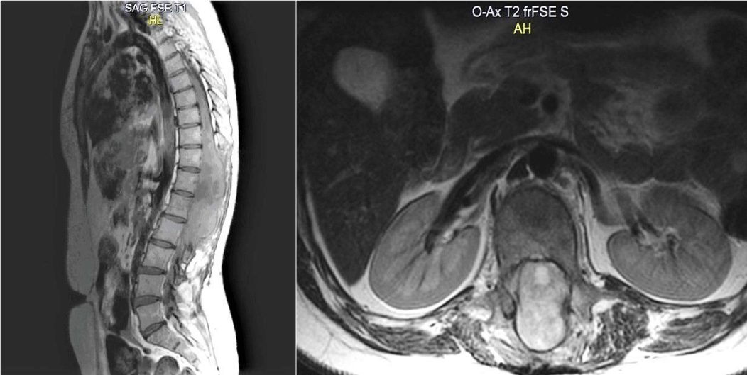
A non-enhanced lumbar spine MRI showed a considerable extradural mass formation extending to several vertebral bodies (Figures 1A,B). A decompression laminectomy Th12-L2 was performed, with the situation improving slightly during the two postoperative weeks.
The pathological specimen was sent to an external facility for microscopic evaluation. Findings suggested small uniform cells with oval nuclei, with cellular rosette formation in the perivascular areas, and a diagnosis of myxopapillary ependymoma of anaplastic grade was made.
At one month after surgery, the patient presented for a follow-up in a debilitated status, while losing ten kilograms in weight, loss of appetite, low-grade but persistent fever (37.5-37.8°C), cough and a slight sense of dyspnea.
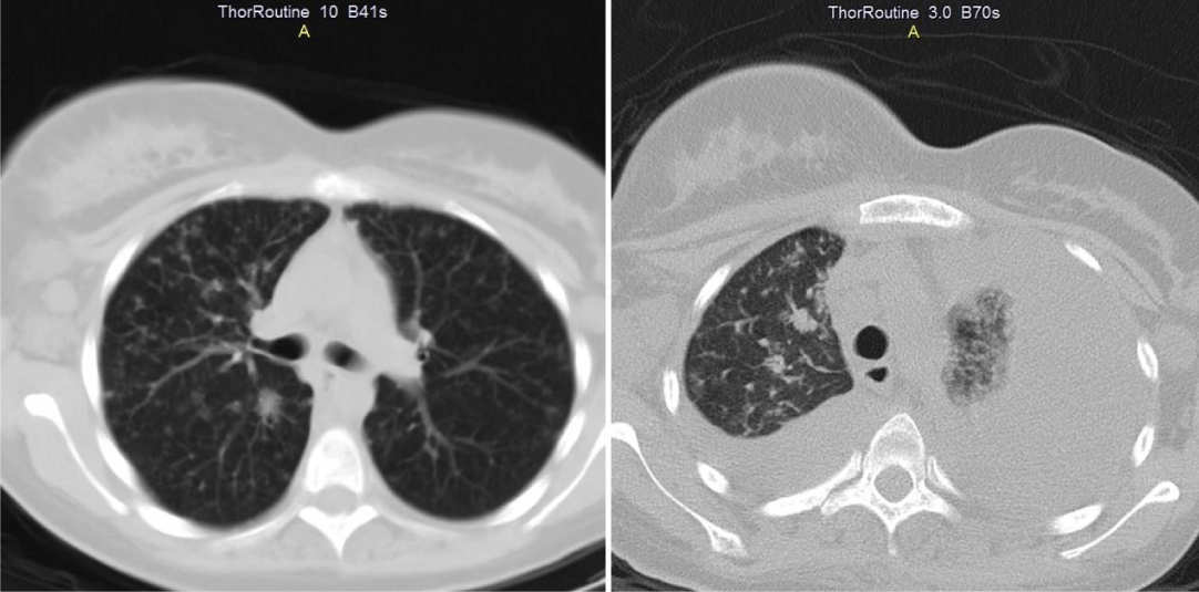
A total body CT was performed, showing miliary mottling in both lungs. The patient was unsuccessfully treated with antibiotics (cefuroxime 2 grams daily intravenous route in combination with moxifloxacin 400 milligram per os daily for a total of seven days) along with supportive therapy (liquids, antipyretics).
Ten days later a control thorax CT showed massive pleural involvement and effusion in the left lung (Figures 2A,B). A Mantoux test was negative, and a pleural puncture was inconclusive.
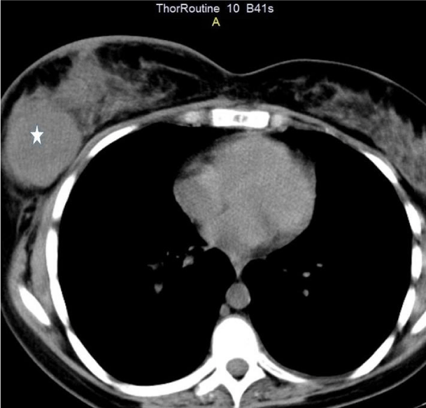
Furthermore, both thorax CTs (initial and of control) showed a growing appearance of a solid mass in her right mammary gland (Figure 3), which was considered a gland metastasis.
The patient was sent for oncological treatment, but her deteriorating situation was not permissive to chemotherapy and she passed away some days later, while receiving palliative care (morphine intramuscular 10 milligrams twice daily along with oxygen via a nasal mask).
The issue of metastasizing neuroepithelial tumors and respective treatment is debatable, since the final outcomes are generally poor. Spinal ependymomas might seed even inside the intracranial space, as some uncommon cases suggest [5].
Sources have previously reported as well cases of caudal ependymoma metastasizing in the lung and other thoracic structures [6,7]. Impressively enough, some authors have succeeded into finding lung metastases forty-six years after the first operation for a spinal ependymoma [8].
Spinal ependymomas represent as a rule benign tumors, with very rare cases of extraneural metastases; pleura is even more rarely involved [9]. Myxopapillary ependymomas are considered as in the WHO classification (World Health Organization) as grade I tumors, but malignant behavior might appear even among this group of tumors [10]. Our case demonstrated that pleural and mammary gland metastases might co-exist, badly influencing the final outcome of a neuroepithelial tumor.
References
- Park CC, Hartmann C, Folkerth R, Loeffler JS, Wen PY, Fine HA, Black PM, Shafman T, Louis DN. Systemic metastasis in glioblastoma may represent the emergence of neoplastic subclones. J Neuropathol Exp Neurol. 2000 Dec;59(12):1044-50. doi: 10.1093/jnen/59.12.1044. PMID: 11138924.
- Smith DR, Hardman JM, Earle KM. Metastasizing neuroectodermal tumors of the central nervous system. J Neurosurg. 1969 Jul;31(1):50-8. doi: 10.3171/jns.1969.31.1.0050. PMID: 4307543.
- Kraetzig T, McLaughlin L, Bilsky MH, Laufer I. Metastases of spinal myxopapillary ependymoma: unique characteristics and clinical management. J Neurosurg Spine. 2018 Feb;28(2):201-208. doi: 10.3171/2017.5.SPINE161164. Epub 2017 Dec 8. PMID: 29219779.
- Kim SI, Lee Y, Kim SK, Kang HJ, Park SH. Aggressive Supratentorial Ependymoma, RELA Fusion-Positive with Extracranial Metastasis: A Case Report. J Pathol Transl Med. 2017 Nov;51(6):588-593. doi: 10.4132/jptm.2017.08.10. Epub 2017 Nov 15. PMID: 29161788; PMCID: PMC5700879.
- Woesler B, Moskopp D, Kuchelmeister K, Schul C, Wassmann H. Intracranial metastasis of a spinal myxopapillary ependymoma. A case report. Neurosurg Rev. 1998;21(1):62-5. doi: 10.1007/BF01111488. PMID: 9584289.
- Rubinstein LJ, Logan WJ. Extraneural metastases in ependymoma of the cauda equina. J Neurol Neurosurg Psychiatry. 1970 Dec;33(6):763-70. doi: 10.1136/jnnp.33.6.763. PMID: 5531896; PMCID: PMC493589.
- Graf M, Blaeker H, Otto HF. Extraneural metastasizing ependymoma of the spinal cord. Pathol Oncol Res. 1999;5(1):56-60. doi: 10.1053/paor.1999.0056. PMID: 10079380.
- Fujimori T, Iwasaki M, Nagamoto Y, Kashii M, Sakaura H, Yoshikawa H. Extraneural metastasis of ependymoma in the cauda equina. Global Spine J. 2013 Mar;3(1):33-40. doi: 10.1055/s-0032-1329888. Epub 2012 Nov 19. PMID: 24436849; PMCID: PMC3854601.
- Rickert CH, Kedziora O, Gullotta F. Ependymoma of the cauda equina. Acta Neurochir (Wien). 1999;141(7):781-2. doi: 10.1007/s007010050376. PMID: 10481792.
- Lee JC, Sharifai N, Dahiya S, Kleinschmidt-DeMasters BK, Rosenblum MK, Reis GF, Samuel D, Siongco AM, Santi M, Storm PB, Ferris SP, Bollen AW, Pekmezci M, Solomon DA, Tihan T, Perry A. Clinicopathologic features of anaplastic myxopapillary ependymomas. Brain Pathol. 2019 Jan;29(1):75-84. doi: 10.1111/bpa.12673. PMID: 30417460; PMCID: PMC7444646.
Content Alerts
SignUp to our
Content alerts.
 This work is licensed under a Creative Commons Attribution 4.0 International License.


