Medicine Group . 2022 June 04;3(6):672-674. doi: 10.37871/jbres1495.
Gastropericardial Fistula as a Life-Threatening Complication after Intragastric Balloon Insertion
Sven Krämer*, Sebastian Wolf, Susanne Wasserberg, Gerhard Brieschenk, Matthias Schrempf, Matthias Anthuber and Dmytro Vlasenko
- Intragastric balloon
- Gastric perforation
- Gastropericardial fistula
- Bariatric surgery
Introduction
Laparoscopic bariatric surgery, while highly effective, is an invasive method for sustainable weight loss [1]. Less than 1% of patients with clinically severe obesity undergo bariatric surgery [2], in part because of concerns regarding surgical complications [3].
This major limitation has prompted calls for nonsurgical treatment options. Endoscopic bariatric therapy to reduce gastric volume via insertion of an Intragastric Balloon (IGB) is an approved alternative for patients with a BMI of 30- 40 kg/m², for whom diet and exercise alone have not been successful [4]. The utility and safety of IGB therapy have been evaluated in the short-term (6 to 12 months), while its suitability for long-term obesity management remains uncertain [5].
Case Report
We present the case of a 71year-old male patient with a history of endoscopic bariatric therapy via IGB insertion in an external hospital (height: 1.78m, weight: 120.0kg, BMI: 38kg/m²). At 12 months after IGB placement the patient presented with an unsatisfactory weight loss of 6 kg and reported recurrent epigastric abdominal pain. No change in bariatric therapy or follow-up was made. Thus, the IGB remained in place.
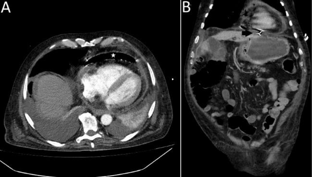
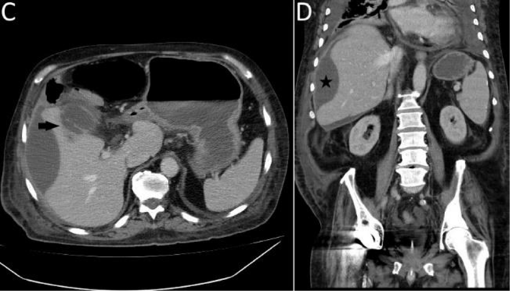
After three years the patient was admitted to a regional emergency department with symptoms indicative of upper gastrointestinal bleedings. Gastroscopy revealed the presence of an IGB. Following its endoscopic removal, endoscopy had a grandstand view on the heart and thus diagnosed a gastropericardial fistula, confirmed by CT scans of the abdomen and chest. The pericardium was gas-filled (Figure 1) and a right perihepatic abscess and a sealed perforated cholecystitis were identified (Figure 2). The patient was septic and presented with cardiorespiratory instability. For further treatment he was transferred to the Intensive Care Unit (ICU) of our university hospital. Initial blood analyses showed a leucocytosis of 15,770 (3,000/ul-10,000/ul), a haemoglobin value of 9.2 g/dl (14.0 g/dl-18.0 g/dl) and a CRP value of 15.6 mg/dl (< 0.5 mg/dl). Subsequently, the patient stabilized and surgery was performed the next day.
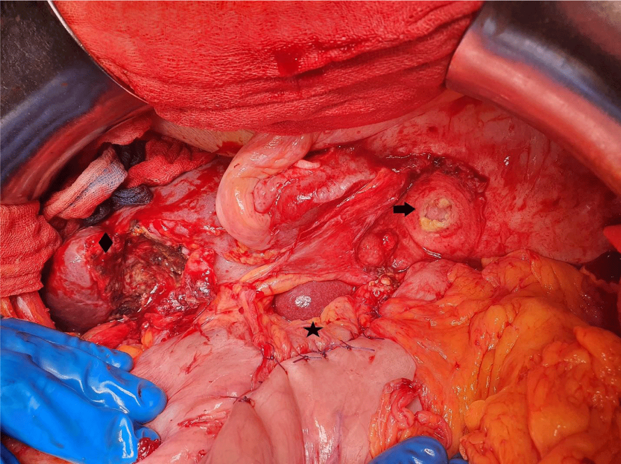
During exploratory laparotomy we detected adhesion of the lesser curvature to the diaphragm and peritonitis of the upper abdomen. Detachment of stomach and fistula from the diaphragm exposed the apex of the beating heart. The pericardial fenestration measured ~ 1 cm with clear serous pericardial effusion. The gastric portion of the fistula was closed with multiple interrupted sutures while the meatus in the pericardium was left open (Figure 3). We then opened the perihepatic abscess and performed a cholecystectomy. The sutures of the stomach and the remaining hole in the diaphragm were covered with an omental patch-plasty. We placed three intraabdominal drains positioning two drains in the right and the left hypochondrial quadrants, respectively. We inserted a third drain into the small pelvis and one chest tube on the right side because of a significant pleural effusion. Postoperatively, the patient remained in the ICU for five days.
Once stabilized, the patient was transferred to the general ward. Abdominal drains were removed 6 days after surgery, while the chest tube was removed a few days later. Subsequent transition to a normal diet was well tolerated.
The patient was discharged into rehabilitation 21 days after surgery in good general condition and without intraabdominal complications.
Discussion
In this case study we report one of the rare but life-threatening complication of IGB placement: a gastropericardial fistula identified three years after IGB insertion.
To the best of our knowledge, this is the first report of a patient with gastropericardial fistula following IGB placement. The pathologic mechanism is a pressure driven penetration through the gastric wall and the diaphragm into the pericardium. Gastric perforation as a complication after IGB insertion has been previously described with endoscopic and laparoscopic approaches achieving successful treatment [6,7]. Complication management should also be as minimally invasive as possible. Several authors reported successful treatment of acute gastric perforation after IGB placement via a laparoscopic approach [6,8]. However, in all these cases a laparoscopic approach has been used for the treatment of the acute gastric perforation only. Due to the complexity of our case including sepsis and multiple intraabdominal problems (gastropericardial fistula, cholecystitis and perihepatic abscess) open surgery was the approach of choice, yielding to patient discharge after 21 days in good general condition and without intraabdominal complications.
The placement of an IGB is a nonsurgical therapy option for weight loss, that has been approved for short-term application (6-12 months) and is more effective than lifestyle interventions alone with regard to weight loss outcome and comorbidity mitigation [4,9]. Uncertainty remains regarding the benefits and safety for long-term outcome. Unlike following bariatric surgery, where reduced gastric volume and malabsorbation persist, IGB removal results in the loss of these effects [1]. In line, low long-term weight loss efficacy and dissatisfaction in patients undergoing IGB therapy have been described [10,11]. Strategies for weight loss maintenance following IGB placement therapy include rigorous adherence to lifestyle modifications, pharmacotherapy, further bariatric measures or sequential IGB placement while maintaining clinical follow-up visits [12,13]. While our patient reported dissatisfactory weight loss of only 6 kg after one year of IGB placement no change in his bariatric therapy or follow up visits was undertaken, likely contributing to a pathophysiological cascade leading to the outcomes described above. Although considered noninvasive, rare but serious complications after IGB insertion have been reported [14]. Therefore, the risk of adverse events after IGB insertion and the need for consistent follow-up visits to avoid severe complications must to be part of the surgical patient education and endoscopic bariatric therapy protocol [14].
The development of a gastropericardial fistula in this case was likely due to the fact that the IGB remained in situ for 3 years, significantly exceeding the approved maximum retention time of 12 months [4].
Conclusion
Although IGB placement is a minimally invasive, endoscopic procedure rare but potentially life-threatening complications must be considered. Adhering to patient safety protocols including informed patient participation, strict follow up, supervision and timely removal of the device can reduce the risk of adverse events. Emphasis should be placed on individualizing bariatric therapy approaches to achieve weight loss maintenance and improve patient health in long-term.
Compliance with Ethical Standards
Conflict of Interest: The authors declare that they have no conflict of interest.
Informed Consent was obtained from the patient.
References
- Park CH, Nam SJ, Choi HS, Kim KO, Kim DH, Kim JW, Sohn W, Yoon JH, Jung SH, Hyun YS, Lee HL; Korean Research Group for Endoscopic Management of Metabolic Disorder and Obesity. Comparative Efficacy of Bariatric Surgery in the Treatment of Morbid Obesity and Diabetes Mellitus: a Systematic Review and Network Meta-Analysis. Obes Surg. 2019 Jul;29(7):2180-2190. doi: 10.1007/s11695-019-03831-6. PMID: 31037599.
- Funk LM, Jolles S, Fischer LE, Voils CI. Patient and Referring Practitioner Characteristics Associated With the Likelihood of Undergoing Bariatric Surgery: A Systematic Review. JAMA Surg. 2015 Oct;150(10):999-1005. doi: 10.1001/jamasurg.2015.1250. PMID: 26222655; PMCID: PMC4992360.
- Lopez EKH, Helm MC, Gould JC, Lak KL. Primary care providers' attitudes and knowledge of bariatric surgery. Surg Endosc. 2020 May;34(5):2273-2278. doi: 10.1007/s00464-019-07018-z. Epub 2019 Jul 31. PMID: 31367984.
- Laing P, Pham T, Taylor LJ, Fang J. Filling the Void: A Review of Intragastric Balloons for Obesity. Dig Dis Sci. 2017 Jun;62(6):1399-1408. doi: 10.1007/s10620-017-4566-2. Epub 2017 Apr 18. PMID: 28421456.
- Saber AA, Shoar S, Almadani MW, Zundel N, Alkuwari MJ, Bashah MM, Rosenthal RJ. Efficacy of First-Time Intragastric Balloon in Weight Loss: a Systematic Review and Meta-analysis of Randomized Controlled Trials. Obes Surg. 2017Feb;27(2):277-287.doi:10.1007/s11695-016-2296-8.PMID:27465936.
- Vinod VC, Younis MU, Mubarik H, Rivas H. A rare case of gastric perforation by a 5-year-old Intra-gastric Balloon in situ: Case report and review of literature. Int J Surg Case Rep. 2020;76:480-483. doi: 10.1016/j.ijscr.2020.10.028. Epub 2020 Oct 12. PMID: 33207414; PMCID: PMC7586043.
- Barrichello Junior SA, Ribeiro IB, Fittipaldi-Fernandez RJ, Hoff AC, de Moura DTH, Minata MK, de Souza TF, Galvão Neto MDP, de Moura EGH. Exclusively endoscopic approach to treating gastric perforation caused by an intragastric balloon: case series and literature review. Endosc Int Open. 2018 Nov;6(11):E1322-E1329. doi: 10.1055/a-0743-5520.Epub2018Nov7.PMID:30410952;PMCID:PMC6221813.
- Lucido FS, Scotti L, Scognamiglio G, Gambardella C, Brusciano L, Del Genio G, Pizza F, Ruggiero R, Parmeggiani D, Nesta G. Gastric perforation by intragastric balloon: Laparoscopic gastric wedge resection can be a strategy? Int J Surg Case Rep. 2020;77S(Suppl):S88-S91. doi: 10.1016/j.ijscr.2020.09.005. Epub 2020 Sep 6. PMID: 33041259; PMCID: PMC7876839.
- Popov VB, Ou A, Schulman AR, Thompson CC. The Impact of Intragastric Balloons on Obesity-Related Co-Morbidities: A Systematic Review and Meta-Analysis. Am J Gastroenterol. 2017 Mar;112(3):429-439. doi: 10.1038/ajg.2016.530.Epub2017Jan24.PMID:28117361.
- Dastis NS, François E, Deviere J, Hittelet A, Ilah Mehdi A, Barea M, Dumonceau JM. Intragastric balloon for weight loss: results in 100 individuals followed for at least 2.5 years. Endoscopy. 2009 Jul;41(7):575-80. doi: 10.1055/s-0029-1214826.Epub2009Jul8.PMID19588283.
- Haddad AE, Rammal MO, Soweid A, Shararra AI, Daniel F, Rahal MA, Shaib Y. Intragastric balloon treatment of obesity: Long-term results and patient satisfaction. Turk J Gastroenterol. 2019 May;30(5):461-466. doi: 10.5152/tjg.2019.17877.PMID:31061001;PMCID:PMC6505645.
- Farina MG, Baratta R, Nigro A, Vinciguerra F, Puglisi C, Schembri R, Virgilio C, Vigneri R, Frittitta L. Intragastric balloon in association with lifestyle and/or pharmacotherapy in the long-term management of obesity. Obes Surg. 2012 Apr;22(4):565-71.doi:10.1007/s11695-011-0514-y.PMID:21901285.
- Alfredo G, Roberta M, Massimiliano C, Michele L, Nicola B, Adriano R. Long-term multiple intragastric balloon treatment--a new strategy to treat morbid obese patients refusing surgery: prospective 6-year follow-up study. Surg Obes Relat Dis. 2014 Mar-Apr;10(2):307-11. doi: 10.1016/j.soard.2013.10.013. Epub 2013 Oct 25. PMID: 24462306.
- Stavrou G, Tsaousi G, Kotzampassi K. Life-threatening visceral complications after intragastric balloon insertion: Is the device, the patient or the doctor to blame? Endosc Int Open. 2019 Feb;7(2):E122-E129. doi: 10.1055/a-0809-4994. Epub 2019 Jan 22. PMID: 30705942; PMCID: PMC6342679.c
Content Alerts
SignUp to our
Content alerts.
 This work is licensed under a Creative Commons Attribution 4.0 International License.


