Medicine Group . 2022 April 29;3(4):413-418. doi: 10.37871/jbres1458.
Simultaneous Echocardiogram and N-Terminal-Probrain Natriuretic Peptide Measurement in Neonates
Lillie Turnbough1, Amber McKenna2 and Shabih Manzar3*
2Resident, Department of Pediatrics, School of Medicine, Louisiana State University Health Sciences Center, 1501 Kings Highway, Shreveport, LA 71103, United States
3Associate Professor, Department of Pediatrics, School of Medicine, Louisiana State University Health Sciences Center, 1501 Kings Highway, Shreveport, LA 71103, United States
- N-terminal-probrain natriuretic peptide (NT-proBNP)
- Echocardiogram
- Neonates
- Low-value care
- High-value care
- Cardiac
- Cost
Abstract
Natriuretic atrial peptides are secreted by the heart in response to the distension of the cardiac chambers, and the N-terminal-probrain natriuretic peptide (NT-proBNP) is used frequently by clinician as an indirect measure of cardiac distension. The direct way of getting the same information is echocardiogram which provides a structural and functional assessment of the heart in real time. This paper discusses the value of obtaining both investigations simultaneously. We examined four cases in which the data on simultaneously done NT-proBNP and echocardiogram was available. We concluded that although NT-proBNP used in conjunction with echocardiogram and clinical evaluation can be effective in monitoring neonatal cardiac hemodynamic status, but using NT-proBNP and echocardiogram at the same time is not necessary. Following clinical suspicion and initial echocardiogram, NT-proBNP can be used to trend the infant's status and guide treatment. As our sample size was small, further research using a multicenter trial would be needed to confirm our conclusions, and that could lead to the development of institutional guidelines benefiting both the hospital and patient alike.
Abbreviations
ASD/PFO: Atrial Septal Defect/Patent Foramen Ovale; PDA: Patent Ductus Arteriosus; AV Canal; Atrioventricular Canal; VSD: Ventricular Septal Defect; LV/RV: Left Ventricle/Right Ventricle
Introduction
Natriuretic atrial peptides are secreted by the heart in response to the distension of the cardiac chambers [1,2]. In neonates, N-Terminal-proBrain Natriuretic Peptide (NT-proBNP) measurement is used as a marker and treatment response indicator for hemodynamically significant Patent Ductus Arteriosus (HsPDA) [3-6]. NT-proBNP has also been used as prediction of BronchoPulmonary Dysplasia (BPD) [7]. However, it is unclear if obtaining NT-proBNP and echocardiogram simultaneously is a high-value care practice. Echocardiogram provides structural and functional assessment of the heart in real time, while NT-proBNP is an indirect measure of cardiac distension. In the era of handheld ultrasound technology, do we have to rely on NT-proBNP for cardiac assessment?
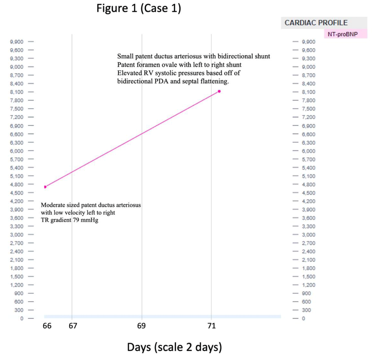
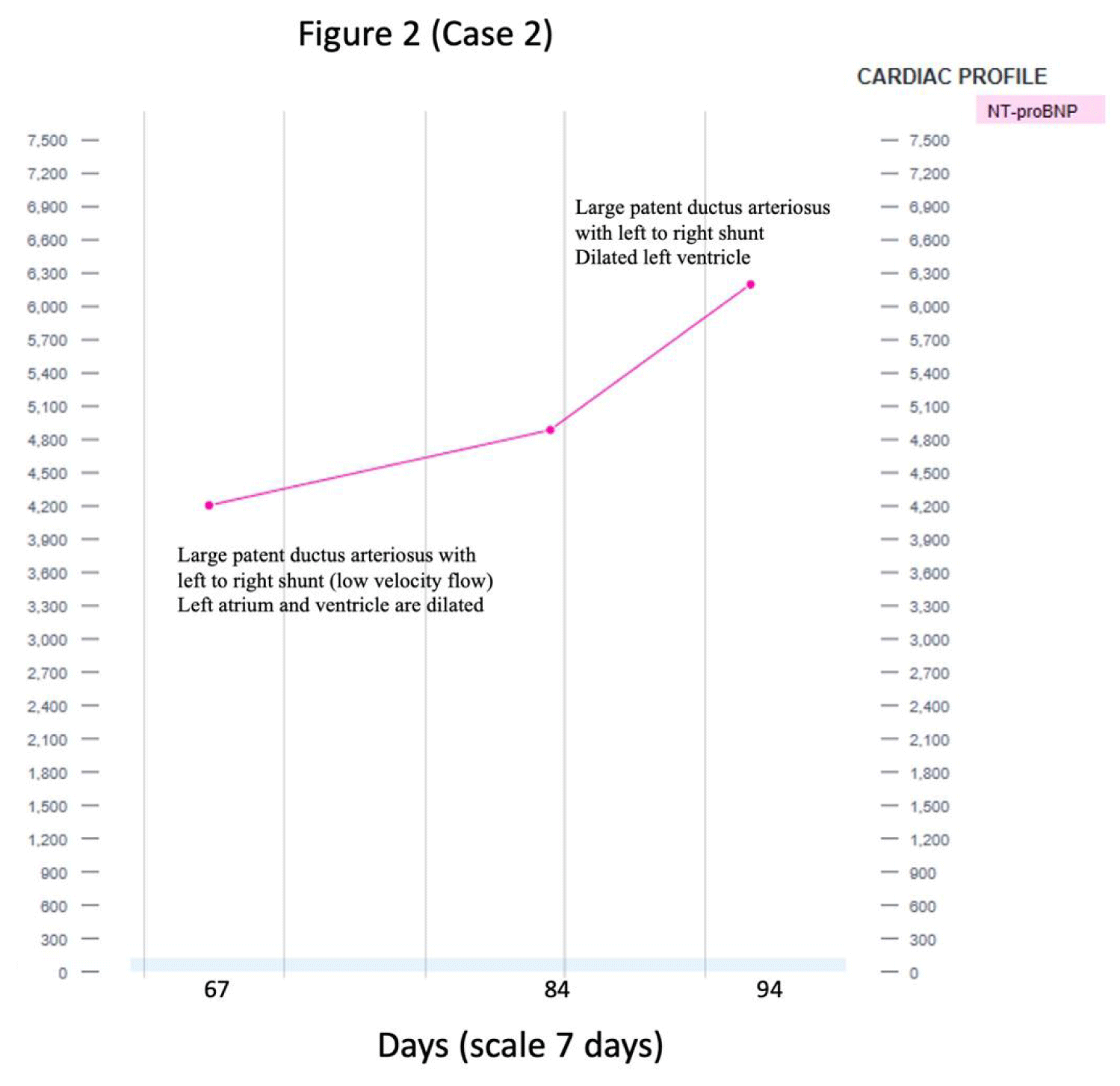
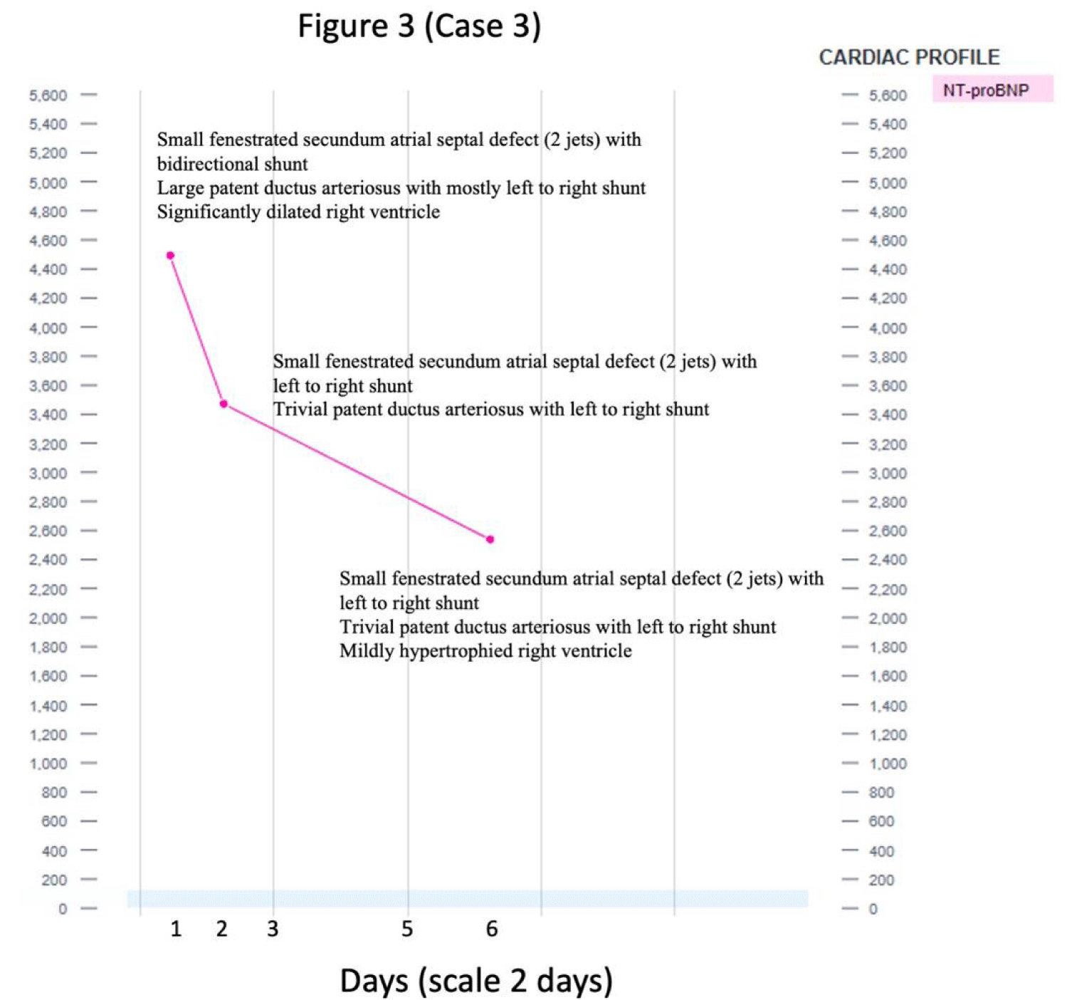
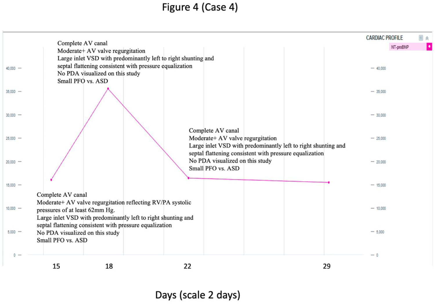
For this article, we examined four cases in which the data on simultaneously done NT-proBNP and echocardiogram was available (Figures 1-4). These cases will aid in highlighting the cost-benefit ratio of simultaneously obtaining the cardiac studies and examine the necessity of this practice.
Cases
In case 1, the NT-proBNP trended up from 4,600 to 8,100 pg/mL while echocardiogram showed improvement in PDA. In brief, the patient was a preterm infant born at 255/7 weeks admitted for extreme prematurity. The first echocardiogram (echo) obtained on day 5 of life demonstrated moderate PDA with the left to right shunting and PFO with the left to right shunt. Repeat echo on day 21 of life demonstrated persistent PDA with large left to right shunt and small left to right atrial shunt. On days 46, 59, and 66 of life, repeat echo showed a moderate sized PDA with low velocity left to right shunt suggesting increased pulmonary artery pressures. On day 72 and 79 of life, PDA and PFO were observed, as was elevated RV systolic pressures based off bidirectional PDA and septal flattening on echo. BNP values on days 66, 72, and 79, were 4,700, 8,120, and 6,277 (Figure 1). In this case, Sildenafil was started on day 79 of life for pulmonary hypertension; BNP and echo portrayed identical information of persistent cardiac abnormality. As noted in (Figure 1) that a rise in NT-proBNP level was not consistent with the echocardiogram. PDA was smaller in the later echocardiogram despite doubling of the NT-proBNP.
In case 2, the NT-proBNP trended up from 4200 to 6300 with no major changes in the cardiac functions (Figure 2). In summary, the patient was a preterm infant born at 231/7 weeks admitted for extreme prematurity. First echo on day 2 of life demonstrated large PDA with bidirectional shunting, increased RV systolic pressure, and moderate Secundum ASD. Repeat echo on day 11 of life demonstrated persistent PDA. Following this report, the patient was initiated on acetaminophen for 6 days for PDA closure. In this case, the echo was used as a supplemental guide for aggressive treatment of PDA. In addition, biweekly echos were conducted, and it was concluded that the baby would require surgical intervention. Patient’s BNP, first measured on day 64 of life, was used for the evaluation of continued LV + LA dilation and volume status. An echo was also ordered for day 64 which showed dilated LA + LV and large PDA with shunt. BNP continued to trend up with no change in intervention. The final echo, following closure of PDA, demonstrated PDA device in good position with minimal residual shunting and mildly dilated LV. As noted in (Figure 2), there was a rise in NT-proBNP level; however, the echo findings remained unchanged.
In case 3, the NT-proBNP trended down from 4500 to 2400 with no significant change in the cardiac functions (Figure 3). In brief, the patient was a term infant born at 376/7 weeks with prenatal ultrasound suggesting a dilated RV and VSD admitted for respiratory distress. First echo, on day 1 of life, demonstrated a small fenestrated secundum ASD with bidirectional shunt, a small apical muscular VSD with low velocity right to left shunt, a large PDA with mostly left to right shunt, and a significantly dilated right ventricle which was concerning for peripheral/cerebral arterio-venous malformation. Follow-up liver and head ultrasounds were normal. Echo on day 4 of life showed stable ASD, a trivial PDA with left to right shunt but some right to left at peak systole, and a mildly hypertrophied right ventricle. Repeat echo on day 11 showed no significant changes. BNP corresponded with improving echo results, with 4493, 2538, and 1758 on days 3, 7 and 11 of life, respectively. As note in (Figure 3), even with the drop in NT-proBNP levels, the echocardiogram findings remained unchanged.
In Case 4, a term neonate with Trisomy 21, the NT-proBNP on day 15 of life was noted to be 15523 with the echocardiogram showing complete AV canal, moderate AV valve regurgitation reflecting RV/PA systolic pressures of at least 62 mm Hg, large inlet VSD with predominantly left to right shunting and septal flattening consistent with pressure equalization and no PDA with small PFO vs. ASD. A follow up NT-proBNP on day 18 of life was noted to be 16460 with similar echocardiogram findings (Figure 4). NT-proBNP on day 22 trended up by 50% with no change in echocardiogram findings. As noted in (Figure 4) NT-proBNP levels had a rise and fall without congruent changes in echo findings.
Discussion
These cases provided some evidence that NT-proBNP did not provide any additional valuable information on the cardiac status of the infant when simultaneously obtained with echocardiogram. To further explore the relationship between NT-proBNP in conjunction with echocardiography, we will discuss the utility and limitations of both NT-proBNP and echo as well as the cost benefit analysis.
The Utility and Limitation of NT-proBNP
NT-proBNP measurements have been used widely for the evaluation of cardiac stress due to multiple conditions [8]. These measurements are considered strong predictors of adverse cardiac events. Recent research has shown that NT-proBNP can be used to guide management of cardiac patients and determine the effectiveness of the interventions to ultimately improve patient outcomes. For example, some institutions use BNP levels as a guide on how aggressively to treat heart failure in adults; i.e. higher BNP levels correspond with more aggressive treatment while down trending results predict clinical improvement. Therefore, BNP levels may be used as a prognostic value [8]. In neonates, BNP/NT-proBNP values are used to diagnose shunts and determine need for intervention in cases of Patent Ductus Arteriosus (PDA) [9]. Furthermore, in select children, NT-proBNP values predict disease severity and are positively correlated with echo evaluations of severity [9].
Even though NT-proBNP values have wide ranged merits, there are limitations to such measurements. BNP concentrations cannot be seen as a measure of cardiac function only. Multiple other disease processes such as liver cirrhosis, pulmonary disorders, endocrine disorders, obesity, anemia, etc. can cause elevations in NT-proBNP [9]. In a recent study in adults, NT-proBNP levels were shown to have a wide range depending on pathophysiology and hemodynamics in both healthy and cardiac patients [8]. Thus, higher values could be seen in healthy patients and lower values in sick patients. When evaluating hemodynamic instability, research suggests that a single NT-proBNP was not sufficient to predict need for mechanical support [9]. While the trends are reported, the field is still lacking solid data to support these theories regarding the utility of obtaining BNP in addition to conventional measures. Due to the sparsity of pediatric research on the topic, it is suggested that the utility BNP/NT-proBNP be limited to the evaluation and monitoring of children with previously diagnosed heart disease [9].
Moreover, NT-proBNP values depend on age. In neonates, the plasma NT-proBNP levels will be considerably high during the first 96 hours and then rapidly decline over the first week. This reduction is typically slowly down trending throughout the first month of life [9]. NT-proBNP concentrations can be affected by multiple gestations (i.e. twins), maternal type 1 diabetes, prematurity, intrauterine growth restrictions, c section following the onset of natural labor, and stress in the antenatal period [9].
The Utility and Limitations of Echocardiography
Echocardiography can be used to monitor the hemodynamic status of an infant and the aid in the management of a PDA [10]. Data from the echo such as volume status and cardiac ejection fraction can aid in the treatment of hemodynamic instability and shock in neonates. In a recent study examining the utility of neonatal echo, approx. 41.5% of echocardiograms performed in the NICU resulted in direct changes in management [10]. However, this contribution was dependent on the timing of the echo. The most management changes guided by echo results were made in the first 7 days of life. Following the first week, the echo results led to less management changes [10]. Echo results can be used as a guide for initiating, repeating, and discontinuing drug administration for PDA treatment.
However, echocardiography has limitations as well. Timing for echo is critical. After day 7 of life, most echos did not result in a change to therapeutic management. For cardiac output measurements, it is usually assumed that left ventricular output is a reliable surrogate for cardiac output. However, in neonates with a PDA, LV output cannot be used as a surrogate. When evaluating hemodynamic instability in the NICU, it is imperative that congenital heart disease is ruled out; however, this is no easy feat. Echo requires technologists who are specifically trained and closely collaborate with cardiology. Due to the need for specially trained techs, interobserver variability is another hurdle in evaluation echos [11].
Cost-Benefit Analysis
In many institutions, it has become cultural and even advised practice to trend BNP/NT-proBNP measurements and echocardiograms. In the days of handheld ultrasound devices and need for judicious use of otherwise costly resources, the simultaneous collection of NT-proBNP levels and echocardiography should be questioned. Echocardiography costs substantially more than BNP levels. For example, in the US, an echo may cost $1,000-$3,000 while a NT-proBNP measurement costs approximately $115. NT-proBNP measurements could significantly reduce the number of echocardiograms and reduce the work up of patients (adult and pediatric) with suspected cardiac anomalies/failure. In doing so, this would also substantially decrease the cost of the patient’s diagnostic work up and care [12]. Other studies have demonstrated that the use of NT-proBNP as a guide for treatment has reduced the time spent inpatient, amount of patients admitted, and the total medical costs [13]. Furthermore, it is suggested in recent research that obtaining NT-proBNP could negate the need for an echo because pathologic findings would be rare in those with normal NT-proBNP levels. Therefore, by implementing a strict NT-proBNP strategy, healthcare institutions could minimize the need for echocardiograms and extensive diagnostic work up which would not only reduce costs but also improve clinical outcomes for patients [12,13].
Conclusion
In conclusion, although NT-proBNP used in conjunction with echocardiogram and clinical evaluation can be an effective in monitoring neonatal cardiac hemodynamic status, using NT-proBNP and echocardiogram at the same time is not necessary. Following clinical suspicion and initial echocardiogram, NT-proBNP can be used to trend the infant's status and guide treatment. As our sample size was small, further research using a multicenter trial would be needed to confirm our conclusion. A study such as that could lead to the development of institutional guidelines benefiting both the hospital and patient alike.
Conflict of Interest Disclosures
The authors have no conflicts of interest relevant to this article to disclose.
Contributor Statements
Dr. Turnbough and Dr. McKenna conceptualized the study concept in collaboration with Dr. Manzar. Each wrote and reviewed the manuscript.
References
- Hall C. Essential biochemistry and physiology of (NT-pro) BNP. Eur J Heart Fail. 2004 Mar 15;6(3):257-60. doi: 10.1016/j.ejheart.2003.12.015. PMID: 14987573.
- Clerico A, Giannoni A, Vittorini S, Passino C. Thirty years of the heart as an endocrine organ: physiological role and clinical utility of cardiac natriuretic hormones. Am J Physiol Heart Circ Physiol. 2011 Jul;301(1):H12-20. doi: 10.1152/ajpheart.00226.2011. Epub 2011 May 6. PMID: 21551272.
- El-Khuffash AF, Amoruso M, Culliton M, Molloy EJ. N-terminal pro-B-type natriuretic peptide as a marker of ductal haemodynamic significance in preterm infants: a prospective observational study. Arch Dis Child Fetal Neonatal Ed. 2007 Sep;92(5):F421-2. doi: 10.1136/adc.2007.119701. PMID: 17712194; PMCID: PMC2675377.
- Choi BM, Lee KH, Eun BL, Yoo KH, Hong YS, Son CS, Lee JW. Utility of rapid B-type natriuretic peptide assay for diagnosis of symptomatic patent ductus arteriosus in preterm infants. Pediatrics. 2005 Mar;115(3):e255-61. doi: 10.1542/peds.2004-1837. Epub 2005 Feb 1. PMID: 15687418.
- Attridge JT, Kaufman DA, Lim DS. B-type natriuretic peptide concentrations to guide treatment of patent ductus arteriosus. Arch Dis Child Fetal Neonatal Ed. 2009 May;94(3):F178-82. doi: 10.1136/adc.2008.147587. Epub 2008 Nov 3. PMID: 18981033.
- Nuntnarumit P, Chongkongkiat P, Khositseth A. N-terminal-pro-brain natriuretic peptide: a guide for early targeted indomethacin therapy for patent ductus arteriosus in preterm Infants. Acta Paediatr. 2011 Sep;100(9):1217-21. doi: 10.1111/j.1651-2227.2011.02304.x. Epub 2011 May 5. PMID: 21457304.
- Rodríguez-Blanco S, Oulego-Erroz I, Alonso-Quintela P, Terroba-Seara S, Jiménez-González A, Palau-Benavides M. N-terminal-probrain natriuretic peptide as a biomarker of moderate to severe bronchopulmonary dysplasia in preterm infants: A prospective observational study. Pediatr Pulmonol. 2018 Aug;53(8):1073-1081. doi: 10.1002/ppul.24053. Epub 2018 May 23. PMID: 29790673.
- Cao Z, Jia Y, Zhu B. BNP and NT-proBNP as Diagnostic Biomarkers for Cardiac Dysfunction in Both Clinical and Forensic Medicine. Int J Mol Sci. 2019 Apr 12;20(8):1820. doi: 10.3390/ijms20081820. PMID: 31013779; PMCID: PMC6515513.
- Cantinotti M, Law Y, Vittorini S, Crocetti M, Marco M, Murzi B, Clerico A. The potential and limitations of plasma BNP measurement in the diagnosis, prognosis, and management of children with heart failure due to congenital cardiac disease: an update. Heart Fail Rev. 2014 Nov;19(6):727-42. doi: 10.1007/s10741-014-9422-2. PMID: 24473828.
- Harabor A, Soraisham AS. Utility of Targeted Neonatal Echocardiography in the Management of Neonatal Illness. J Ultrasound Med. 2015 Jul;34(7):1259-63. doi: 10.7863/ultra.34.7.1259. PMID: 26112629.
- Tissot C, Singh Y. Neonatal functional echocardiography. Curr Opin Pediatr. 2020 Apr;32(2):235-244. doi: 10.1097/MOP.0000000000000887. PMID: 32068595.
- Ferrandis MJ, Ryden I, Lindahl TL, Larsson A. Ruling out cardiac failure: cost-benefit analysis of a sequential testing strategy with NT-proBNP before echocardiography. Ups J Med Sci. 2013 May;118(2):75-9. doi: 10.3109/03009734.2012.751471. Epub 2012 Dec 12. PMID: 23230860; PMCID: PMC3633333.
- Moe GW, Howlett J, Januzzi JL, Zowall H; Canadian Multicenter Improved Management of Patients With Congestive Heart Failure (IMPROVE-CHF) Study Investigators. N-terminal pro-B-type natriuretic peptide testing improves the management of patients with suspected acute heart failure: primary results of the Canadian prospective randomized multicenter IMPROVE-CHF study. Circulation. 2007 Jun 19;115(24):3103-10. doi: 10.1161/CIRCULATIONAHA.106.666255. Epub 2007 Jun 4. PMID: 17548729.
Content Alerts
SignUp to our
Content alerts.
 This work is licensed under a Creative Commons Attribution 4.0 International License.


