> Medicine Group. 2021 October 30;2(10):1005-1014. doi: 10.37871/jbres1344.
-
Subject area(s):
- Neurology
- Brain Disorders
- Depression
- Mental Health
- Psychiatry
- Neurological Disorders
- Neurosurgery
Foville’s Syndrome a Case to Remember: Case Report
Sudikshya Acharya1, Ayush Chandra2,3, Avinash Chandra4* and Basant Pant1
2Tianjin Medical University, Tianjin, P.R. China
3Multiple Sclerosis Society Nepal, Kathmandu, Nepal
4National Academy of Medical Sciences, Bir Hospital, Kathmandu, Nepal
- Foville syndrome
- Infarction
- Pontine region
- Neurology
- Stroke
Abstract
The Foville’s Syndrome is a rare clinical feature of stroke or brain hemorrhage. This is very rare brain stem syndrome and only few cases have been reported worldwide. A case of Foville's syndrome secondary to infarction at the left paramedian pontine region, which was diagnosed and treated at Annapurna Neurological institute and allied Science, Kathmandu, Nepal. A 62 years old gentleman presented with acute headache with sudden onset of vertigo, tinnitus, slurred speech, difficulty while swallowing and numbness and hemiparesis on the right side of the body. The aim of this study was to report a rare case of Foville's syndrome with the infarction at the left paramedian pontine region. The clinical manifestations were well correlated with anatomical involvement. The CT-scan of head, Magnetic Resonance Imaging (MRI), MR-Angiogram (MRA) sequence of cerebral and carotid, etc. helped in the diagnosis of the case along with the other lab investigations.
Introduction
The Foville’s Syndrome is a rare clinical feature of stroke or brain hemorrhage. It is characterized by sixth nerve palsy, facial palsy, facial hypoesthesia, peripheral deafness, Horner’s syndrome, contralateral hemiparesis, ataxia, pain, and thermal hypoesthesia, with lesions in the pontine tegmentum [1]. Very few cases have been reported worldwide. Foville’s syndrome was first described by the French anatomist and psychiatrist Achille-Louis François Foville in 1858 [2]. The aim of our study is to report a rare case of Foville's syndrome secondary to infarction at the left paramedian pontine region, which was diagnosed and treated at Annapurna Neurological institute and allied Science, Kathmandu, Nepal.
Case Presentation
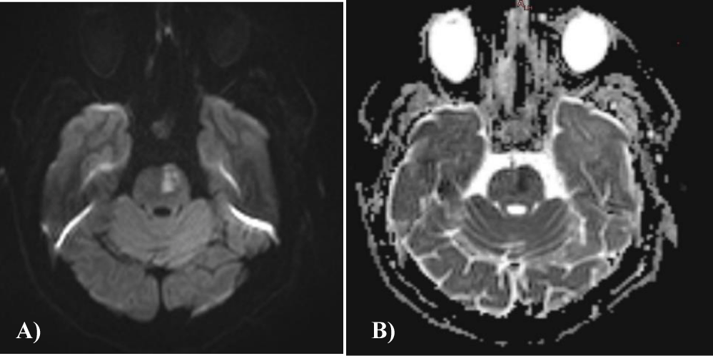
A 62-year-old male presented to the emergency department with a complaint of acute headache with sudden onset of vertigo, tinnitus, slurred speech, difficulty while swallowing and numbness and hemiparesis on the right side of the body. He denied any history of systemic disease, trauma or alcohol consumption. The patient also had complaint of pain in right eye without having any other visual complaints. The results of ophthalmological examination were found normal. On neurological examination with muscle power assessment (MRC Scale) revealed that motor power being 4/5 on limbs on the right side. Hemiparesis also involved right side of face with loss of vibratory sense. On further examination of cranial nerve, which showed patient’s gag reflex was absent on right side. In order to support the diagnosis of this case, several radiological investigations were performed such as CT-scan of head which showed acute infarction at left paramedian pontine region. The Magnetic Resonance Imaging (MRI) DWI/ADC mapping showed small focal diffusion restriction noted in left paramedian pontine (Figure 1A & 1B).
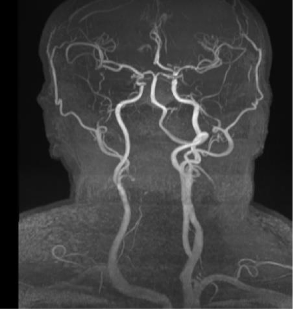
Further, MR-Angiogram (MRA) sequence of cerebral and carotid was done, which showed, slightly reduced caliber of the right Internal Carotid Artery (ICA) throughout its course with 60% of focal stenosis at right petro-cavernous junction as well as stenosed vertebral artery (right more than left) (Figure 2). According to the clinical manifestations and neuroimaging findings, Foville’s syndrome with contralateral hemiparesis resulting from the left paramedian pontine region infarction was diagnosed. The patient was admitted and received supportive treatment. Also, rehabilitation techniques were used to address residual functional deficit through physiotherapy and acupuncture. Serial laboratory investigations including Complete Blood Count (CBC), electrolyte, prothrombin time, activated partial thrombin time, protein C, protein S, urine catecholamine, antinuclear antibody, antiphospholipid antibody, cardiolipin antibody and thyroid function were conducted and their results came in as normal range except for mildly elevated Erythrocyte Sedimentation Rate (ESR) i.e. 65 mm/hr.
Discussion
The foville’s syndrome or defoville’s syndrome was first described by the French anatomist and psychiatrist Achille-Louis Francois Foville in 1858 [2,3]. The characters of this syndrome is sixth nerve palsy, facial palsy, facial hypoesthesia, peripheral deafness, Horner’s syndrome and contralalteral hemiparesis, ataxia, pain and thermal hypoesthesia. The syndrome suggested a lesion in lower pontine tegmentum, which has been reported in association with pontine infarction, hemorrhage, tuberculoma and cerebellar tumors [4,5]. In another variation of this syndrome, superior pons type of foville’s syndrome had also been proposed and been reported in association with the upper pontine region [6], this occurs in association with the upper pons and is caused by aneurysm of basilar artery [7].
In this patient, he had complained of pain on the right eye and had limited abduction and adduction, suggestive of involvement of the right abducens fascicle and medial longitudinal fasciculus, which were different from the classical foville’s syndrome. The patient also presented with the right facial palsy, right facial aesthesia, right hemiparesis and right hemiparaesthesia which represent the involvement of the facial nerve fascicle, trigeminal tract, uncrossed corticosipinal tract and uncrossed spinothalamic tract. Ipsilateral was noted while in emergency department and later in Hospital Outpatient Department (HOD) during the follow-up. Hence, the patient was suggested to be monitored for the possible HOD-associated movement disorders in the future, if need then should be rushed to the HOD.
Conclusion
We would like to conclude, as we reported a rare case of Foville’s syndrome with contralateral hemiparesis due to infarction in left paramedian pontine region. The clinical manifestations were well correlated with anatomical involvement.
Acknowledgement
The authors like to acknowledge the patient and her guardian for providing the required information and consent for this case report.
Authors contribution
All the authors have equally contributed from the time of handling this case to the manuscript formation.
Ethics approval and consent to participate
Ethical approval was taken from Institutional Review Board (IRB) of Annapurna Neurological Institute and Allied Sciences (ANIAS), Kathmandu Nepal. Consent to participation was collected from the patient.
Consent for publication
Patient has provided Consent for publication of data.
Availability of data and material
All the data are available with the corresponding author and can be made available as per required.
References
- Rucker, Janet MD. Eye movement disorders. Journal of Neuro-Ophthalmology. 2008;28:361-362. doi: 10.1097/01.wno.0000342368.61290.8c
- Brogna C, Fiengo L, Türe U. Achille Louis Foville's atlas of brain anatomy and the Defoville syndrome. Neurosurgery. 2012 May;70(5):1265-73; discussion 1273. doi: 10.1227/NEU.0b013e31824008e7. PMID: 22072133.
- Foville A. Note suruneparalysiepeuconnue de certains muscle de l'oeil, etsa liaison avec quelques points de l'anatomieetlaphysiologie de la protuberance annulaire. Bull SocAnat (Paris). 1958;33:373-405.
- Bedi HK, Devpura JC, Bomb BS. Clinical tuberculoma of pons presenting as Foville's syndrome. J Indian Med Assoc. 1973 Aug 16;61(4):184-5. PMID: 4774797.
- Ahmed N, Riazn A, Shuaib A, Siddiqi Z. Foville's syndrome masquerading as Wernicke's encephalopathy. N Z Med J. 2006 Apr 21;119(1232):U1928. PMID: 16633387.
- Takase M, Saeki N, Oka N, Satoh A, Otaki M, Yamaura A. [Superior Foville syndrome after clipping of basilar bifurcation aneurysm--case report (author's transl)]. No Shinkei Geka. 1981;9(3):343-47. Japanese. PMID: 7242818.
- Nakaso K, Nakayasu H, Isoe K, Nakashima K, Takahashi K. [A case of dissecting aneurysm of the basilar artery presented as superior pons type of Foville's syndrome]. Rinsho Shinkeigaku. 1995 Sep;35(9):1040-3. Japanese. PMID: 8565344.
Content Alerts
SignUp to our
Content alerts.
 This work is licensed under a Creative Commons Attribution 4.0 International License.


