> Medicine Group. 2021 May 21;2(5):345-349. doi: 10.37871/jbres1240.
Basic Knowledge on Leprosy: A Short Review
Chamodika Lowe*
- Hansen’s disease
- Infectious
- Leprosy
- Mycobacterium leprae
- Reaction
- Virulence
Abstract
Leprosy is a chronic disease which is caused by the infection of Mycobacterium leprae bacterium and leads to neurological consequences. Regardless of its slow progressiveness, it is an exceptionally serious disease that is continuing to be a challenging health problem around the world. Early diagnosis and treatment is much important in controlling Leprosy. Already published articles and books on Leprosy have been studied and summarized in this review to present a basic understanding on pathogenesis, symptoms, diagnosis and treatment of Leprosy.
Introduction
Leprosy, also called as Hansen’s disease is a chronic infectious disease endemic in tropical countries, that mainly leads to skin and peripheral nerves damage, is caused by the bacterium Mycobacterium leprae. According to WHO (World Health Organization) leprosy is clinically classified as Paucibacillary (PB) if there are ≤ 5 skin lesions and/ or involvement of one nerve trunk only, and Multibacillary (MB) if > 5 skin lesions and/ or >1 nerve trunk involved. It’s a curable disease but early detection is required to prevent transmission and disability [1]. The immune balance alterations between the host and the pathogen result in Leprosy Reactions which are acute incidences affecting the skin and nerves primarily. These reactions which are mainly responsible for the morbidity and neurological disabilities might take place during the disease course, while treating or after treatments. Leprosy reactions are classified into two as Type 1 reaction (T1R/ Reversal/ downgrading) and Type 2 reaction (Erythema Nodosum Leprosum [ENL]) [1].
Type of organism
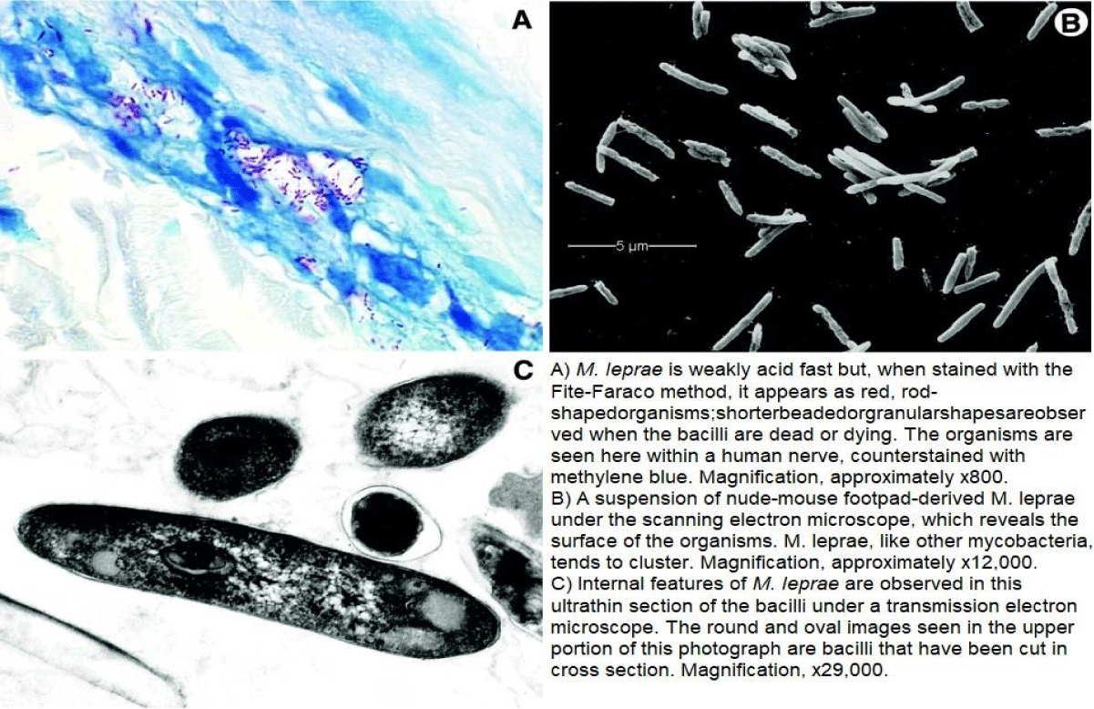
Causative organism, Mycobacterium leprae is a gram-positive, acid-fast-staining, microaerophilic, non-spore-forming, nonmortile bacterium with a straight rod or slightly curved structure [2] (Figure 1).
Transmission
Leprosy is transmitted from an infected to a normal by close, prolonged contact. Transmission is mainly through nasal mucosa and sometimes skin erosions [3].
Other possible transmission paths are blood, breast milk, insect bites and vertical transmission [1].
Pathogenesis
Since M. leprae only infects human and is unable to culture in vitro, research into pathogenesis has been limited. Leprosy bacillus low virulence together with effective host innate immunity may resist development of leprosy. M. leprae will be up taken by Dendritic Cells (DCs) and cause chemokines and cytokines to be produced, regulating inflammation and influencing cause of T-helper1 or T-helper2 response from adaptive cell-mediated immunity. M. leprae infected monocyte-derived DCs downregulates expression of MHC (Major Histocompatibility Complex) class I and II. Production of CD40 ligand-associated interleukin-12 and MHC II is upregulated by M. leprae membrane antigens stimulated DCs suggesting suppression of T-cells and DCs interaction by live bacilli [4].
Symptoms
Cardinal signs are macules which are hypopigmented/ erythematous with sensory loss, peripheral nerves that are thickened, adnexa loss at affected sites in a skin biopsy, positive acid-fast smear of skin [5]. In pure neural cases sensory or motor impairment of nerve/s can be the symptom.
Signs and Symptoms in reactional states
Type 1 reaction’s lesions are composed of the characteristic features erythema, hyperesthesia and edema, followed by scaling and at times ulceration. Neuritis and edema of extremities are seen often to be combined with this type of lesions [1]. Untreated acute neuritis will cause nerves to lose its function permanently leading to peripheral neuropathy [6].
Type 2 reaction results in subcutaneous inflammatory nodules or erythema multiforme lesions distributed symmetrically in any region. Its general symptoms include malaise, arthralgia, myalgia, lymphadenomegaly, edema and fever. There can also be neuritis, uveitis, orchitis, arthritis, dactylitis and kidney or liver damage like internal involvement causing systemic disorders [1,6].
Treatment
Currently leprosy is treated with Multidrug Therapy (MDT) where a combination of dapsone, clofazimine and rifampin are used and the regimens decided depending on the type of leprosy. Ofloxacin and minocycline is also used in treatment of Leprosy as second line drugs. For relapse assessment 10years follow-up is done. Some protection against M. leprae is also provided by BCG vaccine [2].
Treatment/management of reactions
Type 1 reaction is treated with the aim of easing pain, controlling acute inflammation and reversing eye and nerve damage. MDT treatment should be continued while using oral corticosteroids like prednisolone to treat neuritis or moderately inflamed skin plagues [6].
Type 2 reaction’s treatment is mainly targeted at immunosuppression. High doses of corticosteroids such as prednisolone are used in treating more severe case while thalidomide is used to control type 2 reactions and prevent recurrences. Though clofazimine and pentoxifylline have a less effectiveness than both prednisolone and thalidomide, those have also been used in treating type 2 reaction. Two other drugs used here in treating are colchicine and chloroquine [6].
Deformity and it’s Management
Leprosy causes neurological damages or neuropathy that leads to occurrence of lesions frequently. Hence, deformity and disability may result as secondary complications to this neuropathy through sensory impairment and loss of motor function [6].
Deformity and disability can be prevented by minimizing nerve damage through early identification of weakening in nerve function and quick introduction of steroid therapy. Any further damages to a damaged site must be prevented by resting the site and using of antibiotics to treat any secondary infections. Patients must be educated on self-examination, self-care, activities that can risk the damaged areas and protective footwear. Reconstructive surgery might be needed in contractures to improve function [6].
Prevention and control
Leprosy can be prevented by avoiding prolong contact of an infected. Can be controlled making the public aware of leprosy early signs to reduce fear for leprosy and to detect it early, through anti-leprosy campaigns, mass media, posters and stickers. Medical staffs can be trained to reduce fear on leprosy and early diagnose affected. Skin camps can be set up to detect and treat minor cases of skin disorders [7].
Epidemiology
Introduction of MDT in early 1980s has been able to markedly decrease the prevalence of leprosy. Yet, it can still be observed endemic in tropical countries which are specially, developing or underdeveloped. 105 such endemic countries with a high number of cases have been identified from Africa, America, Southeast Asia, Western Mediterranean and Eastern Pacific [1].
Though the elimination target set by WHO has been achieved by Sri-Lanka, there are still new leprosy cases (specially, child cases) being detected showing the presence of leprosy-bacilli in the environment.
Graph
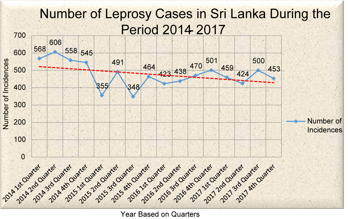
According to the trendline in Graph 1, number of leprosy cases tends be decreased with time. There’s an increment to be seen in 2nd quarters of 2014, 2015 and 2016. Highest number of incidences is seen in 2nd quarter of 2014 while lowest in 3rd quarter of 2015.
In 2014 there are higher rates of cases mostly of children and that shows active transmission of the disease. Several programs such as “Media Seminar on World Leprosy Day” have been conducted in 2014 to modify lifestyles promoting good health. A national action plan has been prepared and has been implemented in several districts via a “model leprosy programme”. Community screening program has been conducted island wide to detect new cases. Anti-leprosy campaigns have been conducted to increase awareness on leprosy and identify patients [9].
All these have led to decrease in new cases in 2015. National action plan implemented in 2014 for some districts has been spread to island wide in 2015. Also the other campaigns have been continued and increased detection rate [10].
Major problem for disease controlling has been identified as discrimination because of the disease and stigma. Main problem by 2016 has been higher MB leprosy cases and late presentation of child cases in high number. Anti-leprosy campaigns have been still continued [11].
As a prophylactic method of prevention pilot study on “Leprosy Postexposure Prophylaxis” (LPEP) was continued in 2017 together with other awareness programs [11].
Conventional Laboratory Diagnosis
Gold standard for leprosy diagnosis is examination of paraffin embedded, neutral buffered formalin fixed sample of full-thickness skin biopsy taken from advancing margin of an active lesion. Here host response histological patterns can be identified in sections stained with hematoxylin and eosin, acid-fast bacilli present in nerves can be identified with carbol fuchsin satain modified Fite-Faraco. Differential diagnosis is need where bacilli are rare [12].
Slit-skin smear is an ancillary procedure used to enumerate semi-quantitatively the acid fast organisms found in infected skin. This is used for follow ups in patients during treatments and after. Only experienced technicians will be able to give reliable results [12].
There are no serological tests for laboratory diagnosis of leprosy but several immunoassays are present to identify the antibodies against M. leprae’s PGL-1 (phenolic glycolipid 1). But these don’t have a sensitivity and specificity for a satisfactory level. Yet can be done for a low cost [12].
Lepromin test is a type of skin test used to test the ability in generation of granulomatous response for mycobacterial antigens. Response development can take time from 48-72 hours to 4 weeks after heat-killed M. leprae is injected intradermally. This test is not specific to leprosy but a negative response is associated with leprosy while positive response associates with bacilli elimination in leprosy [12].
Recent Advances in Diagnosis
Polymerase Chain Reaction (PCR) is a technique used to identify leprosy by amplifying M. leprae-specific sequences and identifies its fragments of DNA. PCR is also used in drug resistance development during treatment. 16S rRNA used for RNA analysis and reverse transcription -PCR have benefited in checking post treatment viability. Clinical and histopathological leprosy diagnosis is provided by PCR. It has a 100% specificity and sensitivity ranging 34 – 80% in PB patients and greater than 90% in MB patients. But this method needs experts to carryout PCR and it is an expensive method [12].
Mass spectrometry- Skin lesion biopsies are used in this method to identify lipid markers [4].
Immunohistochemical Reaction - Monoclonal or polyclonal antibodies is used to detect antigens of leprosy bacilli in this method. Confirmatory tests have to be done since false positive values and false negative valves can appear [1].
Prevention and control
Leprosy can be prevented by avoiding prolong contact of an infected. Can be controlled making the public aware of leprosy early signs to reduce fear for leprosy and to detect it early, through anti-leprosy campaigns, mass media, posters and stickers. Medical staffs can be trained to reduce fear on leprosy and early diagnose affected. Skin camps can be set up to detect and treat minor cases of skin disorders [7].
Quality Control and Quality Assurance
The two parts of quality management are Quality control and quality assurance. And it is also known as the most important fundermentals of laboratory measurements. When quality control fulfill the requirements of quality, quality assurance ensure whether the quality requirements are fulfilled. The key quality systems is constitute with quality control and quality assurance. The guidance of quality assurance and control is the Standard Operation Procedure (SOP). The quality system should be commensurate the business model and business objectives. The commitment and its active involvement of the top management are important to ensure the effectiveness and efficiency, suitability and adequacy of the quality systems [7].
The resources that are most important when improving quality is constitute by the company employees. Every unit employees are responsible to ensure the improvement and efficiency of their work. The motivating environment and the training should be provided by the top management towards the employees to improve their processes. At the end, the quality of the goods and services are a responsibility of everyone [7].
The main sections of quality assurance and quality control are Pre analytical phase, Analytical phase and Post analytical phase [7].
Storage, Sample Collection and Transport is included in Pre analytical phase. As important errors can be occurred in Pre analytical phase so to avoid errors and problems Pre analytical phase should have rigorous control measures [7].
Analytical phase is which sample analysis is occurring at a certain time. Here usually considered laboratory testing, diagnostic procedures, products and processes which ultimately give results. The errors can be reduced by the quality control internally and calibrating of the laboratory equipments [7].
Making of the final report is included in Post - analytical phase. Those reports must be issued to the relavent patients with the correct results as they are important when treating to the patients. And interpretation must also be done in this Post - analytical phase [7].
References
- Lastória JC, Abreu MA. Leprosy: review of the epidemiological, clinical, and etiopathogenic aspects - part 1. An Bras Dermatol. 2014 Mar-Apr;89(2):205-18. doi: 10.1590/abd1806-4841.20142450. PMID: 24770495; PMCID: PMC4008049.
- Scollard DM, Adams LB, Gillis TP, Krahenbuhl JL, Truman RW, Williams DL. The continuing challenges of leprosy. Clin Microbiol Rev. 2006 Apr;19(2):338-81. doi: 10.1128/CMR.19.2.338-381.2006. PMID: 16614253; PMCID: PMC1471987.
- Bhat RM, Prakash C. Leprosy: an overview of pathophysiology. Interdiscip Perspect Infect Dis. 2012;2012:181089. doi: 10.1155/2012/181089. Epub 2012 Sep 4. PMID: 22988457; PMCID: PMC3440852.
- Gulia A, Fried I, Massone C. New insights in the pathogenesis and genetics of leprosy. F1000 Med Rep. 2010 Apr 27;2:30. doi: 10.3410/M2-30. PMID: 20948855; PMCID: PMC2948396.
- Eichelmann K, González González SE, Salas-Alanis JC, Ocampo-Candiani J. Leprosy. An update: definition, pathogenesis, classification, diagnosis, and treatment. Actas Dermosifiliogr. 2013 Sep;104(7):554-63. doi: 10.1016/j.adengl.2012.03.028. Epub 2013 Jul 17. PMID: 23870850.
- Walker SL, Lockwood DN. The clinical and immunological features of leprosy. Br Med Bull. 2006;77-78:103-21. doi: 10.1093/bmb/ldl010. Epub 2006 Nov 7. PMID: 17090777.
- Dewapura DR. Leprosy control in Sri Lanka. World Health Forum. 1994;15(2):173-4. PMID: 8018284.
- Epidemiology unit (2017) Epidermiological Bulletin. Sri Lanka Epidemiology Unit Ministry Of Health. Medical Statistics Unit (2016) Annual Health Bulletin 2014.
- Medical Statistics Unit (2016) Annual Health Bulletin 2014. Sri Lanka Ministry of Health, Nutrition and Indigenous Medicine.
- Medical Statistics Unit (2017) Annual Health Bulletin 2016. Sri Lanka Ministry of Health, Nutrition and Indigenous Medical services.
- Medical Statistics Unit (2017) Annual Health Bulletin 2017. Sri Lanka Ministry of Health, Nutrition and Indigenous Medical services.
- Scollard DM, Adams LB, Gillis TP, Krahenbuhl JL, Truman RW, Williams DL. The continuing challenges of leprosy. Clin Microbiol Rev. 2006 Apr;19(2):338-81. doi: 10.1128/CMR.19.2.338-381.2006. PMID: 16614253; PMCID: PMC1471987.
Content Alerts
SignUp to our
Content alerts.
 This work is licensed under a Creative Commons Attribution 4.0 International License.


