> Medicine Group. 2021 Feb 19;2(2):047-051. doi: 10.37871/jbres1188.
Brain Abscess of Otorhinolaryngologic Origin: Study of 80 Cases in the Neurosurgery Department of Conakry University Hospital Center
Barry Louncény Fatoumata1*, Souare Ibrahima Sory1, Atakla Hugues Ghislain1, Conde Alpha Youssouf1, Djibo Hamani Abdoul Bachir2, Conde Mohamed Lamine2, Youla Seny1, Cisse Fodé Abass2 and Cisse Amara2
2Neurology Department, Conakry University Hospital Center, Conakry, Guinea
- Abscess
- Otorhinolaryngology
- Antibiotic therapy
- Neurosurgery
- Conakry
Abstract
Introduction: Brain abscesses are serious conditions that can be life-threatening if left untreated. The objective of our study was to determine the epidemiological, clinical, paraclinical, therapeutic and evolutionary characteristics of cerebral abscesses of otorhinolaryngological origin in our department.
Methods and Materials: This was a retrospective study of 80 cerebral abscess files of otorhinolaryngological origin collected over a period of 5 years (January 2014-December 2018) at the neurosurgery department of Conakry University Hospital Center.
Results: Abscesses of otorhinolaryngological origin represented 72% of all abscesses. The mean age was 14.7 years with a sex ratio of 4. The clinical picture was dominated by fever (92%), focal signs (55%) and intracranial hypertension (46%). The entrance door was 84% sinus. The frontal site was predominant, 44 cases. Eighty-two percent of patients underwent surgery and 18% were treated with antibiotic therapy alone. The evolution was favorable in 75% of the cases with a mortality rate of 15%.
Conclusion: Brain abscesses are a medical-surgical emergency. The forms of otorhinolaryngologic origin are dominated by sinusitis. Despite the therapeutic difficulties, the prognosis remains acceptable in our study, 15% of deaths.
Introduction
Cerebral abscesses are purulent collections neoformed in the cerebral parenchyma [1]. They are serious and life-threatening conditions that can be life-threatening without adequate treatment [2]. Otolaryngologic (ORL) infections are one of the main causes, especially in older children and adolescents [3-6].
In the developing countries of the South, particularly in Black Africa, brain abscesses remain an ongoing problem [7,8]. In terms of diagnosis, modern imaging techniques are still not available, and in terms of prognosis, the difficulties of treatment and the combination of complications inherent to a delay in diagnosis explain the seriousness of these suppurations [9]. We report our experience in order to evaluate this pathology from an epidemiological, clinical, paraclinical, therapeutic and evolutionary point of view in our department.
Materials and Methods
This is a descriptive retrospective study of 80 brain abscess files of otorhinolaryngologic origin collected over a period of 5 years (January 2014-December 2018) in the neurosurgery archives of Conakry University Hospital Center. The diagnosis was made by CT scan. Follow-up was 6 to 12 months. The parameters studied were frequency, age, sex, clinic, imaging, biology, treatment and progression. We included all patients with a brain abscess during the study period regardless of the entrance door and excluded all patients with a brain abscess of other than otorhinolaryngologic origin. Data were collected and analyzed using SPSS 21.0 software.
Results
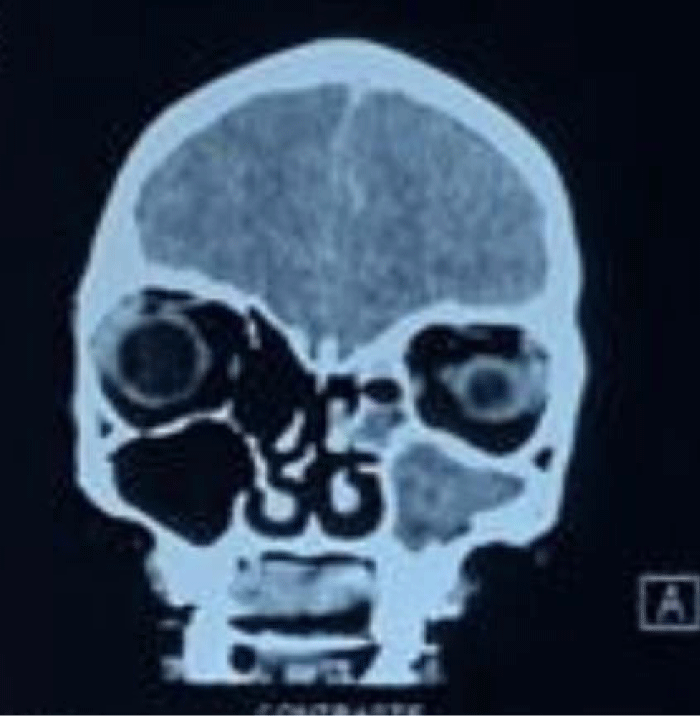
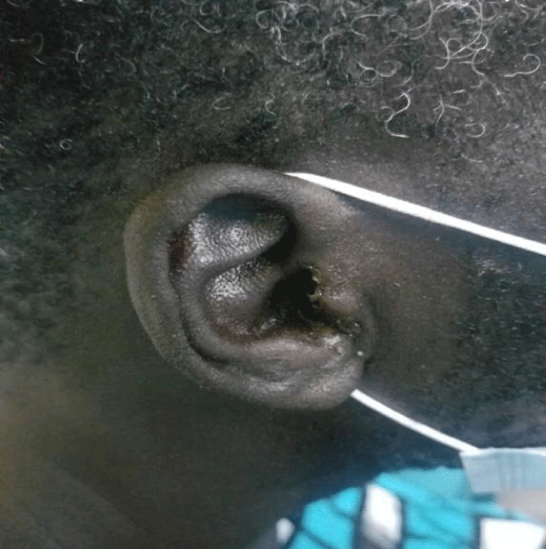
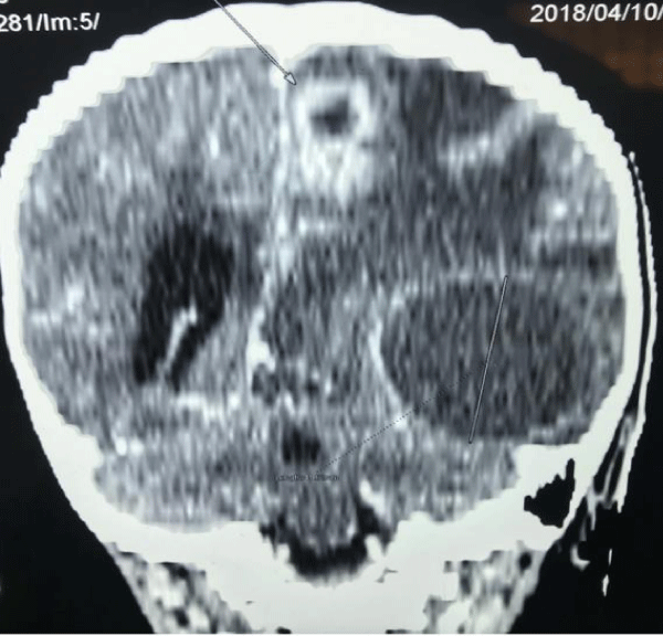
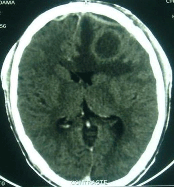
Out of 111 cases of cerebral abscesses collected during the study period, abscesses of otorhinolaryngologic origin represented 80 cases, i.e. 72%. The mean age was 14.7 years (Extremes: 3 months-87 years) with a sex ratio of 4. Sinusitis (Figure 1) was the most common cause of entry, 84%, followed by otitis (Figure 2) 11% and oto-mastoiditis 5%.
The time to diagnosis ranged from 3 days to 5 months. Bergman’s triad was complete in only 45% of cases, clinical data are summarized in (Table 1).
Biology showed hyperleukocytosis in 81%, a positive C-reactive protein in 83%.
Bacteriology was 80% negative; the germ was found in 16 cases (Table 2).
Table 1: Clinical Data. |
|
| Sign | Percentage |
| Intracranial hypertension | 46% |
| Bergman Triad | 45% |
| Fever | 92% |
| Comital crises | 24% |
| Disorder of consciousness | 23% |
| Focal sign | 55% |
Table 2: Bacteriological data. |
|
| Bacteriology | Staff |
| STAPHYLOCOQUES Aureus Saprophyticus Coagulase |
5 2 2 1 |
| Streptococcus pneumoniae | 8 |
| Klebsiella pneumoniae | 1 |
| Enterobacter spp | 1 |
| Proteus mirabilis | 1 |
| Negative bacteriology | 64 |
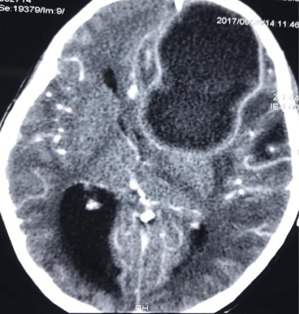
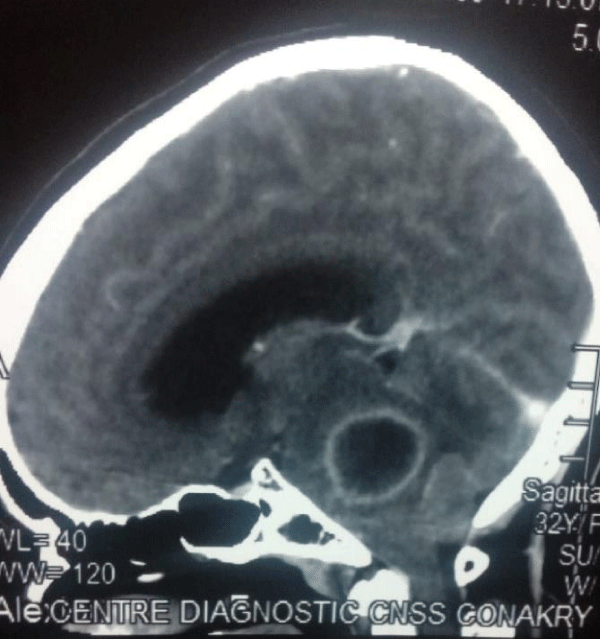
In terms of imaging, 44 abscesses were frontal localization (Figure 3), 21 temporal localization, 8 parietal localization, 1 occipital localization, 2 located in the posterior fossa (Figure 4), multiple in 2 cases (Figure 5) and 2 cases of abscesses associated with the empyema (Figure 6).
Therapeutically, probabilistic tri-antibiotic therapy, combining a 3rd generation cephalosporin (100 mg/kg body weight), metronidazole (40 mg/kg body weight) and 44% combined with gentamycin (4 mg/kg body weight) or thiamphenicole in 29% (50 mg/kg body weight) was started for 3 weeks by parenteral route and continued oral route for 4 to 8 weeks. It was then adapted to the results of the antibiogram. The treatment was 18% medical exclusive for collections measuring less than 2 cm, in 82% of cases surgery was associated with a delay ranging from 24 hours to 3 months. For the majority of cases, it was performed within the first 24 hours, with 78 cases of trepano- punctures and 2 cases of flaps. It was justified on the basis of size and clinical tolerance criteria, particularly for consciousness disorders associated with a compressive collection. The entrance door was treated surgically in 59% of cases, and a wash-drainage device (Albertini drain or endoscopic approach) was put in place in cases of sinusitis through an average meatotomy for 24 to 36 hours. Anticonvulsant treatment (24%), corticosteroid therapy (23%) for intracranial hypertension associated with cerebral edema and heparin therapy (3%) for cerebral thrombophlebitis were also associated. The evolution was marked by recovery without sequelae in 75% of cases, recovery with sequelae in 8% (hemiparesis, comital seizures, convergent strabismus, monocular blindness) of cases, 2% of recurrence, requiring re-puncture and 15% of death.
Discussion
The management and prognosis of brain abscesses have evolved considerably in recent decades with the advent of CT scanning in Guinea, allowing for early diagnosis and a more precise and less aggressive therapeutic procedure.
The frequency of abscesses varies with the level of health and economic development of the regions [7].
The frequency of this pathology is certainly underestimated in developing countries, particularly in sub-Saharan Africa, due to the under-equipment in medical imaging. In Senegalese work at all ages, it was significantly lower than that of other cerebro-meningeal infections such as malaria, purulent meningitis and HIV-associated cryptococcosis [10].
The male predominance found here is classic regardless of the doorway and type of intracranial suppuration [11,12]. Age (Mean: 14.7 years) is consistent with the literature [13-15] and is explained by the higher incidence of sinus and otomastoid infections at this time of life [12,13].
Clinically, fever is inconstant [2,9], present in 92% of our series ranging from 39 to 40°C. Intracranial hypertension varies from 69 to 100% of cases [7], 46% in our series. It is thought to be related to underlying cerebral edema and upper sagittal sinus thrombophlebitis [16]. Signs of focalization are present in 75 to 100% of patients [6,17] compared to 55% in our series. Comital attacks vary from 22.2 to 44% of cases [3,8] and 24% in our series. Consciousness disorders represent 23% in our series compared to 63% in a Senegalese series [14]. Delayed diagnosis would explain these consciousness disorders. Patients are most often admitted at a very advanced stage.
Staphylococcus and Streptococcus predominated in our series, which is similar to the series of Ouiminga, et al. [14] in Senegal but differs from other series where Streptococcus and Haemophilus influenzae represent 30 to 43% [18, 19]. Pus is sterile in 17 to 67% of cases [7,17] compared to 80% in our series. Antibiotic therapy and conditioning of the specimen in case of anaerobic germs can be incriminated [14].
The frontal site was predominant (71%), which is consistent with the data in the literature [20,21]. Although several concomitant abscesses are possible, the cerebral abscess is most often unique. The origin of the “mother” infection seems to condition the location of the abscess, except for dental abscesses. Thus, otologic infections are readily responsible for temporal or occipital abscesses. Sinus infections are most often responsible for the development of frontal abscesses [22].
The treatment is medico-surgical. The medical treatment is based on long-term antibiotic therapy with molecules that diffuse well into the cerebral parenchyma [21]. The antibiotic treatment will be probabilistic from the outset, then secondarily adapted according to the germ detected and the antibiotic susceptibility test. In the absence of a germ, the treatment will be empirical according to the clinical evolution of the patient. The combination of a third-generation cephalosporin with metronidazole seems to us an interesting choice both by venous and oral route. Americans prefer the combination of a third-generation cephalosporin (Ceftriaxone or cefotaxime) and thiamphenicole (1g intravenous every 6 hours) [22]. Intravenous treatment will be prescribed for 15 to 21 days, and the relays oral route for 15 days to 2 months [23]. In our practice, the protocol was 21 days for parenteral therapy and 4 to 8 weeks for oral route bi-antibiotic therapy. Corticosteroid therapy is indicated in cases of life-threatening intracranial hypertension [24] and severe edema, as was the case in 23% of our patients. An anticonvulsant treatment can be combined with antibiotic therapy for its entire duration [12,20] as was the case in 24% of our series. The entrance door must be systematically treated to avoid relapse and recurrence [14,25].
Surgically, the treatment of the abscess itself involves two possible techniques: puncture(s) - aspiration(s) with or without stereotactic guidance, and excision by craniotomy [2]. The choice of procedure will depend on the general condition of the patient, the size and location of the abscess [26]. In our series, all abscesses were treated by unguided puncture-aspiration using the Cushing’s trocar because of the lack of stereotaxis in our technical platform and the invasive nature of excision by craniotomy.
The evolution after treatment has improved considerably with the advent of antibiotics (80 to 100% mortality in the pre-antibiotic era) [27] but also in recent decades with the progress of medical imaging for early diagnosis. Today, a cure without sequelae is the rule in more than half of the cases: 46 to 78% depending on the series [18,19] and 75% in our series. Nevertheless, mortality remains significant: 15% in our series, 11 to 21% in the other series [18,27]. The possible neurological sequelae are variable: 8% of cases in our series, 8 to 33% in the other series [18,28]. They are classified as “minor” sequelae (Monoparesis, epilepsy, visual field deficit, minor behavioural disorders, etc.) and “major” sequelae (Hemiplegia, severe behavioural disorders, etc.) [12]. The timing of the operation is a more important factor than the surgical technique itself. The late surgery observed in our experience is related to diagnostic difficulties. Early diagnosis, adequate treatment and prompt surgery give the patient a better chance of recovery with few neurological complications [21].
Conclusion
Brain abscesses are a medical-surgical emergency. The forms of otorhinolaryngologic origin are dominated by sinusitis, and the difficulties of management from a diagnostic, therapeutic and economic point of view make the seriousness of this disease. Despite these hazards, the prognosis of our patients remains acceptable 15% of deaths. The combination of targeted antibiotic therapy and surgical treatment allows a cure with fewer after-effects.
Acknowledgment
The authors would like to thank the patients and their parents and the ethics committee for consenting to this study.
References
- Dakurah TK, Iddrissu M, Wepeba G, Nuamah I. Hemispheric brain abscess: a review of 46 cases. West Afr J Med. 2006 Apr-Jun;25(2):126-9. doi: 10.4314/wajm.v25i2.28262. PMID: 16918184.
- Emery E, Redondo A, Berthelot JL, Bouali I, Ouahes O, Rey A. Abcès et empyèmes intracrâniens: prise en charge neurochirurgicale [Intracranial abscess and empyema: neurosurgical management]. Ann Fr Anesth Reanim. 1999 May;18(5):567-73. French. doi: 10.1016/s0750-7658(99)80134-8. PMID: 10427394.
- Hakim HE, Malik AC, Aronyk K, Ledi E, Bhargava R. The prevalence of intracranial complications in pediatric frontal sinusitis. Int J Pediatr Otorhinolaryngol. 2006 Aug;70(8):1383-7. doi: 10.1016/j.ijporl.2006.02.003. Epub 2006 Mar 10. PMID: 16530852.
- Kombogiorgas D, Seth R, Athwal R, Modha J, Singh J. Suppurative intracranial complications of sinusitis in adolescence. Single institute experience and review of literature. Br J Neurosurg. 2007 Dec;21(6):603-9. doi: 10.1080/02688690701552856. PMID: 18071989.
- Osma U, Cureoglu S, Hosoglu S. The complications of chronic otitis media: report of 93 cases. J Laryngol Otol. 2000 Feb;114(2):97-100. doi: 10.1258/0022215001905012. PMID: 10748823.
- Passeron H, Sidy Ka A, Diakhaté I, Imbert P. Suppurations intracrâniennes à porte d’entrée otorhinolaryngologique chez l’enfant au Sénégal [Intracranial suppurations of otorhinolaryngological origin in children in Senegal]. Arch Pediatr. 2010 Feb;17(2):132-40. French. doi: 10.1016/j.arcped.2009.11.001. Epub 2009 Nov 29. PMID: 19951836.
- Kabré A, Zabsonré S, Diallo O, Cissé R. Prise en charge médico-chirurgicale des abcès du cerveau à l’ère du scanner en Afrique sub-saharienne: à propos de 112 cas [Management of brain abscesses in era of computed tomography in sub-Saharan Africa: a review of 112 cases]. Neurochirurgie. 2014 Oct;60(5):249-53. French. doi: 10.1016/j.neuchi.2014.06.011. Epub 2014 Sep 20. PMID: 25245921.
- Loembe PM, Okome-Kouakou M, Alliez B. Les suppurations collectées intra-crâniennes en milieu africain [Suppurative intracranial infections in Africa]. Med Trop (Mars). 1997;57(2):186-94. French. PMID: 9304016.
- Gueye M, Badiane SB, Sakho Y, Kone S, Ba MC, Kabre A. Abcès du cerveau et empyèmes extra cérébraux [Brain abscess and extracerebral empyema]. Dakar Med. 1991;36(1):82-7. French. PMID: 1842767.
- Soumaré M, Seydi M, Ndour CT, Fall N, Dieng Y, Sow AI, Diop BM. Profil épidémiologique, clinique et étiologique des affections cérébroméningées observées à la clinique des maladies infectieuses du CHU de Fann à Dakar [Epidemiological, clinical, etiological features of neuromeningeal diseases at the Fann Hospital Infectious Diseases Clinic, Dakar (Senegal)]. Med Mal Infect. 2005 Jul-Aug;35(7-8):383-9. French. doi: 10.1016/j.medmal.2005.03.009. PMID: 15975752.
- Gelabert-González M, Serramito-García R, García-Allut A, Cutrín-Prieto J. Management of brain abscess in children. J Paediatr Child Health. 2008 Dec;44(12):731-5. doi: 10.1111/j.1440-1754.2008.01415.x. Epub 2008 Nov 18. PMID: 19054291.
- Page C, Lehmann P, Jeanjean P, Strunski V, Legars D. Abcès et empyèmes intracrâniens d’origine O.R.L [Intra cranial abscess and empyemas from E.N.T. origin]. Ann Otolaryngol Chir Cervicofac. 2005 Jun;122(3):120-6. French. doi: 10.1016/s0003-438x(05)82336-9. PMID: 16142090.
- Migirov L, Duvdevani S, Kronenberg J. Otogenic intracranial complications: a review of 28 cases. Acta Otolaryngol. 2005 Aug;125(8):819-22. doi: 10.1080/00016480510038590. PMID: 16158527.
- H.A.K. Ouiminga, A.B.Thiam, N.Ndoye, H.Fatigba, M.Thioub, S.Memou, M. Gaye Sakho, A.Korchi, J.Mendy, M.C.Ba, S.B.Badiane. Les empyèmes intracrâniens : aspects épidémiologiques, cliniques, paracliniques et thérapeutiques. Étude rétrospective de 100 observations. Neurochirurgie 60 (2014) 299–303.
- Hakim HE, Malik AC, Aronyk K, Ledi E, Bhargava R. The prevalence of intracranial complications in pediatric frontal sinusitis. Int J Pediatr Otorhinolaryngol. 2006 Aug;70(8):1383-7. doi: 10.1016/j.ijporl.2006.02.003. Epub 2006 Mar 10. PMID: 16530852.
- Nathoo N, Narotam PK, Nadvi S, van Dellen JR. Taming an old enemy: a profile of intracranial suppuration. World Neurosurg. 2012 Mar-Apr;77(3-4):484-90. doi: 10.1016/j.wneu.2011.04.023. Epub 2011 Nov 7. PMID: 22120393.
- Broalet E, N’dri Oka D, Eholie SP, Guillao-Lasme EB, Varlet G, Bazeze V. Abcès et empyèmes intracrâniens chez l’enfant, observés à Abidjan. Afr J Neurol Sci 2002;21(1):38–41.
- Chakroun M, Abid F, Jmal A, Ben Romdhane F, Hattab MN, Bouzouaia N. Les abcès cérébraux. Etude de 24 cas. Médecine du Maghreb 2002;97:15-9.
- Donaldson G, Webster D, Crandon IW. Brain abscess at the University Hospital of the West Indies. West Indian Med J. 2000 Sep;49(3):212-5. PMID: 11076212.
- Albu S, Tomescu E, Bassam S, Merca Z. Intracranial complications of sinusitis. Acta Otorhinolaryngol Belg. 2001;55(4):265-72. PMID: 11859644.
- Younis RT, Lazar RH, Anand VK. Intracranial complications of sinusitis: a 15-year review of 39 cases. Ear Nose Throat J. 2002 Sep;81(9):636-8, 640-2, 644. PMID: 12353440.
- Leys D. Abcès cérébraux et empyèmes intracrâniens. Encyclopédie Médico-Chirurgicale 17-485-A-10. p.1-7.
- Heran NS, Steinbok P, Cochrane DD. Conservative neurosurgical management of intracranial epidural abscesses in children. Neurosurgery. 2003 Oct;53(4):893-7; discussion 897-8. doi: 10.1227/01.neu.0000084163.51521.58. PMID: 14519222.
- Kojima A, Yamaguchi N, Okui S. Supra- and infratentorial subdural empyema secondary to septicemia in a patient with liver abscess--case report. Neurol Med Chir (Tokyo). 2004 Feb;44(2):90-3. doi: 10.2176/nmc.44.90. PMID: 15018332.
- Ali A, Kurien M, Mathews SS, Mathew J. Complications of acute infective rhinosinusitis: experience from a developing country. Singapore Med J. 2005 Oct;46(10):540-4. PMID: 16172774.
- Dolan RW, Chowdhury K. Diagnosis and treatment of intracranial complications of paranasal sinus infections. J Oral Maxillofac Surg. 1995 Sep;53(9):1080-7. doi: 10.1016/0278-2391(95)90128-0. PMID: 7643279.
- Singh B, Van Dellen J, Ramjettan S, Maharaj TJ. Sinogenic intracranial complications. J Laryngol Otol. 1995 Oct;109(10):945-50. doi: 10.1017/s0022215100131731. PMID: 7499946.
- Maniglia AJ, Goodwin WJ, Arnold JE, Ganz E. Intracranial abscesses secondary to nasal, sinus, and orbital infections in adults and children. Arch Otolaryngol Head Neck Surg. 1989 Dec;115(12):1424-9. doi: 10.1001/archotol.1989.01860360026011. PMID: 2573380.
Content Alerts
SignUp to our
Content alerts.
 This work is licensed under a Creative Commons Attribution 4.0 International License.


