> Medicine. 2020 September 16;1(5):150-153. doi: 10.37871/jbres1134.
-
Subject area(s):
- Veterinary Science
- Toxicology
- Biology
Canine Myiasis and its Causal Agents in India
Amandeep Singh*
- Myiasis
- Pet dogs
- Dipteran larva
- Parasitosis
Myiasis, the invasion of tissues of live humans and vertebrate animals with dipteran larvae is common throughout the tropical regions of the world. It is a real welfare problem of worldwide distribution and a matter of great concern among medical and veterinary fields. The reports indicate that dogs are the most common canine species affected by myiasis. The present study was conducted to ascertain prevalence of myiasis among pet dogs and to identify myiasis-causing flies in India. The study resulted in the identification of three species of myiasis causing flies among pet dogs. The Old World Screw-worm fly- Chrysomya bezziana was reported to be the predominant fly species followed by Musca domestica and Sarcophaga ruficornis. The overall infestation rate was high in rainy season followed by summer, spring, autumn and least in winter. Incidence of myiasis was found to be higher among adult males than the females. The most frequently infested body regions were the face, neck and perianal region among males and face, neck and vulvar lips among females. Most infestations were found in the wounds at front body parts of the adult males, suggesting the common occurrence of myiasis in consequence of competitive fighting.
Myiasis is the infestation of live human and other vertebrate animals with dipterous larvae which at least for certain period feed on the host’s dead or living tissue, liquid body substances or ingested food [1]. The dipterous larvae, often called ‘maggots’, complete or at least for a certain period continue their normal development on or in the vertebrate body. The gravid flies are attracted to open wounds or natural body openings such as eye, ear, nose, vagina, anus etc. to lay their eggs. Oviposition is encouraged by foul smelling purulent discharge from diseased tissue which is followed by development of larvae. After hatching, the larva invades the broken skin and burrow in the dermal layers or pre-existing wounds resulting into enlargement of wounds [2]. They cause lesions and make the domestic animals deficient of blood, debilitated and in severe cases may result into death, if the vital organs like brain or lungs are invaded. The maggots irritate the animals; they do not feed properly and become poor in health. Parasitized animal often become restless and rubs the affected areas. Severe infestation may lead to death within few days probably due to septicaemia [1]. There is a widespread incidence of myiasis among domestic animals throughout the world in tropical countries including India which fulfills all the favorable conditions for the exuberant growth of myiasis causing flies and their larvae. Numerous reports are available on incidence of myiasis in domestic animals in various parts of the world. The earliest studies of myiasis among domestic animals had been reported from India from 1920 to 1977 [3-6]. Some of the studies from the other parts of world include sheep-strike in Great Britain by Lucilia sericata [7]; blowfly strike by Lucilia cuprina among sheep in New Zealand [8]; a review on the incidence of myiasis in man and animals in Africa [9]; myiasis in man and domestic animals from Australia by Chrysomya bezziana in 95 per cent of the cases [10] and by Chrysomya bezziana from India [11-13]. Similarly myiasis had been reported among pet dogs in Hong Kong due to the Oldworld Screwworm fly- Chrysomya bezziana [14,15]; from Brazil by Dermatobia hominis [16] and by Cordylobia anthropophaga from Nigeria [17]. However the present study is the only one of its kind from the State of Punjab (India) as not even single report was available in the literature about the incidence of myiasis among pet dogs. The objective of the present study was to ascertain the fly species responsible for causing myiasis among pet dogs in the State of Punjab (India), their seasonal occurrence and predisposing factors for this disease condition.
To collect the data for the present study, investigations were made on dogs from the veterinary hospitals and veterinary clinics located in the rural and urban areas in different districts of Punjab (India). Extensive surveys have been made to work out the incidence of myiasis among dogs in relation to its frequency and seasonal occurrence. Necessary information was collected about different parameters by interviewing the veterinary officers, veterinary pharmacists, private practitioners as well as animal owners. The veterinary hospitals, clinics and dairy farms were visited regularly for a period of three years and the dogs were scanned for maggot wounds on their body parts. The maggots collected from wounds were preserved in glass vials containing 70% alcohol. These maggots were then processed for preparing permanent mounts of the taxonomically important body regions like anterior spiracles, posterior spiracles and cephalopharyngeal skeleton. The three types of permanent mounts of maggots were examined under suitable magnifications for observing their characteristics, morphological features on their body segments and structure of their anterior and posterior spiracles. Identification was done by following the keys given by Zumpt [1]. Some of the live maggots were also collected in a jar for rearing them up to adult stage for easy identification. Adults were identified with the help of the keys available in literature [18,19].
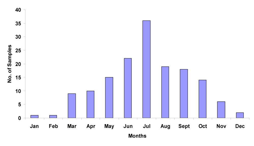
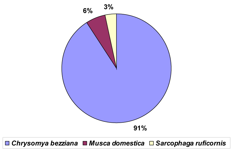
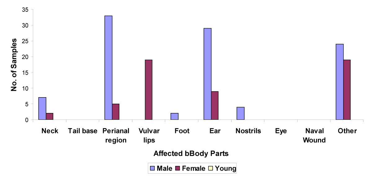
Myiasis has been found to have widespread occurrence among pet dogs throughout the year. A total of 153 cases of myiasis were reported in the pet dogs from the different districts of Punjab (India). Five seasons are recognized in the state of Punjab (India). These are: Summer (mid-April to the end of June), rainy (July to September), autumn (September to November), winter (December to February) and spring (March to mid-April) [20]. The incidence was found to be minimum during the winter season (December-February), followed by summer (April to June) and maximum during the rainy season (July-September) (Figure 1). The incidence of myiasis was highest (41.1%) during rainy season when temperature range (30-35ºC) and relative humidity (60-70%) were optimum for the abundant growth of myiasis causing flies and their larvae [21]. In dry hot season (April to June) and cold season (Dec-Feb) infestation incidences were low viz. 22.2% and 2.6% respectively. The old world screwworm fly- Chrysomya bezziana was found to be the predominating fly involved in causing myiasis among pet dogs as the maggots of this fly were encountered in the wounds on majority of animals (90.8%) (Figure 2). The present study confirmed the findings of the various previous records as Chrysomya bezziana had been reported to be the predominant fly responsible for causing myiasis among domestic animals [3,4,10-15]. In addition to Chrysomya bezziana, the maggots of the common house fly - Musca domestica were also found infesting the wounds of 5.8% animals. Least number (3.2%) of animals were found to be infested with the larvae of Sarcophaga ruficornis. Among dogs the wounds on body regions like neck, perianal region, vulvar lips and ear canal are the most susceptible sites for the onset of myiasis (Figure 3). The gums were found to be least affected because of the reflex act of the animals for waiving off the flies. Not even a single case of myiasis in young ones was reported.
Significant differences were found among the three fly species Chrysomya bezziana, Musca domestica and Sarcophaga ruficornis (p < 0.01) as causal agents of myiasis among pet dogs in the State of Punjab (India). The findings that the maggots of Chrysomya bezziana were encountered in the wounds on majority of animals can be attributed to the fact that the fly is well known obligatory myiasis causing species [1,10]. Due to its obligatory nature, eggs and larvae of the fly must grow within the body of live animals for the completion of its life cycle. Since temperature range for hatching of eggs and larval development of C. bezziana is 27-37ºC which is provided by the live animal only and not the carcasses [21]. The findings that the larvae of Musca domestica and Sarcophaga ruficornis were seen in the wounds of less number of animals, can be explained by the fact that the flies are facultative myiasis causing species [1]. It is not essential for these flies to lay their eggs within the body of live animals for the completion of their life cycle. The optimum temperature ranges for hatching of eggs and larval development for the flies Musca domestica and Sarcophaga ruficornis are 17-32ºC and 20-35ºC respectively which is prevalent in carcasses [22,23]. It may be rather a matter of chance that the fly happened to frequent live animal, that lead to oviposition and hence establishment of its larvae into the wounds [24]. Among dogs, the wounds on body regions like neck, perianal region, vulvar lips and ear canal are the most susceptible sites for the onset of myiasis (Figure 3) as the animal cannot lick these regions, whereas most of the other body regions are often licked and cleaned up from time to time. Moreover, the front parts the body of adult males like face, neck and nose are often injured in consequence of competitive fighting. Several other studies had reported the similar findings [6,12]. The body parts like vulvar lips and perianal region are more vulnerable to the attack of flies because these regions frequently come in contact with the ground while sitting and become soiled with excreta. These parts may easily get injured and thus become attractive for the myiasis causing flies. Highest cases of infestation of vulvar lips among females may also be attributed to the injury to the external genitalia during parturition [24]. Purulent discharge due to diseases like otitis externa contributes towards the infestation of ear canal [2]. Non reporting of myiasis among young pet dogs may be attributed to the fact that the young ones, kept as pets in homes, are given special care.
It is concluded that that Chrysomya bezziana is the most important myiasis-causing fly among the dogs sampled here, sometimes leading to severe damages. Inter-dog aggression, fights due to territorial behavior, diseases like otitis externa and weeping skin wounds with subsequent larval infestation are the key factors for occurrence of myiasis among dogs. The preventive measures like maintenance of neat and clean surroundings, control of fly populations, and covering of wounds can be helpful in prevention of onset of myiasis. Since the myiasis is a zoonotic disease and threats the human health, more studies are needed on the assessment of causative agents of myiasis. Undoubtedly, faunistic and ecological studies on these flies will help us to have better understanding about them and design effective control programs.
- Zumpt F. Myiasis in man and animals in the old world. Butterworths, London. 1965; 267. https://tinyurl.com/y2ohtjol
- Singh A, Singh Z. Incidence of Myiasis among humans- a review. Parasitol Res. 2015; 114: 3189-3199. DOI: 10.1007/S00436- 015- 4620-y
- Patton, WS. Cutaneous myiasis in man and animals in India. Indian Med Gaz. 1920; 12: 455-456. https://tinyurl.com/yxpnypge
- Rao MAN, Pillay MR. Some notes on cutaneous myiasis in animals in Madras Presidency. The Indian J Vet Sci Ani Husb. 1936; 6: 261-265. https://tinyurl.com/y3mtox2o
- Sen Gupta CM, Menon PB, Basu BC. Studies on myiasis and its treatment. Indian Vet J. 195127: 341 349. https://tinyurl.com/y3o7uu28
- Pattanayak PC, Misra SC. Studies on the epidemology, biology and control of bovine cutaneous myiasis flies. Orissa Vet J. 1977; 11: 52-57
- Haddow AJ, Thompson RCM. Sheep myiasis in South-west Scotland, with special reference to the species involved. Parasitol. 1937; 29: 96-99. DOI: https://doi.org/10.1017/S0031182000026238
- Macfarlane WV. Blowfly strike in Marlborough, New Zealand. New Zealand J Sci Tech. 1942; 23: 205
- Zumpt F. Myiasis in Man and Animals in Africa. SAJ Clin Sci. 1951; 2: 38-69. https://tinyurl.com/y6mnrfwb
- Norris KR, Murray MD. Notes on the screw-worm fly, Chrysomya bezziana as a pest of cattle in New Guinea. Technical paper of the division of entomology. CSIRO Australia. 1964; 6. https://tinyurl.com/y2qtcdtp
- Kumar R, Ruprah NS. Incidence and etiology of cutaneous myiasis in domestic animals at Hisar. Indian Vet J. 1984; 61: 918-921. https://tinyurl.com/yy5532xo
- Joseph SA, Karunamoorthy G, Lalitha CM. A study on the incidence of myiasis in domestic animals of Tamil Nadu, India. Cherion. 1987; 16: 175-178. https://tinyurl.com/yxog56c9
- Handa A. Cutaneous myiasis: Etiology, histopathology and treatment in sheep. M V Sc Thesis, CCS Haryana Agricultural University, Hisar. 1995
- Chemonges NS. Chrysomya bezziana in pet dogs in Hong Kong: A potential threat to Australia. Australian Vet J. 2003; 81: 202-205. DOI: 10.1111/j.1751-0813.2003.tb11471.x
- McNae JC, Lewis SJ. Retrospective Study of Old World screw-worm fly (Chrysomya bezziana) myiasis in 59 dogs in Hong Kong over a one year period. Australian Vet J. 2004; 82: 211-214. DOI: 10.1111/j.1751-0813.2004.tb12677.x
- Ribeiro BCC, SanavriaA, Helena H, Monteiro MS, Oliveira MO, de SouzaFS. An inquiry of cases of myiasis by Dermatobia hominis in dogs (Canis familiaris) of the Northern and Western zones of Rio de Janeiro city in 2000. Brazilian J Vet Res Ani Sci. 2003; 40: 144-150. DOI: https://dx.doi.org/10.1590/S1413-95962002000400002
- Ogo NI, Onovoh E, Ayodele DR, Ajayi OO, Chukwu CO, Sugun M, et al. Cutaneous myiasis in Jos metropolis of Plateau State, Nigeria, associated with Cordylobia anthropophaga. Vet Arhiv. 2009; 79: 293-299.
- Senior-White R, Aubertain D, Smart J. Fauna of British India including remainder of the oriental region, Diptera, Vol. VI Family Calliphoridae, Taylor and Francis. London. 1940.
- Singh I. Taxonomic studies on the family Calliphoridae (Diptera: Cyclorrhapha) from the north-west India. Ph.D. Thesis, Punjabi University, Patiala. 2002.
- Mavi HS, Tiwana DS. Geography of Punjab. India: National Book Trust. 1993. https://tinyurl.com/yypb3h5a
- Spradbery JP. A manual for the diagnosis of screw-worm fly. Department of Agriculture, Fisheries and Forestry, Australia. 2002. https://tinyurl.com/y6ojka6j
- Wang Y, Yang L, Y Zhang, Tao L, Wang J. Development of Musca domestica at constant temperatures and the first case report of its application for estimating the minimum postmortem interval. Forensic Sci Int. 2018; 285: 172-180. DOI: 10.1016/j.forsciint.2018.02.004
- Fakoorziba MR, Assareh M, Keshavarzi D, Soltani A ‚ Fard MDM, Zarenezhad M. New Record of Sarcophaga ruficornis, Fabricius, 1794 (Diptera: Sarcophagidae) from Iran, A Flesh Fly Species of Medical and Forensic Importance. J Forensic Sci Criminal Inves. 2017; 3: 555602. DOI: 10.19080/JFSCI.2017.03.555602
- Singh A, Singh D. A study on the incidence of myiasis among dairy animals in the State of Punjab, India. IOSR J Agri Vet Sci. 2016; 9: 30-34. https://tinyurl.com/y5cwk4xv
Content Alerts
SignUp to our
Content alerts.
 This work is licensed under a Creative Commons Attribution 4.0 International License.


